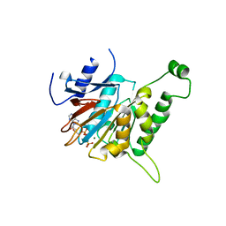
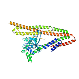
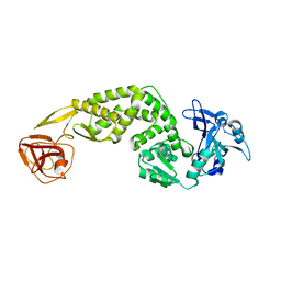
5YK2
 
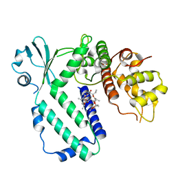 | |
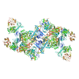
7EIZ
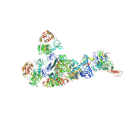 | | Coupling of N7-methyltransferase and 3'-5' exoribonuclease with SARS-CoV-2 polymerase reveals mechanisms for capping and proofreading | | 分子名称: | Helicase, MAGNESIUM ION, Non-structural protein 10, ... | | 著者 | Yan, L, Yang, Y.X, Li, M.Y, Zhang, Y, Zheng, L.T, Ge, J, Huang, Y.C, Liu, Z.Y, Wang, T, Gao, S, Zhang, R, Huang, Y.Y, Guddat, L.W, Gao, Y, Rao, Z.H, Lou, Z.Y. | | 登録日 | 2021-04-01 | | 公開日 | 2021-09-22 | | 最終更新日 | 2023-07-26 | | 実験手法 | ELECTRON MICROSCOPY | | 主引用文献 | Coupling of N7-methyltransferase and 3'-5' exoribonuclease with SARS-CoV-2 polymerase reveals mechanisms for capping and proofreading
Cell, 184, 2021
|
|
5ZZ8
 | | Structure of the Herpes simplex virus type 2 C-capsid with capsid-vertex-specific component | | 分子名称: | Major capsid protein, UL17, UL25, ... | | 著者 | Wang, J.L, Yuan, S, Zhu, D.J, Tang, H, Wang, N, Chen, W.Y, Gao, Q, Li, Y.H, Wang, J.Z, Liu, H.R, Zhang, X.Z, Rao, Z.H, Wang, X.X. | | 登録日 | 2018-05-31 | | 公開日 | 2018-10-10 | | 最終更新日 | 2019-11-06 | | 実験手法 | ELECTRON MICROSCOPY (3.75 Å) | | 主引用文献 | Structure of the herpes simplex virus type 2 C-capsid with capsid-vertex-specific component.
Nat Commun, 9, 2018
|
|
6AKT
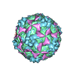 | | Cryo-EM structure of CVA10 A-particle | | 分子名称: | VP1, VP2, VP3 | | 著者 | Zhu, L, Sun, Y, Fan, J.Y, Zhu, B, Cao, L, Gao, Q, Zhang, Y.J, Liu, H.R, Rao, Z.H, Wang, X.X. | | 登録日 | 2018-09-03 | | 公開日 | 2019-01-16 | | 最終更新日 | 2024-03-27 | | 実験手法 | ELECTRON MICROSCOPY (2.8 Å) | | 主引用文献 | Structures of Coxsackievirus A10 unveil the molecular mechanisms of receptor binding and viral uncoating.
Nat Commun, 9, 2018
|
|
6AKS
 | | Cryo-EM structure of CVA10 mature virus | | 分子名称: | SPHINGOSINE, VP1, VP2, ... | | 著者 | Zhu, L, Sun, Y, Fan, J.Y, Zhu, B, Cao, L, Gao, Q, Zhang, Y.J, Liu, H.R, Rao, Z.H, Wang, X.X. | | 登録日 | 2018-09-03 | | 公開日 | 2019-01-16 | | 最終更新日 | 2024-03-27 | | 実験手法 | ELECTRON MICROSCOPY (3 Å) | | 主引用文献 | Structures of Coxsackievirus A10 unveil the molecular mechanisms of receptor binding and viral uncoating.
Nat Commun, 9, 2018
|
|
7EGQ
 | | Co-transcriptional capping machineries in SARS-CoV-2 RTC: Coupling of N7-methyltransferase and 3'-5' exoribonuclease with polymerase reveals mechanisms for capping and proofreading | | 分子名称: | Helicase, MAGNESIUM ION, Non-structural protein 10, ... | | 著者 | Yan, L.M, Yang, Y.X, Li, M.Y, Zhang, Y, Zheng, L.T, Ge, J, Huang, Y.C, Liu, Z.Y, Wang, T, Gao, S, Zhang, R, Huang, Y.Y, Guddat, L.W, Gao, Y, Rao, Z.H, Lou, Z.Y. | | 登録日 | 2021-03-25 | | 公開日 | 2021-07-21 | | 最終更新日 | 2024-06-05 | | 実験手法 | ELECTRON MICROSCOPY (3.35 Å) | | 主引用文献 | Coupling of N7-methyltransferase and 3'-5' exoribonuclease with SARS-CoV-2 polymerase reveals mechanisms for capping and proofreading.
Cell, 184, 2021
|
|
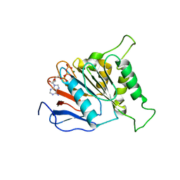
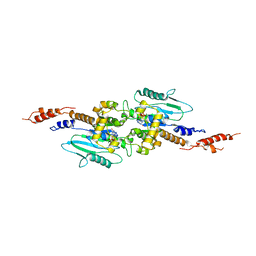
3PDV
 | |
6J19
 | | ATPase | | 分子名称: | ADENOSINE-5'-TRIPHOSPHATE, ESAT-6-like protein EsxB, ESX-1 secretion system protein EccCb1, ... | | 著者 | Wang, S.H, Li, J, Rao, Z.H. | | 登録日 | 2018-12-28 | | 公開日 | 2019-12-04 | | 最終更新日 | 2023-11-22 | | 実験手法 | X-RAY DIFFRACTION (1.978 Å) | | 主引用文献 | Structural insights into substrate recognition by the type VII secretion system.
Protein Cell, 11, 2020
|
|
6J18
 | | ATPase | | 分子名称: | ADENOSINE-5'-TRIPHOSPHATE, ESX-5 secretion system protein EccC5, MAGNESIUM ION | | 著者 | Wang, S.H, Li, J, Rao, Z.H. | | 登録日 | 2018-12-28 | | 公開日 | 2019-12-04 | | 最終更新日 | 2023-11-22 | | 実験手法 | X-RAY DIFFRACTION (2 Å) | | 主引用文献 | Structural insights into substrate recognition by the type VII secretion system.
Protein Cell, 11, 2020
|
|
6JD5
 | | ATPase | | 分子名称: | ADENOSINE-5'-TRIPHOSPHATE, ESX conserved component EccC2. ESX-2 type VII secretion system protein. Possible membrane protein, MAGNESIUM ION | | 著者 | Wang, S.H, Li, J, Rao, Z.H. | | 登録日 | 2019-01-31 | | 公開日 | 2019-12-04 | | 最終更新日 | 2023-11-22 | | 実験手法 | X-RAY DIFFRACTION (2.2 Å) | | 主引用文献 | Structural insights into substrate recognition by the type VII secretion system.
Protein Cell, 11, 2020
|
|
6J17
 | | ATPase | | 分子名称: | ADENOSINE-5'-TRIPHOSPHATE, ESX-3 secretion system protein EccC3, MAGNESIUM ION | | 著者 | Wang, S.H, Li, J, Rao, Z.H. | | 登録日 | 2018-12-28 | | 公開日 | 2019-12-04 | | 最終更新日 | 2023-11-22 | | 実験手法 | X-RAY DIFFRACTION (1.975 Å) | | 主引用文献 | Structural insights into substrate recognition by the type VII secretion system.
Protein Cell, 11, 2020
|
|
6J72
 | | Crystal structure of IniA from Mycobacterium smegmatis with GTP bound | | 分子名称: | GUANOSINE-5'-TRIPHOSPHATE, Isoniazid inducible gene protein IniA, L(+)-TARTARIC ACID, ... | | 著者 | Wang, M.F, Guo, X.Y, Hu, J.J, Li, J, Rao, Z.H. | | 登録日 | 2019-01-16 | | 公開日 | 2019-09-11 | | 最終更新日 | 2024-03-27 | | 実験手法 | X-RAY DIFFRACTION (2.2 Å) | | 主引用文献 | Mycobacterial dynamin-like protein IniA mediates membrane fission.
Nat Commun, 10, 2019
|
|
6JD4
 | | ATPase | | 分子名称: | ADENOSINE-5'-TRIPHOSPHATE, ESX-1 secretion system protein EccCb1, MAGNESIUM ION | | 著者 | Wang, S.H, Li, J, Rao, Z.H. | | 登録日 | 2019-01-31 | | 公開日 | 2019-12-04 | | 最終更新日 | 2023-11-22 | | 実験手法 | X-RAY DIFFRACTION (2.1 Å) | | 主引用文献 | Structural insights into substrate recognition by the type VII secretion system.
Protein Cell, 11, 2020
|
|
6J73
 | | Crystal structure of IniA from Mycobacterium smegmatis | | 分子名称: | Isoniazid inducible gene protein IniA | | 著者 | Wang, M.F, Guo, X.Y, Hu, J.J, Li, J, Rao, Z.H. | | 登録日 | 2019-01-16 | | 公開日 | 2019-09-11 | | 最終更新日 | 2024-03-27 | | 実験手法 | X-RAY DIFFRACTION (3.211 Å) | | 主引用文献 | Mycobacterial dynamin-like protein IniA mediates membrane fission.
Nat Commun, 10, 2019
|
|
5YEW
 | | Structural basis for GTP hydrolysis and conformational change of mitofusin 1 in mediating mitochondrial fusion | | 分子名称: | BERYLLIUM TRIFLUORIDE ION, GUANOSINE-5'-DIPHOSPHATE, MAGNESIUM ION, ... | | 著者 | Yan, L, Qi, Y, Huang, X, Yu, C. | | 登録日 | 2017-09-20 | | 公開日 | 2018-01-31 | | 最終更新日 | 2024-03-06 | | 実験手法 | X-RAY DIFFRACTION (3.2 Å) | | 主引用文献 | Structural basis for GTP hydrolysis and conformational change of MFN1 in mediating membrane fusion
Nat. Struct. Mol. Biol., 25, 2018
|
|
3T35
 | | Arabidopsis thaliana dynamin-related protein 1A in postfission state | | 分子名称: | Dynamin-related protein 1A, LINKER, GUANOSINE-5'-DIPHOSPHATE | | 著者 | Yan, L.M, Ma, Y.Y, Sun, Y.N, Lou, Z.Y. | | 登録日 | 2011-07-24 | | 公開日 | 2012-06-06 | | 最終更新日 | 2023-11-01 | | 実験手法 | X-RAY DIFFRACTION (3.592 Å) | | 主引用文献 | Structural basis for mechanochemical role of Arabidopsis thaliana dynamin-related protein in membrane fission
J Mol Cell Biol, 3, 2011
|
|
7FA1
 | | Crystal Structure of N-terminus of the non-structural protein 2 from SARS coronavirus | | 分子名称: | Non-structural protein 2, ZINC ION | | 著者 | Li, Y.Y, Ren, Z.L, Bao, Z.H, Ming, Z.H, Yan, L.M, Lou, Z.Y, Rao, Z.H. | | 登録日 | 2021-07-05 | | 公開日 | 2022-08-10 | | 最終更新日 | 2024-02-28 | | 実験手法 | X-RAY DIFFRACTION (1.6 Å) | | 主引用文献 | The Life of SARS-CoV-2 Inside Cells: Replication-Transcription Complex Assembly and Function.
Annu.Rev.Biochem., 91, 2022
|
|
7FAC
 | | Crystal Structure of C-terminus of the non-structural protein 2 from SARS coronavirus | | 分子名称: | Non-structural protein 2, ZINC ION | | 著者 | Li, Y.Y, Ren, Z.L, Bao, Z.H, Ming, Z.H, Yan, L.M, Lou, Z.Y, Rao, Z.H. | | 登録日 | 2021-07-06 | | 公開日 | 2022-08-10 | | 最終更新日 | 2024-02-28 | | 実験手法 | X-RAY DIFFRACTION (2.71 Å) | | 主引用文献 | The Life of SARS-CoV-2 Inside Cells: Replication-Transcription Complex Assembly and Function.
Annu.Rev.Biochem., 91, 2022
|
|
3T34
 | | Arabidopsis thaliana dynamin-related protein 1A (AtDRP1A) in prefission state | | 分子名称: | Dynamin-related protein 1A, LINKER, GUANOSINE-5'-DIPHOSPHATE, ... | | 著者 | Yan, L.M, Ma, Y.Y, Sun, Y.N, Lou, Z.Y. | | 登録日 | 2011-07-24 | | 公開日 | 2012-06-06 | | 最終更新日 | 2023-11-01 | | 実験手法 | X-RAY DIFFRACTION (2.405 Å) | | 主引用文献 | Structural basis for mechanochemical role of Arabidopsis thaliana dynamin-related protein in membrane fission
J Mol Cell Biol, 3, 2011
|
|
5F5M
 | | Crystal structure of Marburg virus nucleoprotein core domain | | 分子名称: | Nucleoprotein | | 著者 | Guo, Y, Liu, B.C, Liu, X, Li, G.B, Wang, W.M, Dong, S.S, Wang, W.J. | | 登録日 | 2015-12-04 | | 公開日 | 2017-05-31 | | 最終更新日 | 2024-03-20 | | 実験手法 | X-RAY DIFFRACTION (2.902 Å) | | 主引用文献 | Structural Insight into Nucleoprotein Conformation Change Chaperoned by VP35 Peptide in Marburg Virus
J. Virol., 91, 2017
|
|
5F5O
 | | Crystal structure of Marburg virus nucleoprotein core domain bound to VP35 regulation peptide | | 分子名称: | Nucleoprotein, Peptide from Polymerase cofactor VP35, SULFATE ION | | 著者 | Guo, Y, Liu, B.C, Liu, X, Li, G.B, Wang, W.M, Dong, S.S, Wang, W.J. | | 登録日 | 2015-12-04 | | 公開日 | 2017-05-31 | | 最終更新日 | 2024-03-20 | | 実験手法 | X-RAY DIFFRACTION (2.2 Å) | | 主引用文献 | Structural Insight into Nucleoprotein Conformation Change Chaperoned by VP35 Peptide in Marburg Virus
J. Virol., 91, 2017
|
|
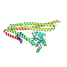
4JOD
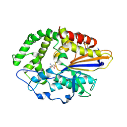 | |
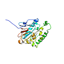
4JOB
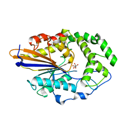 | |
4JOC
 | |
4JR6
 | | Crystal structure of DsbA from Mycobacterium tuberculosis (reduced) | | 分子名称: | Possible conserved membrane or secreted protein, SULFATE ION | | 著者 | Wang, L. | | 登録日 | 2013-03-21 | | 公開日 | 2013-07-17 | | 最終更新日 | 2017-11-15 | | 実験手法 | X-RAY DIFFRACTION (1.902 Å) | | 主引用文献 | Structure analysis of the extracellular domain reveals disulfide bond forming-protein properties of Mycobacterium tuberculosis Rv2969c.
Protein Cell, 4, 2013
|
|


