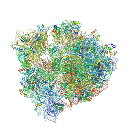
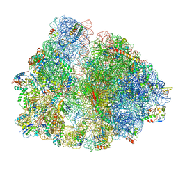
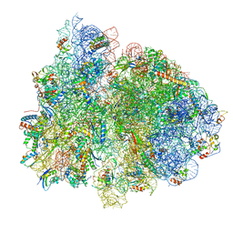
7PIR
 
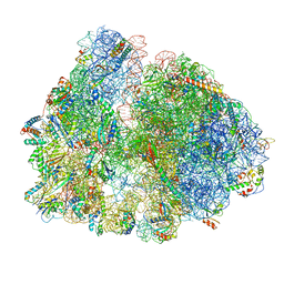 | | 70S ribosome with A*- and P/E-site tRNAs in pseudouridimycin-treated Mycoplasma pneumoniae cells | | 分子名称: | 16S ribosomal RNA, 23S ribosomal RNA, 30S ribosomal protein S10, ... | | 著者 | Xue, L, Lenz, S, Rappsilber, J, Mahamid, J. | | 登録日 | 2021-08-23 | | 公開日 | 2022-05-25 | | 最終更新日 | 2022-10-19 | | 実験手法 | ELECTRON MICROSCOPY (12.1 Å) | | 主引用文献 | Visualizing translation dynamics at atomic detail inside a bacterial cell.
Nature, 610, 2022
|
|
6HY4
 | |
6NBG
 | | 2.05 Angstrom Resolution Crystal Structure of Hypothetical Protein KP1_5497 from Klebsiella pneumoniae. | | 分子名称: | CHLORIDE ION, Glucosamine-6-phosphate deaminase, PHOSPHATE ION | | 著者 | Minasov, G, Shuvalova, L, Kiryukhina, O, Dubrovska, I, Satchell, K.J.F, Joachimiak, A, Center for Structural Genomics of Infectious Diseases (CSGID) | | 登録日 | 2018-12-07 | | 公開日 | 2018-12-19 | | 最終更新日 | 2023-06-14 | | 実験手法 | X-RAY DIFFRACTION (2.05 Å) | | 主引用文献 | A Structural Systems Biology Approach to High-Risk CG23 Klebsiella pneumoniae.
Microbiol Resour Announc, 12, 2023
|
|
6NKJ
 | | 1.3 Angstrom Resolution Crystal Structure of UDP-N-acetylglucosamine 1-carboxyvinyltransferase from Streptococcus pneumoniae in Complex with (2R)-2-(phosphonooxy)propanoic acid. | | 分子名称: | (2R)-2-(phosphonooxy)propanoic acid, 1,2-ETHANEDIOL, CHLORIDE ION, ... | | 著者 | Minasov, G, Shuvalova, L, Dubrovska, I, Kiryukhina, O, Grimshaw, S, Kwon, K, Anderson, W.F, Satchell, K.J.F, Joachimiak, A, Center for Structural Genomics of Infectious Diseases (CSGID) | | 登録日 | 2019-01-07 | | 公開日 | 2019-01-16 | | 最終更新日 | 2023-10-11 | | 実験手法 | X-RAY DIFFRACTION (1.3 Å) | | 主引用文献 | 1.3 Angstrom Resolution Crystal Structure of UDP-N-acetylglucosamine 1-carboxyvinyltransferase from Streptococcus pneumoniae in Complex with (2R)-2-(phosphonooxy)propanoic acid.
To Be Published
|
|
3DYD
 | | Human Tyrosine Aminotransferase | | 分子名称: | PYRIDOXAL-5'-PHOSPHATE, Tyrosine aminotransferase | | 著者 | Karlberg, T, Moche, M, Andersson, J, Arrowsmith, C.H, Berglund, H, Collins, R, Dahlgren, L.G, Edwards, A.M, Flodin, S, Flores, A, Graslund, S, Hammarstrom, M, Johansson, A, Johansson, I, Kotenyova, T, Lehtio, L, Nilsson, M.E, Nordlund, P, Nyman, T, Olesen, K, Persson, C, Sagemark, J, Thorsell, A.G, Tresaugues, L, Van Den Berg, S, Weigelt, J, Welin, M, Wikstrom, M, Wisniewska, M, Schuler, H, Structural Genomics Consortium (SGC) | | 登録日 | 2008-07-27 | | 公開日 | 2008-08-19 | | 最終更新日 | 2023-08-30 | | 実験手法 | X-RAY DIFFRACTION (2.3 Å) | | 主引用文献 | Human Tyrosine Aminotransferase
To be Published
|
|
6NMC
 | | CryoEM structure of the LbCas12a-crRNA-2xAcrVA1 complex | | 分子名称: | AcrVA1, Cpf1, MAGNESIUM ION, ... | | 著者 | Chang, L, Li, Z, Zhang, H. | | 登録日 | 2019-01-10 | | 公開日 | 2019-06-12 | | 最終更新日 | 2024-03-20 | | 実験手法 | ELECTRON MICROSCOPY (4.24 Å) | | 主引用文献 | Structural Basis for the Inhibition of CRISPR-Cas12a by Anti-CRISPR Proteins.
Cell Host Microbe, 25, 2019
|
|
1RAX
 | |
7WQV
 | | Crystal structure of a neutralizing monoclonal antibody (Ab08) in complex with SARS-CoV-2 receptor-binding domain (RBD) | | 分子名称: | (4S)-2-METHYL-2,4-PENTANEDIOL, 2-acetamido-2-deoxy-beta-D-glucopyranose, Ab08, ... | | 著者 | Zha, J, Meng, L, Zhang, X, Li, D. | | 登録日 | 2022-01-26 | | 公開日 | 2023-01-25 | | 最終更新日 | 2023-11-29 | | 実験手法 | X-RAY DIFFRACTION (2.8 Å) | | 主引用文献 | A Spike-destructing human antibody effectively neutralizes Omicron-included SARS-CoV-2 variants with therapeutic efficacy.
Plos Pathog., 19, 2023
|
|
6MUV
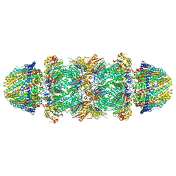 | | The structure of the Plasmodium falciparum 20S proteasome in complex with two PA28 activators | | 分子名称: | 20S proteasome alpha-1 subunit, 20S proteasome alpha-2 subunit, 20S proteasome alpha-3 subunit, ... | | 著者 | Metcalfe, R.D, Xie, S.C, Hanssen, E, Gillett, D.L, Leis, A.P, Tilley, L, Griffin, M.D.W. | | 登録日 | 2018-10-23 | | 公開日 | 2019-08-07 | | 最終更新日 | 2024-03-13 | | 実験手法 | ELECTRON MICROSCOPY (3.8 Å) | | 主引用文献 | The structure of the PA28-20S proteasome complex from Plasmodium falciparum and implications for proteostasis.
Nat Microbiol, 4, 2019
|
|
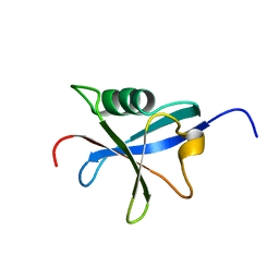
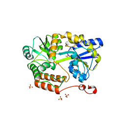
3G9Q
 | | Crystal structure of the FhuD fold-family BSU3320, a periplasmic binding protein component of a Fep/Fec-like ferrichrome ABC transporter from Bacillus subtilis. Northeast Structural Genomics Consortium Target SR577A | | 分子名称: | Ferrichrome-binding protein | | 著者 | Forouhar, F, Neely, H, Seetharaman, J, Janjua, H, Mao, L, Xiao, R, Ciccosanti, C, Foote, E.L, Lee, D, Nair, R, Acton, T.B, Rost, B, Montelione, G.T, Tong, L, Hunt, J.F, Northeast Structural Genomics Consortium (NESG) | | 登録日 | 2009-02-13 | | 公開日 | 2009-03-03 | | 最終更新日 | 2017-11-01 | | 実験手法 | X-RAY DIFFRACTION (2.6 Å) | | 主引用文献 | Crystal structure of the FhuD fold-family BSU3320, a periplasmic binding protein component of a Fep/Fec-like ferrichrome ABC transporter from Bacillus subtilis. Northeast Structural Genomics Consortium Target SR577A.
To be Published
|
|
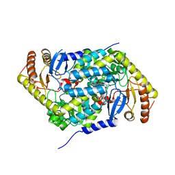
6N0I
 | | 2.60 Angstrom Resolution Crystal Structure of Elongation Factor G 2 from Pseudomonas putida. | | 分子名称: | DI(HYDROXYETHYL)ETHER, Elongation factor G 2, SULFATE ION | | 著者 | Minasov, G, Shuvalova, L, Wawrzak, Z, Cardona-Correa, A, Anderson, W.F, Satchell, K.J.F, Joachimiak, A, Center for Structural Genomics of Infectious Diseases (CSGID) | | 登録日 | 2018-11-07 | | 公開日 | 2018-11-14 | | 最終更新日 | 2023-10-11 | | 実験手法 | X-RAY DIFFRACTION (2.6 Å) | | 主引用文献 | 2.60 Angstrom Resolution Crystal Structure of Elongation Factor G 2 from Pseudomonas putida.
To Be Published
|
|
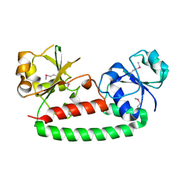
6N13
 | | UbcH7-Ub Complex with R0RBR Parkin and phosphoubiquitin | | 分子名称: | E3 ubiquitin-protein ligase parkin, Ubiquitin-conjugating enzyme E2 L3, ZINC ION, ... | | 著者 | Condos, T.E.C, Dunkerley, K.M, Freeman, E.A, Barber, K.R, Aguirre, J.D, Chaugule, V.K, Xiao, Y, Konermann, L, Walden, H, Shaw, G.S. | | 登録日 | 2018-11-08 | | 公開日 | 2018-11-28 | | 最終更新日 | 2020-01-08 | | 実験手法 | SOLUTION NMR | | 主引用文献 | Synergistic recruitment of UbcH7~Ub and phosphorylated Ubl domain triggers parkin activation.
EMBO J., 37, 2018
|
|
6HRH
 | | Structure of human erythroid-specific 5'-aminolevulinate synthase, ALAS2 | | 分子名称: | 5-aminolevulinate synthase, erythroid-specific, mitochondrial, ... | | 著者 | Bailey, H.J, Shrestha, L, Rembeza, E, Newman, J, Kupinska, K, Diaz-saez, L, Kennedy, E, Burgess-Brown, N, von Delft, F, Arrowsmith, C, Edwards, A, Bountra, C, Yue, W.W, Structural Genomics Consortium (SGC) | | 登録日 | 2018-09-27 | | 公開日 | 2018-11-07 | | 最終更新日 | 2024-01-24 | | 実験手法 | X-RAY DIFFRACTION (2.3 Å) | | 主引用文献 | Structure of human erythroid-specific 5'-aminolevulinate synthase, ALAS2
To Be Published
|
|
6N84
 | | MBP-fusion protein of transducin-alpha residues 327-350 | | 分子名称: | Maltose/maltodextrin-binding periplasmic protein,Guanine nucleotide-binding protein G(t) subunit alpha-2, SULFATE ION, alpha-D-glucopyranose-(1-4)-alpha-D-glucopyranose | | 著者 | Srivastava, D, Gakhar, L, Artemyev, N.O. | | 登録日 | 2018-11-28 | | 公開日 | 2019-07-10 | | 最終更新日 | 2024-01-10 | | 実験手法 | X-RAY DIFFRACTION (1.75 Å) | | 主引用文献 | Structural underpinnings of Ric8A function as a G-protein alpha-subunit chaperone and guanine-nucleotide exchange factor.
Nat Commun, 10, 2019
|
|
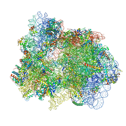
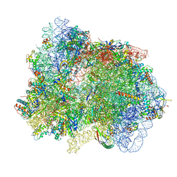
6N1D
 | | X-ray Crystal complex showing Spontaneous Ribosomal Translocation of mRNA and tRNAs into a Chimeric Hybrid State | | 分子名称: | 16S rRNA, 23S rRNA, 30S ribosomal protein S10, ... | | 著者 | Noller, H.F, Donohue, J.P, Lancaster, L, Zhou, J. | | 登録日 | 2018-11-08 | | 公開日 | 2019-04-03 | | 最終更新日 | 2023-10-11 | | 実験手法 | X-RAY DIFFRACTION (3.2 Å) | | 主引用文献 | Spontaneous ribosomal translocation of mRNA and tRNAs into a chimeric hybrid state.
Proc. Natl. Acad. Sci. U.S.A., 116, 2019
|
|
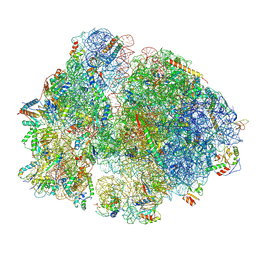
7PI8
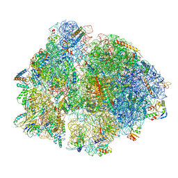 | | 70S ribosome with P-site tRNA in spectinomycin-treated Mycoplasma pneumoniae cells | | 分子名称: | 16S ribosomal RNA, 23S ribosomal RNA, 30S ribosomal protein S10, ... | | 著者 | Xue, L, Lenz, S, Rappsilber, J, Mahamid, J. | | 登録日 | 2021-08-19 | | 公開日 | 2022-05-25 | | 最終更新日 | 2022-10-19 | | 実験手法 | ELECTRON MICROSCOPY (8.9 Å) | | 主引用文献 | Visualizing translation dynamics at atomic detail inside a bacterial cell.
Nature, 610, 2022
|
|
7PIC
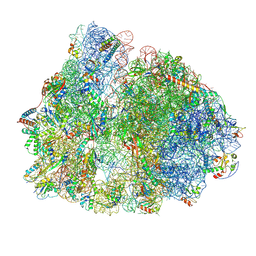 | | 70S ribosome with P/E-site tRNA in spectinomycin-treated Mycoplasma pneumoniae cells | | 分子名称: | 16S ribosomal RNA, 23S ribosomal RNA, 30S ribosomal protein S10, ... | | 著者 | Xue, L, Lenz, S, Rappsilber, J, Mahamid, J. | | 登録日 | 2021-08-19 | | 公開日 | 2022-05-25 | | 最終更新日 | 2022-10-19 | | 実験手法 | ELECTRON MICROSCOPY (9.1 Å) | | 主引用文献 | Visualizing translation dynamics at atomic detail inside a bacterial cell.
Nature, 610, 2022
|
|
7PIA
 | | 70S ribosome with A/P- and P/E-site tRNAs in spectinomycin-treated Mycoplasma pneumoniae cells | | 分子名称: | 16S ribosomal RNA, 23S ribosomal RNA, 30S ribosomal protein S10, ... | | 著者 | Xue, L, Lenz, S, Rappsilber, J, Mahamid, J. | | 登録日 | 2021-08-19 | | 公開日 | 2022-05-25 | | 最終更新日 | 2022-10-19 | | 実験手法 | ELECTRON MICROSCOPY (13.6 Å) | | 主引用文献 | Visualizing translation dynamics at atomic detail inside a bacterial cell.
Nature, 610, 2022
|
|
7PIS
 | | 70S ribosome with EF-G, A*- and P/E-site tRNAs in pseudouridimycin-treated Mycoplasma pneumoniae cells | | 分子名称: | 16S ribosomal RNA, 23S ribosomal RNA, 30S ribosomal protein S10, ... | | 著者 | Xue, L, Lenz, S, Rappsilber, J, Mahamid, J. | | 登録日 | 2021-08-23 | | 公開日 | 2022-05-25 | | 最終更新日 | 2022-10-19 | | 実験手法 | ELECTRON MICROSCOPY (15 Å) | | 主引用文献 | Visualizing translation dynamics at atomic detail inside a bacterial cell.
Nature, 610, 2022
|
|
7PIQ
 | | 70S ribosome with A- and P-site tRNAs in pseudouridimycin-treated Mycoplasma pneumoniae cells | | 分子名称: | 16S ribosomal RNA, 23S ribosomal RNA, 30S ribosomal protein S10, ... | | 著者 | Xue, L, Lenz, S, Rappsilber, J, Mahamid, J. | | 登録日 | 2021-08-23 | | 公開日 | 2022-05-25 | | 最終更新日 | 2022-10-19 | | 実験手法 | ELECTRON MICROSCOPY (9.7 Å) | | 主引用文献 | Visualizing translation dynamics at atomic detail inside a bacterial cell.
Nature, 610, 2022
|
|
1L3N
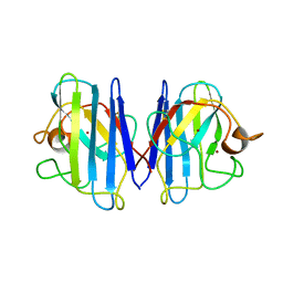 | | The Solution Structure of Reduced Dimeric Copper Zinc SOD: the Structural Effects of Dimerization | | 分子名称: | COPPER (I) ION, ZINC ION, superoxide dismutase [Cu-Zn] | | 著者 | Banci, L, Bertini, I, Cramaro, F, Del Conte, R, Viezzoli, M.S. | | 登録日 | 2002-02-28 | | 公開日 | 2002-05-08 | | 最終更新日 | 2021-10-27 | | 実験手法 | SOLUTION NMR | | 主引用文献 | The solution structure of reduced dimeric copper zinc superoxide dismutase. The structural effects of dimerization
Eur.J.Biochem., 269, 2002
|
|
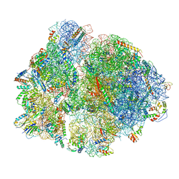
7PHA
 | | 70S ribosome with EF-Tu-tRNA and P-site tRNA in chloramphenicol-treated Mycoplasma pneumoniae cells | | 分子名称: | 16S ribosomal RNA, 23S ribosomal RNA, 30S ribosomal protein S10, ... | | 著者 | Xue, L, Lenz, S, Rappsilber, J, Mahamid, J. | | 登録日 | 2021-08-16 | | 公開日 | 2022-05-25 | | 最終更新日 | 2022-10-19 | | 実験手法 | ELECTRON MICROSCOPY (8.5 Å) | | 主引用文献 | Visualizing translation dynamics at atomic detail inside a bacterial cell.
Nature, 610, 2022
|
|
7PIT
 | | 70S ribosome with EF-G, A/P- and P/E-site tRNAs in pseudouridimycin-treated Mycoplasma pneumoniae cells | | 分子名称: | 16S ribosomal RNA, 23S ribosomal RNA, 30S ribosomal protein S10, ... | | 著者 | Xue, L, Lenz, S, Rappsilber, J, Mahamid, J. | | 登録日 | 2021-08-23 | | 公開日 | 2022-05-25 | | 最終更新日 | 2022-11-23 | | 実験手法 | ELECTRON MICROSCOPY (5.7 Å) | | 主引用文献 | Visualizing translation dynamics at atomic detail inside a bacterial cell.
Nature, 610, 2022
|
|
7PIO
 | | 70S ribosome with P-site tRNA in pseudouridimycin-treated Mycoplasma pneumoniae cells | | 分子名称: | 16S ribosomal RNA, 23S ribosomal RNA, 30S ribosomal protein S10, ... | | 著者 | Xue, L, Lenz, S, Rappsilber, J, Mahamid, J. | | 登録日 | 2021-08-23 | | 公開日 | 2022-05-25 | | 最終更新日 | 2022-10-19 | | 実験手法 | ELECTRON MICROSCOPY (9.5 Å) | | 主引用文献 | Visualizing translation dynamics at atomic detail inside a bacterial cell.
Nature, 610, 2022
|
|
6MYV
 | | Sialidase26 co-crystallized with DANA-Gc | | 分子名称: | 2,6-anhydro-3,5-dideoxy-5-[(hydroxyacetyl)amino]-D-glycero-L-altro-non-2-enonic acid, Sialidase26 | | 著者 | Zaramela, L.S, Martino, C, Alisson-Silva, F, Rees, S.D, Diaz, S.L, Chuzel, L, Ganatra, M.B, Taron, C.H, Zuniga, C, Huang, J, Siegel, D, Chang, G, Varki, A, Zengler, K. | | 登録日 | 2018-11-02 | | 公開日 | 2019-10-02 | | 最終更新日 | 2023-10-11 | | 実験手法 | X-RAY DIFFRACTION (2.2 Å) | | 主引用文献 | Gut bacteria responding to dietary change encode sialidases that exhibit preference for red meat-associated carbohydrates.
Nat Microbiol, 4, 2019
|
|


