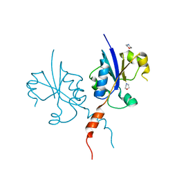
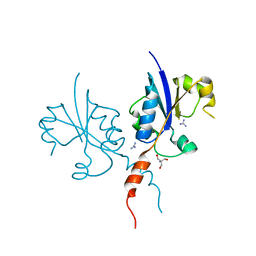
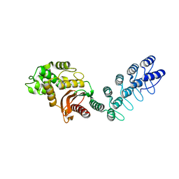
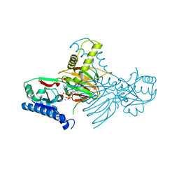
7Q9W
 
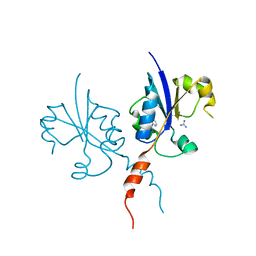 | | Crystal structure of the C-terminal catalytic domain of Plasmodium falciparum CTP:phosphocholine cytidylyltransferase with 4-(aminomethyl)pyridin-2-amine | | 分子名称: | 4-(aminomethyl)pyridin-2-amine, Cholinephosphate cytidylyltransferase, Guanidinium | | 著者 | Duclovel, C, Gelin, M, Krimm, I, Cerdan, R, Guichou, J.-F. | | 登録日 | 2021-11-15 | | 公開日 | 2022-11-30 | | 最終更新日 | 2024-01-31 | | 実験手法 | X-RAY DIFFRACTION (1.8 Å) | | 主引用文献 | Crystallographic screening using ultra-low-molecular-weight ligands to guide drug design of PfCCT inhibitors.
To Be Published
|
|
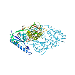
7QA7
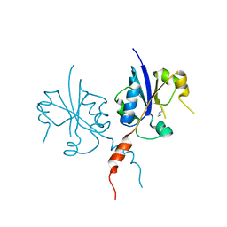 | | Crystal structure of the C-terminal catalytic domain of Plasmodium falciparum CTP:phosphocholine cytidylyltransferase with Cyclopropanemethylamine | | 分子名称: | Cholinephosphate cytidylyltransferase, cyclopropylmethanamine | | 著者 | Duclovel, C, Gelin, M, Krimm, I, Cerdan, R, Guichou, J.-F. | | 登録日 | 2021-11-16 | | 公開日 | 2022-11-30 | | 最終更新日 | 2024-01-31 | | 実験手法 | X-RAY DIFFRACTION (2.36 Å) | | 主引用文献 | Crystallographic screening using ultra-low-molecular-weight ligands to guide drug design of PfCCT inhibitors
To Be Published
|
|
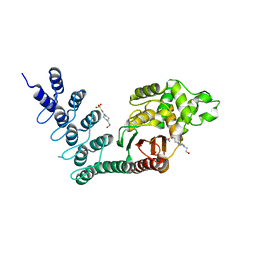
7Q9V
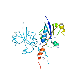 | | Crystal structure of the C-terminal catalytic domain of Plasmodium falciparum CTP:phosphocholine cytidylyltransferase with 1-(1,3-oxazol-4-yl)methanamine | | 分子名称: | 1,3-oxazol-4-ylmethanamine, Cholinephosphate cytidylyltransferase, Guanidinium | | 著者 | Duclovel, C, Gelin, M, Krimm, I, Cerdan, R, Guichou, J.-F. | | 登録日 | 2021-11-15 | | 公開日 | 2022-11-30 | | 最終更新日 | 2024-01-31 | | 実験手法 | X-RAY DIFFRACTION (1.5 Å) | | 主引用文献 | Crystallographic screening using ultra-low-molecular-weight ligands to guide drug design of PfCCT inhibitors
To Be Published
|
|
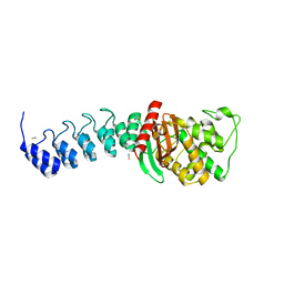
7QAD
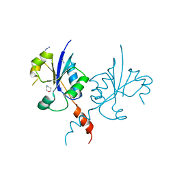 | | Crystal structure of the C-terminal catalytic domain of Plasmodium falciparum CTP:phosphocholine cytidylyltransferase with 1,4-Oxazepane hydrochloride | | 分子名称: | 1,4-oxazepane, Cholinephosphate cytidylyltransferase, Guanidinium | | 著者 | Duclovel, C, Gelin, M, Krimm, I, Cerdan, R, Guichou, J.-F. | | 登録日 | 2021-11-16 | | 公開日 | 2022-11-30 | | 最終更新日 | 2024-01-31 | | 実験手法 | X-RAY DIFFRACTION (2.11 Å) | | 主引用文献 | Crystallographic screening using ultra-low-molecular-weight ligands to guide drug design of PfCCT inhibitors.
To Be Published
|
|
7QD3
 | | Crystal structure of the C-terminal catalytic domain of Plasmodium falciparum CTP:phosphocholine cytidylyltransferase with morpholine | | 分子名称: | Cholinephosphate cytidylyltransferase, morpholine | | 著者 | Duclovel, C, Gelin, M, Krimm, I, Cerdan, R, Guichou, J.-F. | | 登録日 | 2021-11-26 | | 公開日 | 2022-12-14 | | 最終更新日 | 2024-01-31 | | 実験手法 | X-RAY DIFFRACTION (2.43 Å) | | 主引用文献 | Crystallographic screening using ultra-low-molecular-weight ligands to guide drug design of PfCCT inhibitors
To Be Published
|
|
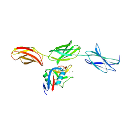
2Y6H
 | | X-2 L110F CBM4-2 Carbohydrate Binding Module from a Thermostable Rhodothermus marinus Xylanase | | 分子名称: | CALCIUM ION, XYLANASE | | 著者 | von Schantz, L, Hakansson, M, Logan, D.T, Walse, B, Osterlin, J, Nordberg-Karlsson, E, Ohlin, M. | | 登録日 | 2011-01-21 | | 公開日 | 2012-03-07 | | 最終更新日 | 2024-05-01 | | 実験手法 | X-RAY DIFFRACTION (1.08 Å) | | 主引用文献 | Structural basis for carbohydrate-binding specificity--a comparative assessment of two engineered carbohydrate-binding modules.
Glycobiology, 22, 2012
|
|
5AHM
 | | IMP-bound form of the DeltaCBS mutant of IMPDH from Pseudomonas aeruginosa | | 分子名称: | INOSINE-5'-MONOPHOSPHATE DEHYDROGENASE, INOSINIC ACID, PHOSPHATE ION | | 著者 | Labesse, G, Alexandre, T, Gelin, M, Haouz, A, Munier-Lehmann, H. | | 登録日 | 2015-02-06 | | 公開日 | 2015-07-15 | | 最終更新日 | 2024-01-10 | | 実験手法 | X-RAY DIFFRACTION (1.74 Å) | | 主引用文献 | Crystallographic Studies of Two Variants of Pseudomonas Aeruginosa Impdh with Impaired Allosteric Regulation
Acta Crystallogr.,Sect.D, 71, 2015
|
|
7QVO
 | | Crystal structure of the C-terminal catalytic domain of Plasmodium falciparum CTP:phosphocholine cytidylyltransferase with guanidine | | 分子名称: | 2-AMINO-2-HYDROXYMETHYL-PROPANE-1,3-DIOL, Cholinephosphate cytidylyltransferase, Guanidinium | | 著者 | Duclovel, C, Gelin, M, Krimm, I, Cerdan, R, Guichou, J.-F. | | 登録日 | 2022-01-22 | | 公開日 | 2023-01-25 | | 最終更新日 | 2024-02-07 | | 実験手法 | X-RAY DIFFRACTION (1.62 Å) | | 主引用文献 | Crystallographic screening using ultra-low-molecular-weight ligands to guide drug design of PfCCT inhibitors
To Be Published
|
|
7QVN
 | | Crystal structure of the C-terminal catalytic domain of Plasmodium falciparum CTP:phosphocholine cytidylyltransferase with(1H-pyrazol-5-yl)methanol | | 分子名称: | 1~{H}-pyrazol-5-ylmethanol, Cholinephosphate cytidylyltransferase, Guanidinium | | 著者 | Duclovel, C, Gelin, M, Krimm, I, Cerdan, R, Guichou, J.-F. | | 登録日 | 2022-01-22 | | 公開日 | 2023-01-25 | | 最終更新日 | 2024-02-07 | | 実験手法 | X-RAY DIFFRACTION (2.21 Å) | | 主引用文献 | Crystallographic screening using ultra-low-molecular-weight ligands to guide drug design of PfCCT inhibitors
To Be Published
|
|
7QBJ
 | | bacterial IMPDH chimera | | 分子名称: | Inosine-5'-monophosphate dehydrogenase | | 著者 | Labesse, G, Gelin, M, Munier-Lehmann, H, Gedeon, A, Haouz, A. | | 登録日 | 2021-11-19 | | 公開日 | 2023-05-31 | | 最終更新日 | 2024-02-07 | | 実験手法 | X-RAY DIFFRACTION (2.27 Å) | | 主引用文献 | Insight into the role of the Bateman domain at the molecular and physiological levels through engineered IMP dehydrogenases.
Protein Sci., 32, 2023
|
|
7QEM
 | | bacterial IMPDH chimera | | 分子名称: | INOSINIC ACID, Inosine-5'-monophosphate dehydrogenase | | 著者 | Labesse, G, Gelin, M, Gedeon, A, Haouz, A, Munier-Lehmann, H. | | 登録日 | 2021-12-03 | | 公開日 | 2023-06-14 | | 最終更新日 | 2024-02-07 | | 実験手法 | X-RAY DIFFRACTION (3.09 Å) | | 主引用文献 | Insight into the role of the Bateman domain at the molecular and physiological levels through engineered IMP dehydrogenases.
Protein Sci., 32, 2023
|
|
7QDX
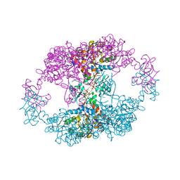 | | bacterial IMPDH chimera | | 分子名称: | ADENOSINE-5'-TRIPHOSPHATE, Inosine-5'-monophosphate dehydrogenase, MAGNESIUM ION | | 著者 | Labesse, G, Gelin, M, Munier-Lehmann, H, Gedeon, A, Haouz, A. | | 登録日 | 2021-11-30 | | 公開日 | 2023-06-14 | | 最終更新日 | 2024-02-07 | | 実験手法 | X-RAY DIFFRACTION (2.9 Å) | | 主引用文献 | Insight into the role of the Bateman domain at the molecular and physiological levels through engineered IMP dehydrogenases.
Protein Sci., 32, 2023
|
|
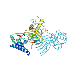
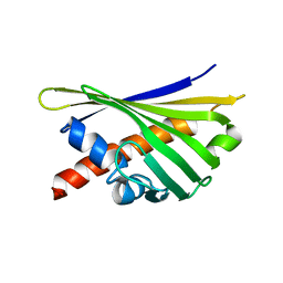
6I2Q
 | | Crystal structure of the wild-type SucA domain of Mycobacterium smegmatis KGD (alpha-ketoglutarate decarboxylase), in complex with GarA | | 分子名称: | CALCIUM ION, Glycogen accumulation regulator GarA, MAGNESIUM ION, ... | | 著者 | Wagner, T, Bellinzoni, M, Alzari, P.M. | | 登録日 | 2018-11-01 | | 公開日 | 2019-05-22 | | 最終更新日 | 2024-01-24 | | 実験手法 | X-RAY DIFFRACTION (2.15 Å) | | 主引用文献 | Structural insights into the functional versatility of an FHA domain protein in mycobacterial signaling.
Sci.Signal., 12, 2019
|
|
1RCF
 | | STRUCTURE OF THE TRIGONAL FORM OF RECOMBINANT OXIDIZED FLAVODOXIN FROM ANABAENA 7120 AT 1.40 ANGSTROMS RESOLUTION | | 分子名称: | FLAVIN MONONUCLEOTIDE, FLAVODOXIN, SULFATE ION | | 著者 | Burkhart, B, Ramakrishnan, B, Yan, H, Reedstrom, R, Markley, J, Straus, N, Sundaralingam, M. | | 登録日 | 1994-10-31 | | 公開日 | 1995-01-26 | | 最終更新日 | 2024-02-14 | | 実験手法 | X-RAY DIFFRACTION (1.4 Å) | | 主引用文献 | Structure of the trigonal form of recombinant oxidized flavodoxin from Anabaena 7120 at 1.40 A resolution.
Acta Crystallogr.,Sect.D, 51, 1995
|
|
5AQ9
 | | DARPin-based Crystallization Chaperones exploit Molecular Geometry as a Screening Dimension in Protein Crystallography | | 分子名称: | MALTOSE-BINDING PERIPLASMIC PROTEIN, OFF7_DB08V4 | | 著者 | Batyuk, A, Wu, Y, Honegger, A, Heberling, M, Plueckthun, A. | | 登録日 | 2015-09-21 | | 公開日 | 2016-03-23 | | 最終更新日 | 2024-01-10 | | 実験手法 | X-RAY DIFFRACTION (1.86 Å) | | 主引用文献 | Darpin-Based Crystallization Chaperones Exploit Molecular Geometry as a Screening Dimension in Protein Crystallography
J.Mol.Biol., 428, 2016
|
|
1TDQ
 | | Structural basis for the interactions between tenascins and the C-type lectin domains from lecticans: evidence for a cross-linking role for tenascins | | 分子名称: | Aggrecan core protein, CALCIUM ION, Tenascin-R | | 著者 | Lundell, A, Olin, A.I, Moergelin, M, al-Karadaghi, S, Aspberg, A, Logan, D.T. | | 登録日 | 2004-05-24 | | 公開日 | 2004-08-31 | | 最終更新日 | 2021-03-31 | | 実験手法 | X-RAY DIFFRACTION (2.6 Å) | | 主引用文献 | Structural basis for interactions between tenascins and lectican C-type lectin domains: evidence for a crosslinking role for tenascins
Structure, 12, 2004
|
|
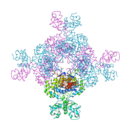
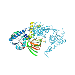
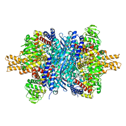
7R4K
 | | Crystal structure of human mitochondrial NAD kinase | | 分子名称: | MAGNESIUM ION, NAD kinase 2, mitochondrial, ... | | 著者 | Labesse, G, Mary, C, Gelin, M, Lionne, C. | | 登録日 | 2022-02-08 | | 公開日 | 2022-07-06 | | 最終更新日 | 2024-05-01 | | 実験手法 | X-RAY DIFFRACTION (3.33 Å) | | 主引用文献 | Crystal structure of human NADK2 reveals a dimeric organization and active site occlusion by lysine acetylation.
Mol.Cell, 82, 2022
|
|
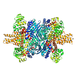
7R4J
 | | Crystal structure of human mitochondrial NAD kinase | | 分子名称: | CALCIUM ION, NAD kinase 2, mitochondrial | | 著者 | Labesse, G, Mary, C, Gelin, M, Lionne, C. | | 登録日 | 2022-02-08 | | 公開日 | 2022-07-06 | | 最終更新日 | 2024-05-01 | | 実験手法 | X-RAY DIFFRACTION (2.95 Å) | | 主引用文献 | Crystal structure of human NADK2 reveals a dimeric organization and active site occlusion by lysine acetylation.
Mol.Cell, 82, 2022
|
|
7R4M
 | | Crystal structure of mitochondrial NAD kinase | | 分子名称: | CALCIUM ION, NAD kinase 2, mitochondrial, ... | | 著者 | Labesse, G, Mary, C, Gelin, M, Lionne, C. | | 登録日 | 2022-02-08 | | 公開日 | 2022-07-06 | | 最終更新日 | 2024-05-01 | | 実験手法 | X-RAY DIFFRACTION (2.29 Å) | | 主引用文献 | Crystal structure of human NADK2 reveals a dimeric organization and active site occlusion by lysine acetylation.
Mol.Cell, 82, 2022
|
|
7R4L
 | | Crystal structure of human mitochondrial NAD kinase | | 分子名称: | FE (III) ION, NAD kinase 2, mitochondrial, ... | | 著者 | Labesse, G, Mary, C, Gelin, M, Lionne, C. | | 登録日 | 2022-02-08 | | 公開日 | 2022-07-06 | | 最終更新日 | 2024-05-01 | | 実験手法 | X-RAY DIFFRACTION (2.6 Å) | | 主引用文献 | Crystal structure of human NADK2 reveals a dimeric organization and active site occlusion by lysine acetylation.
Mol.Cell, 82, 2022
|
|
5AQ8
 | | DARPin-based Crystallization Chaperones exploit Molecular Geometry as a Screening Dimension in Protein Crystallography | | 分子名称: | 4-(2-HYDROXYETHYL)-1-PIPERAZINE ETHANESULFONIC ACID, OFF7_DB12V4, THIOCYANATE ION | | 著者 | Batyuk, A, Wu, Y, Honegger, A, Heberling, M, Plueckthun, A. | | 登録日 | 2015-09-21 | | 公開日 | 2016-03-23 | | 最終更新日 | 2024-01-10 | | 実験手法 | X-RAY DIFFRACTION (1.62 Å) | | 主引用文献 | Darpin-Based Crystallization Chaperones Exploit Molecular Geometry as a Screening Dimension in Protein Crystallography
J.Mol.Biol., 428, 2016
|
|
5AQA
 | | DARPin-based Crystallization Chaperones exploit Molecular Geometry as a Screening Dimension in Protein Crystallography | | 分子名称: | OFF7_DB04V3, THIOCYANATE ION | | 著者 | Batyuk, A, Wu, Y, Honegger, A, Heberling, M, Plueckthun, A. | | 登録日 | 2015-09-21 | | 公開日 | 2016-03-23 | | 最終更新日 | 2024-01-10 | | 実験手法 | X-RAY DIFFRACTION (2.6 Å) | | 主引用文献 | Darpin-Based Crystallization Chaperones Exploit Molecular Geometry as a Screening Dimension in Protein Crystallography
J.Mol.Biol., 428, 2016
|
|
8AR7
 | |
8AR8
 | |
8QHH
 | |


