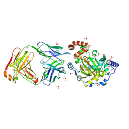
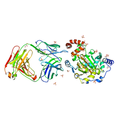
4GO1
 
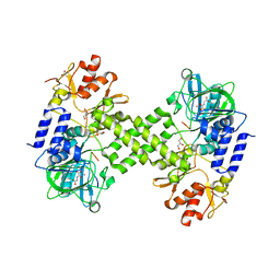
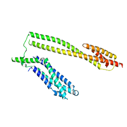
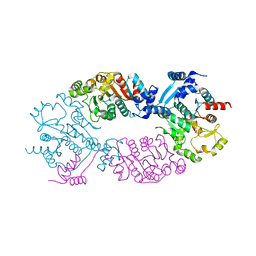 | | Crystal Structure of full length transcription repressor LsrR from E. coli. | | 分子名称: | GLYCEROL, Transcriptional regulator LsrR | | 著者 | Wu, M, Tao, Y, Liu, X, Zang, J. | | 登録日 | 2012-08-17 | | 公開日 | 2013-04-24 | | 最終更新日 | 2023-11-08 | | 実験手法 | X-RAY DIFFRACTION (3 Å) | | 主引用文献 | Structural Basis for Phosphorylated Autoinducer-2 Modulation of the Oligomerization State of the Global Transcription Regulator LsrR from Escherichia coli
J.Biol.Chem., 288, 2013
|
|
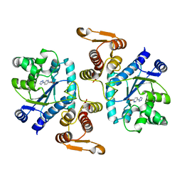
3RY7
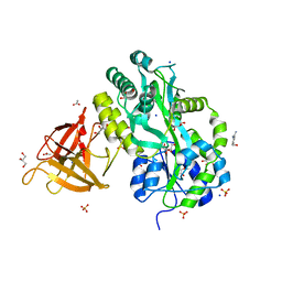
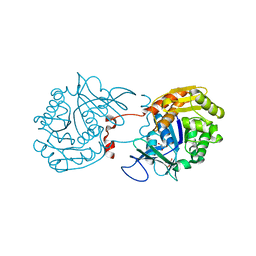 | | Crystal Structure of Sa239 | | 分子名称: | GLYCEROL, Ribokinase | | 著者 | Li, J, Wu, M, Wang, L, Zang, J. | | 登録日 | 2011-05-11 | | 公開日 | 2012-04-25 | | 最終更新日 | 2023-11-01 | | 実験手法 | X-RAY DIFFRACTION (2.15 Å) | | 主引用文献 | Crystal structure of Sa239 reveals the structural basis for the activation of ribokinase by monovalent cations.
J.Struct.Biol., 177, 2012
|
|
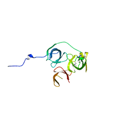
4Y33
 | | Crystal of NO66 in complex with Ni(II)and N-oxalylglycine (NOG) | | 分子名称: | Bifunctional lysine-specific demethylase and histidyl-hydroxylase NO66, N-OXALYLGLYCINE, NICKEL (II) ION | | 著者 | Wang, C, Zhang, Q, Zang, J. | | 登録日 | 2015-02-10 | | 公開日 | 2015-10-07 | | 最終更新日 | 2024-03-20 | | 実験手法 | X-RAY DIFFRACTION (2.7 Å) | | 主引用文献 | Structure of the JmjC domain-containing protein NO66 complexed with ribosomal protein Rpl8.
Acta Crystallogr.,Sect.D, 71, 2015
|
|
4Y4R
 | | Crystal structure of ribosomal oxygenase NO66 dimer mutant | | 分子名称: | ACETATE ION, Bifunctional lysine-specific demethylase and histidyl-hydroxylase NO66, NICKEL (II) ION | | 著者 | Wang, C, Hang, T, Zang, J. | | 登録日 | 2015-02-11 | | 公開日 | 2015-10-07 | | 最終更新日 | 2024-03-20 | | 実験手法 | X-RAY DIFFRACTION (3.3 Å) | | 主引用文献 | Structure of the JmjC domain-containing protein NO66 complexed with ribosomal protein Rpl8.
Acta Crystallogr.,Sect.D, 71, 2015
|
|
4Y3O
 | | Crystal structure of Ribosomal oxygenase NO66 in complex with substrate Rpl8 peptide and Ni(II) and cofactor N-oxalyglycine | | 分子名称: | ACETATE ION, Bifunctional lysine-specific demethylase and histidyl-hydroxylase NO66, GLYCEROL, ... | | 著者 | Wang, C, Zhang, Q, Zang, J. | | 登録日 | 2015-02-10 | | 公開日 | 2015-10-07 | | 最終更新日 | 2024-03-20 | | 実験手法 | X-RAY DIFFRACTION (2.2 Å) | | 主引用文献 | Structure of the JmjC domain-containing protein NO66 complexed with ribosomal protein Rpl8.
Acta Crystallogr.,Sect.D, 71, 2015
|
|
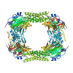
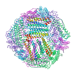
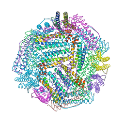
5GOU
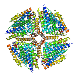 | | Structure of a 16-mer protein nanocage fabricated from its 24-mer analogue by subunit interface redesign | | 分子名称: | Ferritin heavy chain | | 著者 | Zhang, S, Zang, J, Wang, W, Wang, H, Zhao, G. | | 登録日 | 2016-07-29 | | 公開日 | 2017-02-22 | | 最終更新日 | 2023-11-08 | | 実験手法 | X-RAY DIFFRACTION (2.91 Å) | | 主引用文献 | Conversion of the Native 24-mer Ferritin Nanocage into Its Non-Native 16-mer Analogue by Insertion of Extra Amino Acid Residues.
Angew. Chem. Int. Ed. Engl., 55, 2016
|
|
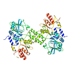
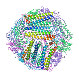
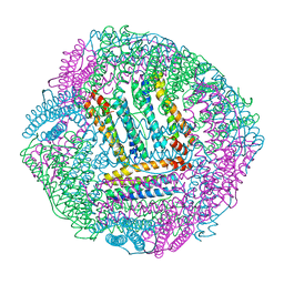
5GN8
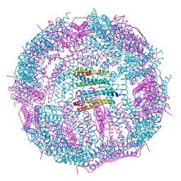 | | Structure of a 48-mer protein nanocage fabricated from its 24-mer analogue by subunit interface redesign | | 分子名称: | CALCIUM ION, Ferritin heavy chain | | 著者 | Zhang, S, Zang, J, Zhao, G, Mikami, B. | | 登録日 | 2016-07-19 | | 公開日 | 2016-12-14 | | 最終更新日 | 2023-11-08 | | 実験手法 | X-RAY DIFFRACTION (2.805 Å) | | 主引用文献 | "Silent" Amino Acid Residues at Key Subunit Interfaces Regulate the Geometry of Protein Nanocages
ACS Nano, 10, 2016
|
|
5J6P
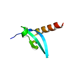
 | | Crystal Structure of Mis18(17-118) from Schizosaccharomyces pombe | | 分子名称: | Kinetochore protein mis18, ZINC ION | | 著者 | Wang, C, Shao, C, Zhang, M, Zhang, X, Zang, J. | | 登録日 | 2016-04-05 | | 公開日 | 2017-11-01 | | 最終更新日 | 2024-03-20 | | 実験手法 | X-RAY DIFFRACTION (2.6 Å) | | 主引用文献 | Crystal Structure of Mis18(17-118) from Schizosaccharomyces pombe
To Be Published
|
|
5WWL
 | | Crystal structure of the Schizogenesis pombe kinetochore Mis12C subcomplex | | 分子名称: | Centromere protein mis12, Kinetochore protein nnf1 | | 著者 | Wang, C, Zhou, X, Wu, M, Zhang, X, Zang, J. | | 登録日 | 2017-01-02 | | 公開日 | 2017-11-15 | | 最終更新日 | 2024-03-20 | | 実験手法 | X-RAY DIFFRACTION (2.4 Å) | | 主引用文献 | Phosphorylation of CENP-C by Aurora B facilitates kinetochore attachment error correction in mitosis.
Proc. Natl. Acad. Sci. U.S.A., 114, 2017
|
|
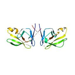
1WT9
 | | crystal structure of Aa-X-bp-I, a snake venom protein with the activity of binding to coagulation factor X from Agkistrodon acutus | | 分子名称: | CALCIUM ION, agkisacutacin A chain, anticoagulant protein-B | | 著者 | Zhu, Z, Liu, S, Mo, X, Yu, X, Liang, Z, Zang, J, Zhao, W, Teng, M, Niu, L. | | 登録日 | 2004-11-18 | | 公開日 | 2006-03-07 | | 最終更新日 | 2011-07-13 | | 実験手法 | X-RAY DIFFRACTION (2.01 Å) | | 主引用文献 | Characterizations and Crystal structures of two snake venom proteins with the activity of binding coagulation factor X from Agkistrodon acutus
To be Published
|
|
1Y17
 | | crystal structure of Aa-X-bp-II, a snake venom protein with the activity of binding to coagulation factor X from Agkistrodon acutus | | 分子名称: | CALCIUM ION, anticoagulant protein A, anticoagulant protein-B | | 著者 | Zhu, Z, Liu, S, Mo, X, Yu, X, Liang, Z, Zang, J, Zhao, W, Teng, M, Niu, L. | | 登録日 | 2004-11-17 | | 公開日 | 2006-03-07 | | 最終更新日 | 2011-07-13 | | 実験手法 | X-RAY DIFFRACTION (2.4 Å) | | 主引用文献 | Characterizations and Crystal structures of two snake venom proteins with the activity of binding coagulation factor X from Agkistrodon acutus
To be Published
|
|
3MP8
 | | Crystal structure of Sgf29 tudor domain | | 分子名称: | 4-(HYDROXYMETHYL)BENZAMIDINE, ACETIC ACID, GLYCEROL, ... | | 著者 | Li, J, Wu, M, Ruan, J, Zang, J. | | 登録日 | 2010-04-26 | | 公開日 | 2011-05-04 | | 最終更新日 | 2023-11-01 | | 実験手法 | X-RAY DIFFRACTION (1.92 Å) | | 主引用文献 | Sgf29 binds histone H3K4me2/3 and is required for SAGA complex recruitment and histone H3 acetylation
Embo J., 30, 2011
|
|
3MP6
 | | Complex Structure of Sgf29 and dimethylated H3K4 | | 分子名称: | H3K4me2 peptide, Maltose-binding periplasmic protein,LINKER,SAGA-associated factor 29, alpha-D-glucopyranose-(1-4)-alpha-D-glucopyranose | | 著者 | Li, J, Wu, M, Ruan, J, Zang, J. | | 登録日 | 2010-04-25 | | 公開日 | 2011-05-04 | | 最終更新日 | 2023-11-01 | | 実験手法 | X-RAY DIFFRACTION (1.48 Å) | | 主引用文献 | Sgf29 binds histone H3K4me2/3 and is required for SAGA complex recruitment and histone H3 acetylation
Embo J., 30, 2011
|
|
3MP1
 | | Complex structure of Sgf29 and trimethylated H3K4 | | 分子名称: | ACETATE ION, H3K4me3 peptide, Maltose-binding periplasmic protein,LINKER,SAGA-associated factor 29, ... | | 著者 | Li, J, Ruan, J, Wu, M, Xue, X, Zang, J. | | 登録日 | 2010-04-24 | | 公開日 | 2011-05-04 | | 最終更新日 | 2023-11-01 | | 実験手法 | X-RAY DIFFRACTION (2.6 Å) | | 主引用文献 | Sgf29 binds histone H3K4me2/3 and is required for SAGA complex recruitment and histone H3 acetylation
Embo J., 30, 2011
|
|
3LWE
 | | The crystal structure of MPP8 | | 分子名称: | M-phase phosphoprotein 8 | | 著者 | Li, Z, Li, Z, Ruan, J, Xu, C, Tong, Y, Pan, P.W, Tempel, W, Crombet, L, Min, J, Zang, J, Structural Genomics Consortium (SGC) | | 登録日 | 2010-02-23 | | 公開日 | 2010-03-02 | | 最終更新日 | 2023-09-06 | | 実験手法 | X-RAY DIFFRACTION (2.05 Å) | | 主引用文献 | Structural basis for specific binding of human MPP8 chromodomain to histone H3 methylated at lysine 9.
Plos One, 6, 2011
|
|
3LD8
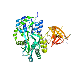
 | | Structure of JMJD6 and Fab Fragments | | 分子名称: | Bifunctional arginine demethylase and lysyl-hydroxylase JMJD6, FE (III) ION, GLYCEROL, ... | | 著者 | Hong, X, Zang, J, White, J, Kappler, J.W, Wang, C, Zhang, G. | | 登録日 | 2010-01-12 | | 公開日 | 2010-08-04 | | 最終更新日 | 2012-06-20 | | 実験手法 | X-RAY DIFFRACTION (2.7 Å) | | 主引用文献 | Interaction of JMJD6 with single-stranded RNA.
Proc.Natl.Acad.Sci.USA, 107, 2010
|
|
3LDB
 | | Structure of JMJD6 complexd with ALPHA-KETOGLUTARATE and Fab Fragment. | | 分子名称: | 2-OXOGLUTARIC ACID, Bifunctional arginine demethylase and lysyl-hydroxylase JMJD6, FE (III) ION, ... | | 著者 | Hong, X, Zang, J, White, J, Kappler, J.W, Wang, C, Zhang, G. | | 登録日 | 2010-01-12 | | 公開日 | 2010-08-04 | | 最終更新日 | 2023-09-06 | | 実験手法 | X-RAY DIFFRACTION (2.7 Å) | | 主引用文献 | Interaction of JMJD6 with single-stranded RNA.
Proc.Natl.Acad.Sci.USA, 107, 2010
|
|
3N2Y
 | |
5Y5W
 | |
7EKA
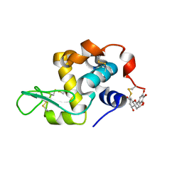 | | crystal structure of epigallocatechin binding with alpha-lactalbumin | | 分子名称: | 2-(3,4,5-TRIHYDROXY-PHENYL)-CHROMAN-3,5,7-TRIOL, Alpha-lactalbumin | | 著者 | Ma, J, Yao, Q, Chen, X, Zang, J. | | 登録日 | 2021-04-05 | | 公開日 | 2023-11-08 | | 実験手法 | X-RAY DIFFRACTION (1.2 Å) | | 主引用文献 | Weak Binding of Epigallocatechin to alpha-Lactalbumin Greatly Improves Its Stability and Uptake by Caco-2 Cells.
J.Agric.Food Chem., 69, 2021
|
|
8IQX
 | |
8IQW
 | |
8IQV
 | |
8IQZ
 | |
8IR0
 | |


