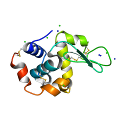
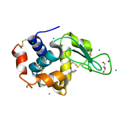
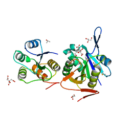
3T6U
 
 | |
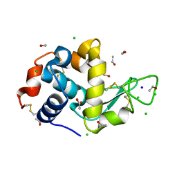
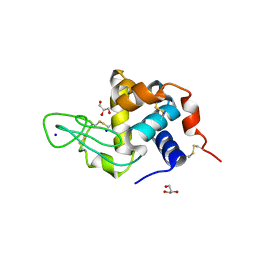
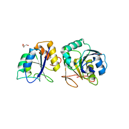
3RNX
 | |
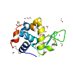
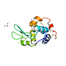
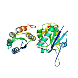
3RW8
 | |
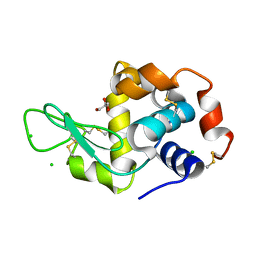
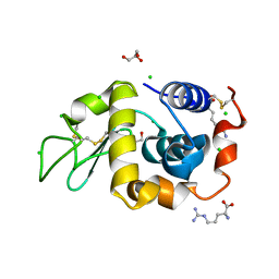
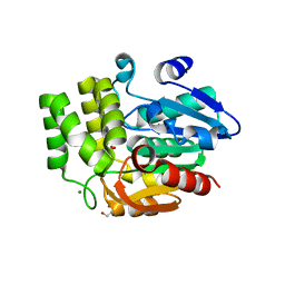
3SP3
 | |
4HSF
 | |
4II8
 | | Lysozyme with Benzyl alcohol | | 分子名称: | CHLORIDE ION, GLYCEROL, Lysozyme C, ... | | 著者 | Sharma, P, Ashish | | 登録日 | 2012-12-20 | | 公開日 | 2013-12-25 | | 最終更新日 | 2023-11-08 | | 実験手法 | X-RAY DIFFRACTION (1.88 Å) | | 主引用文献 | Characterization of heat induced spherulites of lysozyme reveals new insight on amyloid initiation
Sci Rep, 6, 2016
|
|
4DC4
 | | Lysozyme Trimer | | 分子名称: | CHLORIDE ION, GLYCEROL, Lysozyme C, ... | | 著者 | Sharma, P, Ashish | | 登録日 | 2012-01-17 | | 公開日 | 2013-01-23 | | 最終更新日 | 2023-11-08 | | 実験手法 | X-RAY DIFFRACTION (2.654 Å) | | 主引用文献 | Characterization of heat induced spherulites of lysozyme reveals new insight on amyloid initiation
Sci Rep, 6, 2016
|
|
4D9Z
 | | Lysozyme at 318K | | 分子名称: | CHLORIDE ION, GLYCEROL, Lysozyme C, ... | | 著者 | Sharma, P, Ashish | | 登録日 | 2012-01-12 | | 公開日 | 2013-01-16 | | 最終更新日 | 2023-11-08 | | 実験手法 | X-RAY DIFFRACTION (1.709 Å) | | 主引用文献 | Characterization of heat induced spherulites of lysozyme reveals new insight on amyloid initiation
Sci Rep, 6, 2016
|
|
4EOF
 | | Lysozyme in the presence of arginine | | 分子名称: | ACETATE ION, ARGININE, CHLORIDE ION, ... | | 著者 | Sharma, P, Ashish | | 登録日 | 2012-04-14 | | 公開日 | 2013-04-17 | | 最終更新日 | 2023-11-08 | | 実験手法 | X-RAY DIFFRACTION (1.83 Å) | | 主引用文献 | Characterization of heat induced spherulites of lysozyme reveals new insight on amyloid initiation.
Sci Rep, 6, 2016
|
|
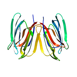
2O92
 | | Crystal structure of a signalling protein (SPG-40) complex with tetrasaccharide at 3.0A resolution | | 分子名称: | 2-acetamido-2-deoxy-beta-D-glucopyranose-(1-4)-2-acetamido-2-deoxy-beta-D-glucopyranose-(1-4)-2-acetamido-2-deoxy-beta-D-glucopyranose-(1-4)-2-acetamido-2-deoxy-beta-D-glucopyranose, Chitinase-3-like protein 1, alpha-D-mannopyranose-(1-4)-2-acetamido-2-deoxy-beta-D-glucopyranose-(1-4)-2-acetamido-2-deoxy-beta-D-glucopyranose | | 著者 | Sharma, P, Singh, N, Sharma, S, Kaur, P, Singh, T.P. | | 登録日 | 2006-12-13 | | 公開日 | 2006-12-26 | | 最終更新日 | 2023-10-25 | | 実験手法 | X-RAY DIFFRACTION (3 Å) | | 主引用文献 | Crystal structure of a signalling protein (SPG-40) complex with tetrasaccharide at 3.0A resolution
To be Published
|
|
2OLH
 | | Crystal structure of a signalling protein (SPG-40) complex with cellobiose at 2.78 A resolution | | 分子名称: | Chitinase-3-like protein 1, alpha-D-mannopyranose-(1-4)-2-acetamido-2-deoxy-beta-D-glucopyranose-(1-4)-2-acetamido-2-deoxy-beta-D-glucopyranose, beta-D-glucopyranose-(1-4)-beta-D-glucopyranose | | 著者 | Sharma, P, Singh, N, Sharma, S, Bhushan, A, Kaur, P, Singh, T.P. | | 登録日 | 2007-01-19 | | 公開日 | 2007-02-13 | | 最終更新日 | 2023-10-25 | | 実験手法 | X-RAY DIFFRACTION (2.78 Å) | | 主引用文献 | Crystal structure of a signalling protein (SPG-40) complex with cellobiose at 2.78 A resolution
To be Published
|
|
2PI6
 | | Crystal structure of the sheep signalling glycoprotein (SPS-40) complex with 2-methyl-2-4-pentanediol at 1.65A resolution reveals specific binding characteristics of SPS-40 | | 分子名称: | (4S)-2-METHYL-2,4-PENTANEDIOL, Chitinase-3-like protein 1, ETHANOL, ... | | 著者 | Sharma, P, Singh, N, Sharma, S, Kaur, P, Betzel, C, Singh, T.P. | | 登録日 | 2007-04-13 | | 公開日 | 2007-05-01 | | 最終更新日 | 2023-10-25 | | 実験手法 | X-RAY DIFFRACTION (1.65 Å) | | 主引用文献 | Tryptophan as a three-way switch in regulating the function of the secretory signalling glycoprotein (SPS-40) from mammary glands: structure of SPS-40 complexed with 2-methylpentane-2,4-diol at 1.6 A resolution.
Acta Crystallogr.,Sect.D, 65, 2009
|
|
2R2K
 | | Crystal structure of the complex of camel peptidoglycan recognition protein with disaccharide at 3.2A resolution | | 分子名称: | 2-acetamido-2-deoxy-beta-D-glucopyranose-(1-4)-2-acetamido-2-deoxy-beta-D-glucopyranose, L(+)-TARTARIC ACID, Peptidoglycan recognition protein | | 著者 | Sharma, P, Jain, R, Singh, N, Sharma, S, Bhushan, A, Kaur, P, Singh, T.P. | | 登録日 | 2007-08-26 | | 公開日 | 2007-09-18 | | 最終更新日 | 2023-10-25 | | 実験手法 | X-RAY DIFFRACTION (3.25 Å) | | 主引用文献 | Crystal structure of the complex of camel peptidoglycan recognition protein with disaccharide at 3.2A resolution
To be Published
|
|
4FNN
 | | Crystal structure of the complex of CPGRP-S with stearic acid at 2.2 A RESOLUTION | | 分子名称: | Peptidoglycan recognition protein 1, STEARIC ACID | | 著者 | Dube, D, Sharma, P, Sinha, M, Kaur, P, Sharma, S, Singh, T.P. | | 登録日 | 2012-06-20 | | 公開日 | 2012-07-25 | | 最終更新日 | 2023-09-13 | | 実験手法 | X-RAY DIFFRACTION (2.24 Å) | | 主引用文献 | Structural basis of the binding of fatty acids to peptidoglycan recognition protein, PGRP-S through second binding site.
Arch.Biochem.Biophys., 529, 2013
|
|
3UIL
 | | Crystal Structure of the complex of PGRP-S with lauric acid at 2.2 A resolution | | 分子名称: | GLYCEROL, LAURIC ACID, Peptidoglycan recognition protein 1 | | 著者 | Dube, D, Sharma, P, Sinha, M, Kaur, P, Sharma, S, Singh, T.P. | | 登録日 | 2011-11-05 | | 公開日 | 2012-07-11 | | 最終更新日 | 2023-11-01 | | 実験手法 | X-RAY DIFFRACTION (2.2 Å) | | 主引用文献 | Structural basis of the binding of fatty acids to peptidoglycan recognition protein, PGRP-S through second binding site
Arch.Biochem.Biophys., 529, 2013
|
|
3UMQ
 | | Crystal structure of peptidoglycan recognition protein-S complexed with butyric acid at 2.2 A resolution | | 分子名称: | GLYCEROL, Peptidoglycan recognition protein 1, butanoic acid | | 著者 | Pandey, N, Sharma, P, Sinha, M, Bhushan, A, Kaur, P, Sharma, S, Singh, T.P. | | 登録日 | 2011-11-14 | | 公開日 | 2012-07-04 | | 最終更新日 | 2023-11-01 | | 実験手法 | X-RAY DIFFRACTION (2.2 Å) | | 主引用文献 | Structural basis of the binding of fatty acids to peptidoglycan recognition protein, PGRP-S through second binding site
Arch.Biochem.Biophys., 529, 2013
|
|
3USX
 | | Crystal structure of PGRP-S complexed with Myristic Acid at 2.28 A resolution | | 分子名称: | GLYCEROL, MYRISTIC ACID, Peptidoglycan recognition protein 1 | | 著者 | Yamini, S, Sharma, P, Sinha, M, Kaur, P, Sharma, S, Singh, T.P. | | 登録日 | 2011-11-24 | | 公開日 | 2012-01-11 | | 最終更新日 | 2023-11-08 | | 実験手法 | X-RAY DIFFRACTION (2.28 Å) | | 主引用文献 | Structural basis of the binding of fatty acids to peptidoglycan recognition protein, PGRP-S through second binding site
Arch.Biochem.Biophys., 529, 2013
|
|
5GVY
 | | Crystal structure of SALT protein from Oryza sativa | | 分子名称: | Salt stress-induced protein, alpha-D-mannopyranose | | 著者 | Sharma, P, Sagar, A, Kaur, N, Sharma, I, Kirat, K, Ashish, F.N.U, Pati, P.K. | | 登録日 | 2016-09-07 | | 公開日 | 2017-09-13 | | 最終更新日 | 2023-11-08 | | 実験手法 | X-RAY DIFFRACTION (1.662 Å) | | 主引用文献 | Structural insights into rice SalTol QTL located SALT protein.
Sci Rep, 10, 2020
|
|
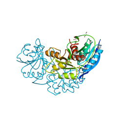
8SHH
 | | Crystal structure of EvdS6 decarboxylase in ligand free state | | 分子名称: | DI(HYDROXYETHYL)ETHER, NICOTINAMIDE-ADENINE-DINUCLEOTIDE, dTDP-glucose 4,6-dehydratase | | 著者 | Sharma, P, Frigo, L, Dulin, C.C, Bachmann, B.O, Iverson, T.M. | | 登録日 | 2023-04-14 | | 公開日 | 2023-08-09 | | 最終更新日 | 2024-05-22 | | 実験手法 | X-RAY DIFFRACTION (1.93 Å) | | 主引用文献 | EvdS6 is a bifunctional decarboxylase from the everninomicin gene cluster.
J.Biol.Chem., 299, 2023
|
|
8SK0
 | | Crystal structure of EvdS6 decarboxylase in ligand bound state | | 分子名称: | CITRATE ANION, DI(HYDROXYETHYL)ETHER, GLYCEROL, ... | | 著者 | Sharma, P, Frigo, L, Dulin, C.C, Bachmann, B.O, Iverson, T.M. | | 登録日 | 2023-04-18 | | 公開日 | 2023-08-09 | | 最終更新日 | 2024-05-22 | | 実験手法 | X-RAY DIFFRACTION (1.51 Å) | | 主引用文献 | EvdS6 is a bifunctional decarboxylase from the everninomicin gene cluster.
J.Biol.Chem., 299, 2023
|
|
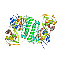
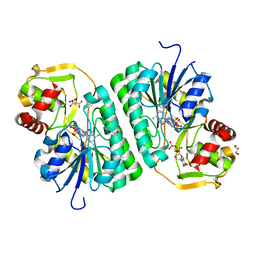
4Q0M
 | | Crystal structure of Pyrococcus furiosus L-asparaginase | | 分子名称: | (4S)-2-METHYL-1,4,5,6-TETRAHYDROPYRIMIDINE-4-CARBOXYLIC ACID, GLYCEROL, L-asparaginase, ... | | 著者 | Sharma, P, Tomar, R, Singh, S, Yadav, S.P.S, Ashish, Kundu, B. | | 登録日 | 2014-04-02 | | 公開日 | 2014-12-10 | | 最終更新日 | 2023-11-08 | | 実験手法 | X-RAY DIFFRACTION (2.226 Å) | | 主引用文献 | Structural and functional insights into an archaeal L-asparaginase obtained through the linker-less assembly of constituent domains.
Acta Crystallogr.,Sect.D, 70, 2014
|
|
4RA9
 | | Crystal Structure of Conjoint Pyrococcus Furiosus L-asparaginase with Citrate | | 分子名称: | CITRATE ANION, GLYCEROL, ISOPROPYL ALCOHOL, ... | | 著者 | Sharma, P, Tomar, R, Singh, S, Yadav, S.P.S, Ashish, Kundu, B. | | 登録日 | 2014-09-09 | | 公開日 | 2014-12-10 | | 最終更新日 | 2023-11-08 | | 実験手法 | X-RAY DIFFRACTION (2.049 Å) | | 主引用文献 | Structural and functional insights into an archaeal L-asparaginase obtained through the linker-less assembly of constituent domains.
Acta Crystallogr.,Sect.D, 70, 2014
|
|
4RA6
 | | Crystal structure of linker less Pyrococcus furiosus L-asparaginase | | 分子名称: | GLYCEROL, L-asparaginase | | 著者 | Sharma, P, Tomar, R, Ashish, Kundu, B. | | 登録日 | 2014-09-09 | | 公開日 | 2014-12-10 | | 最終更新日 | 2023-11-08 | | 実験手法 | X-RAY DIFFRACTION (2.503 Å) | | 主引用文献 | Structural and functional insights into an archaeal L-asparaginase obtained through the linker-less assembly of constituent domains.
Acta Crystallogr.,Sect.D, 70, 2014
|
|
4NJE
 | | Crystal structure of Pyrococcus furiosus L-asparaginase with ligand | | 分子名称: | ASPARTIC ACID, L-asparaginase | | 著者 | Sharma, P, Tomar, R, Singh, S, Yadav, S.P.S, Ashish, Kundu, B. | | 登録日 | 2013-11-09 | | 公開日 | 2014-12-10 | | 最終更新日 | 2023-11-08 | | 実験手法 | X-RAY DIFFRACTION (2.5 Å) | | 主引用文献 | Structural and functional insights into an archaeal L-asparaginase obtained through the linker-less assembly of constituent domains.
Acta Crystallogr.,Sect.D, 70, 2014
|
|
6UH8
 | | Crystal structure of DAD2 N242I mutant | | 分子名称: | 2-(N-MORPHOLINO)-ETHANESULFONIC ACID, Decreased Apical Dominance 2, GLYCEROL, ... | | 著者 | Sharma, P, Hamiaux, C, Snowden, K.C. | | 登録日 | 2019-09-27 | | 公開日 | 2020-02-26 | | 最終更新日 | 2023-10-11 | | 実験手法 | X-RAY DIFFRACTION (1.58 Å) | | 主引用文献 | Flexibility of the petunia strigolactone receptor DAD2 promotes its interaction with signaling partners.
J.Biol.Chem., 295, 2020
|
|


