1QIE
 
 | |
1QII
 | |
1QIH
 | |
1QID
 | |
1QIJ
 | |
1QIK
 | |
1QIM
 | |
1QUB
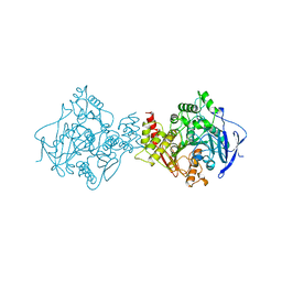
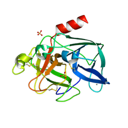
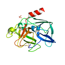
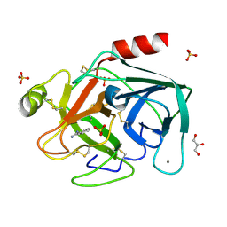
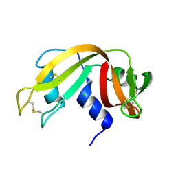
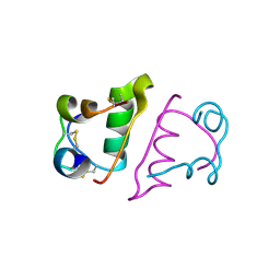
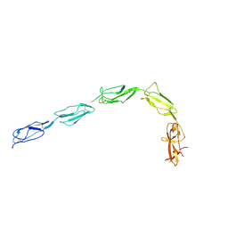 | | CRYSTAL STRUCTURE OF THE GLYCOSYLATED FIVE-DOMAIN HUMAN BETA2-GLYCOPROTEIN I PURIFIED FROM BLOOD PLASMA | | 分子名称: | 2-acetamido-2-deoxy-beta-D-glucopyranose, 2-acetamido-2-deoxy-beta-D-glucopyranose-(1-4)-2-acetamido-2-deoxy-beta-D-glucopyranose, PROTEIN (human beta2-Glycoprotein I), ... | | 著者 | Bouma, B, de Groot, P.G, van den Elsen, J.M.H, Ravelli, R.B.G, Schouten, A, Simmelink, M.J.A, Derksen, R.H.W.M, Kroon, J, Gros, P. | | 登録日 | 1999-07-01 | | 公開日 | 1999-10-08 | | 最終更新日 | 2024-10-30 | | 実験手法 | X-RAY DIFFRACTION (2.7 Å) | | 主引用文献 | Adhesion mechanism of human beta(2)-glycoprotein I to phospholipids based on its crystal structure.
EMBO J., 18, 1999
|
|
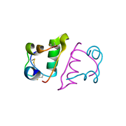
1ZZJ
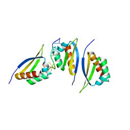 | | Structure of the third KH domain of hnRNP K in complex with 15-mer ssDNA | | 分子名称: | 5'-D(*TP*TP*CP*CP*CP*CP*TP*CP*CP*CP*CP*AP*TP*TP*T)-3', Heterogeneous nuclear ribonucleoprotein K | | 著者 | Backe, P.H, Messias, A.C, Ravelli, R.B, Sattler, M, Cusack, S. | | 登録日 | 2005-06-14 | | 公開日 | 2005-08-09 | | 最終更新日 | 2024-03-13 | | 実験手法 | X-RAY DIFFRACTION (2.3 Å) | | 主引用文献 | X-Ray Crystallographic and NMR Studies of the Third KH Domain of hnRNP K in Complex with Single-Stranded Nucleic Acids
STRUCTURE, 13, 2005
|
|
2BLQ
 | |
2BN3
 | |
2BLY
 | |
2BLV
 | |
2BLO
 | |
2BLW
 | |
2BLX
 | |
2BLP
 | |
2BLZ
 | |
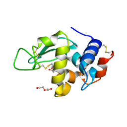
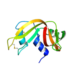
1ZZK
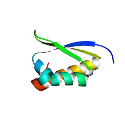 | | Crystal Structure of the third KH domain of hnRNP K at 0.95A resolution | | 分子名称: | Heterogeneous nuclear ribonucleoprotein K | | 著者 | Backe, P.H, Messias, A.C, Ravelli, R.B, Sattler, M, Cusack, S. | | 登録日 | 2005-06-14 | | 公開日 | 2005-08-09 | | 最終更新日 | 2024-03-13 | | 実験手法 | X-RAY DIFFRACTION (0.95 Å) | | 主引用文献 | X-Ray Crystallographic and NMR Studies of the Third KH Domain of hnRNP K in Complex with Single-Stranded Nucleic Acids
STRUCTURE, 13, 2005
|
|
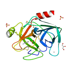
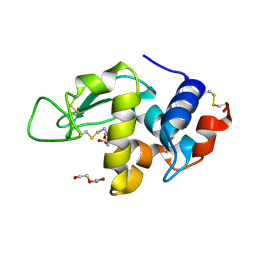
1ZZI
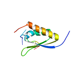 | | Crystal Structure Analysis of the third KH domain of hnRNP K in complex with ssDNA | | 分子名称: | 5'-D(*CP*TP*CP*CP*CP*C)-3', Heterogeneous nuclear ribonucleoprotein K | | 著者 | Backe, P.H, Messias, A.C, Ravelli, R.B, Sattler, M, Cusack, S. | | 登録日 | 2005-06-14 | | 公開日 | 2005-08-09 | | 最終更新日 | 2024-03-13 | | 実験手法 | X-RAY DIFFRACTION (1.8 Å) | | 主引用文献 | X-Ray Crystallographic and NMR Studies of the Third KH Domain of hnRNP K in Complex with Single-Stranded Nucleic Acids
STRUCTURE, 13, 2005
|
|
1Z2B
 | | Tubulin-colchicine-vinblastine: stathmin-like domain complex | | 分子名称: | (2ALPHA,2'BETA,3BETA,4ALPHA,5BETA)-VINCALEUKOBLASTINE, 2-MERCAPTO-N-[1,2,3,10-TETRAMETHOXY-9-OXO-5,6,7,9-TETRAHYDRO-BENZO[A]HEPTALEN-7-YL]ACETAMIDE, GUANOSINE-5'-DIPHOSPHATE, ... | | 著者 | Gigant, B, Wang, C, Ravelli, R.B.G, Roussi, F, Steinmetz, M.O, Curmi, P.A, Sobel, A, Knossow, M. | | 登録日 | 2005-03-08 | | 公開日 | 2005-05-31 | | 最終更新日 | 2023-08-23 | | 実験手法 | X-RAY DIFFRACTION (4.1 Å) | | 主引用文献 | Structural basis for the regulation of tubulin by vinblastine.
Nature, 435, 2005
|
|
2BN1
 | |
8AEY
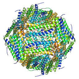 | | 3 A CRYO-EM STRUCTURE OF MYCOBACTERIUM TUBERCULOSIS FERRITIN FROM TIMEPIX3 detector | | 分子名称: | Ferritin BfrB | | 著者 | Zhang, Y, van Schayck, J.P, Knoops, K, Peters, P.J, Ravelli, R.B.G. | | 登録日 | 2022-07-14 | | 公開日 | 2023-01-18 | | 最終更新日 | 2024-07-24 | | 実験手法 | ELECTRON MICROSCOPY (3.05 Å) | | 主引用文献 | Integration of an Event-driven Timepix3 Hybrid Pixel Detector into a Cryo-EM Workflow
Microsc Microanal, 2023
|
|
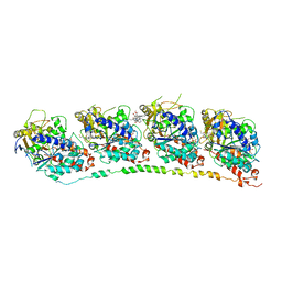
7O6E
 | | 2.12 A cryo-EM structure of Mycobacterium tuberculosis Ferritin | | 分子名称: | Ferritin BfrB | | 著者 | Gijsbers, A, Zhang, Y, Gao, Y, Peters, P.J, Ravelli, R.B.G. | | 登録日 | 2021-04-10 | | 公開日 | 2021-05-19 | | 最終更新日 | 2024-07-10 | | 実験手法 | ELECTRON MICROSCOPY (2.1 Å) | | 主引用文献 | Mycobacterium tuberculosis ferritin: a suitable workhorse protein for cryo-EM development.
Acta Crystallogr D Struct Biol, 77, 2021
|
|
6Y1A
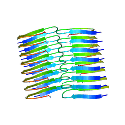 | | Amyloid fibril structure of islet amyloid polypeptide | | 分子名称: | AMINO GROUP, Islet amyloid polypeptide | | 著者 | Roeder, C, Kupreichyk, T, Gremer, L, Schaefer, L.U, Pothula, K.R, Ravelli, R.B.G, Willbold, D, Hoyer, W, Schroder, G.F. | | 登録日 | 2020-02-11 | | 公開日 | 2020-03-04 | | 最終更新日 | 2025-04-09 | | 実験手法 | ELECTRON MICROSCOPY (4.2 Å) | | 主引用文献 | Cryo-EM structure of islet amyloid polypeptide fibrils reveals similarities with amyloid-beta fibrils.
Nat.Struct.Mol.Biol., 27, 2020
|
|


