4MZD
 
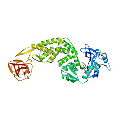
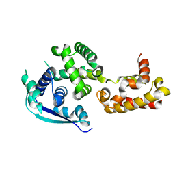
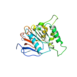
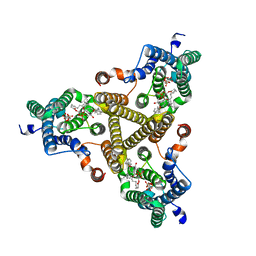
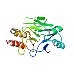
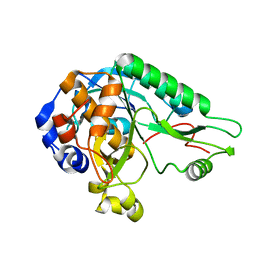 | | High resolution crystal structure of the nisin leader peptidase NisP from Lactococcus lactis | | 分子名称: | Nisin leader peptide-processing serine protease NisP | | 著者 | Rao, Z.H, Xu, Y.Y, Li, X, Yang, W. | | 登録日 | 2013-09-30 | | 公開日 | 2014-06-11 | | 最終更新日 | 2024-02-28 | | 実験手法 | X-RAY DIFFRACTION (1.1 Å) | | 主引用文献 | Structure of the nisin leader peptidase NisP revealing a C-terminal autocleavage activity.
Acta Crystallogr.,Sect.D, 70, 2014
|
|
5YK1
 | |
5YK2
 | |
5YK0
 | |
7FAC
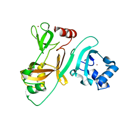
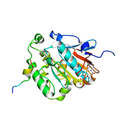
 | | Crystal Structure of C-terminus of the non-structural protein 2 from SARS coronavirus | | 分子名称: | Non-structural protein 2, ZINC ION | | 著者 | Li, Y.Y, Ren, Z.L, Bao, Z.H, Ming, Z.H, Yan, L.M, Lou, Z.Y, Rao, Z.H. | | 登録日 | 2021-07-06 | | 公開日 | 2022-08-10 | | 最終更新日 | 2024-02-28 | | 実験手法 | X-RAY DIFFRACTION (2.71 Å) | | 主引用文献 | The Life of SARS-CoV-2 Inside Cells: Replication-Transcription Complex Assembly and Function.
Annu.Rev.Biochem., 91, 2022
|
|
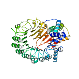
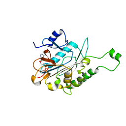
7FA1
 | | Crystal Structure of N-terminus of the non-structural protein 2 from SARS coronavirus | | 分子名称: | Non-structural protein 2, ZINC ION | | 著者 | Li, Y.Y, Ren, Z.L, Bao, Z.H, Ming, Z.H, Yan, L.M, Lou, Z.Y, Rao, Z.H. | | 登録日 | 2021-07-05 | | 公開日 | 2022-08-10 | | 最終更新日 | 2024-02-28 | | 実験手法 | X-RAY DIFFRACTION (1.6 Å) | | 主引用文献 | The Life of SARS-CoV-2 Inside Cells: Replication-Transcription Complex Assembly and Function.
Annu.Rev.Biochem., 91, 2022
|
|
5HYW
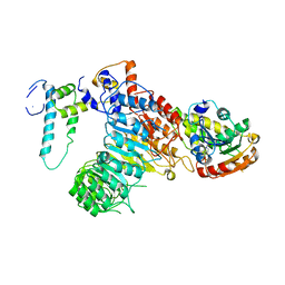
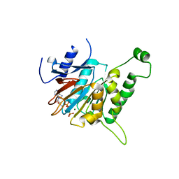
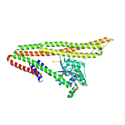
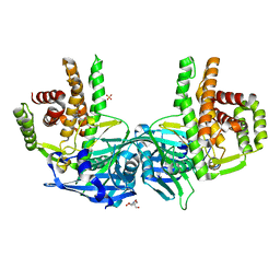
 | | The crystal structure of the D3-ASK1 complex | | 分子名称: | F-box/LRR-repeat MAX2 homolog, SKP1-like protein 1A | | 著者 | Yao, R.F, Ming, Z.H, Yan, L.M, Rao, Z.H, Lou, Z.Y, Xie, D.X. | | 登録日 | 2016-02-02 | | 公開日 | 2016-08-03 | | 最終更新日 | 2023-11-08 | | 実験手法 | X-RAY DIFFRACTION (3.01 Å) | | 主引用文献 | DWARF14 is a non-canonical hormone receptor for strigolactone
Nature, 536, 2016
|
|
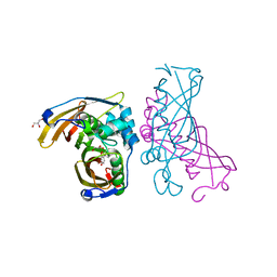
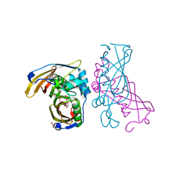
5HZG
 | | The crystal structure of the strigolactone-induced AtD14-D3-ASK1 complex | | 分子名称: | (2Z)-2-methylbut-2-ene-1,4-diol, F-box/LRR-repeat MAX2 homolog, SKP1-like protein 1A, ... | | 著者 | Yao, R.F, Ming, Z.H, Yan, L.M, Rao, Z.H, Lou, Z.Y, Xie, D.X. | | 登録日 | 2016-02-02 | | 公開日 | 2016-08-03 | | 最終更新日 | 2016-08-31 | | 実験手法 | X-RAY DIFFRACTION (3.3 Å) | | 主引用文献 | DWARF14 is a non-canonical hormone receptor for strigolactone
Nature, 536, 2016
|
|
4Z9P
 | | Crystal structure of Ebola virus nucleoprotein core domain at 1.8A resolution | | 分子名称: | Nucleoprotein | | 著者 | Guo, Y, Dong, S.S, Yang, P, Li, G.B, Liu, B.C, Yang, C, Rao, Z.H. | | 登録日 | 2015-04-11 | | 公開日 | 2015-05-20 | | 最終更新日 | 2024-03-20 | | 実験手法 | X-RAY DIFFRACTION (1.792 Å) | | 主引用文献 | Insight into the Ebola virus nucleocapsid assembly mechanism: crystal structure of Ebola virus nucleoprotein core domain at 1.8 A resolution.
Protein Cell, 6, 2015
|
|
3BU7
 | | Crystal Structure and Biochemical Characterization of GDOsp, a Gentisate 1,2-Dioxygenase from Silicibacter Pomeroyi | | 分子名称: | FE (II) ION, Gentisate 1,2-dioxygenase | | 著者 | Chen, J, Wang, M.Z, Zhu, G.Y, Zhang, X.C, Rao, Z.H. | | 登録日 | 2008-01-02 | | 公開日 | 2008-08-12 | | 最終更新日 | 2023-08-30 | | 実験手法 | X-RAY DIFFRACTION (2.8 Å) | | 主引用文献 | Crystal structure and mutagenic analysis of GDOsp, a gentisate 1,2-dioxygenase from Silicibacter pomeroyi.
Protein Sci., 17, 2008
|
|
6J19
 | | ATPase | | 分子名称: | ADENOSINE-5'-TRIPHOSPHATE, ESAT-6-like protein EsxB, ESX-1 secretion system protein EccCb1, ... | | 著者 | Wang, S.H, Li, J, Rao, Z.H. | | 登録日 | 2018-12-28 | | 公開日 | 2019-12-04 | | 最終更新日 | 2023-11-22 | | 実験手法 | X-RAY DIFFRACTION (1.978 Å) | | 主引用文献 | Structural insights into substrate recognition by the type VII secretion system.
Protein Cell, 11, 2020
|
|
6J18
 | | ATPase | | 分子名称: | ADENOSINE-5'-TRIPHOSPHATE, ESX-5 secretion system protein EccC5, MAGNESIUM ION | | 著者 | Wang, S.H, Li, J, Rao, Z.H. | | 登録日 | 2018-12-28 | | 公開日 | 2019-12-04 | | 最終更新日 | 2023-11-22 | | 実験手法 | X-RAY DIFFRACTION (2 Å) | | 主引用文献 | Structural insights into substrate recognition by the type VII secretion system.
Protein Cell, 11, 2020
|
|
6JD5
 | | ATPase | | 分子名称: | ADENOSINE-5'-TRIPHOSPHATE, ESX conserved component EccC2. ESX-2 type VII secretion system protein. Possible membrane protein, MAGNESIUM ION | | 著者 | Wang, S.H, Li, J, Rao, Z.H. | | 登録日 | 2019-01-31 | | 公開日 | 2019-12-04 | | 最終更新日 | 2023-11-22 | | 実験手法 | X-RAY DIFFRACTION (2.2 Å) | | 主引用文献 | Structural insights into substrate recognition by the type VII secretion system.
Protein Cell, 11, 2020
|
|
6J17
 | | ATPase | | 分子名称: | ADENOSINE-5'-TRIPHOSPHATE, ESX-3 secretion system protein EccC3, MAGNESIUM ION | | 著者 | Wang, S.H, Li, J, Rao, Z.H. | | 登録日 | 2018-12-28 | | 公開日 | 2019-12-04 | | 最終更新日 | 2023-11-22 | | 実験手法 | X-RAY DIFFRACTION (1.975 Å) | | 主引用文献 | Structural insights into substrate recognition by the type VII secretion system.
Protein Cell, 11, 2020
|
|
6J72
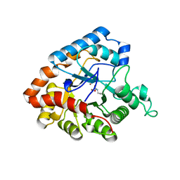
 | | Crystal structure of IniA from Mycobacterium smegmatis with GTP bound | | 分子名称: | GUANOSINE-5'-TRIPHOSPHATE, Isoniazid inducible gene protein IniA, L(+)-TARTARIC ACID, ... | | 著者 | Wang, M.F, Guo, X.Y, Hu, J.J, Li, J, Rao, Z.H. | | 登録日 | 2019-01-16 | | 公開日 | 2019-09-11 | | 最終更新日 | 2024-03-27 | | 実験手法 | X-RAY DIFFRACTION (2.2 Å) | | 主引用文献 | Mycobacterial dynamin-like protein IniA mediates membrane fission.
Nat Commun, 10, 2019
|
|
4IF2
 | |
8J8K
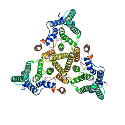 | | Membrane bound PRTase, C3 symmetry, acceptor bound | | 分子名称: | Decaprenyl-phosphate phosphoribosyltransferase, MONO-TRANS, OCTA-CIS DECAPRENYL-PHOSPHATE | | 著者 | Wu, F.Y, Gao, S, Zhang, L, Rao, Z.H. | | 登録日 | 2023-05-01 | | 公開日 | 2024-02-07 | | 最終更新日 | 2024-04-17 | | 実験手法 | ELECTRON MICROSCOPY (3.36 Å) | | 主引用文献 | Structural analysis of phosphoribosyltransferase-mediated cell wall precursor synthesis in Mycobacterium tuberculosis.
Nat Microbiol, 9, 2024
|
|
8J8J
 | | Membrane bound PRTase, C3 symmetry, donor bound | | 分子名称: | (1R)-2-{[(S)-{[(2S)-2,3-dihydroxypropyl]oxy}(hydroxy)phosphoryl]oxy}-1-[(hexadecanoyloxy)methyl]ethyl (9Z)-octadec-9-enoate, 1-O-pyrophosphono-5-O-phosphono-alpha-D-ribofuranose, Decaprenyl-phosphate phosphoribosyltransferase, ... | | 著者 | Wu, F.Y, Gao, S, Zhang, L, Rao, Z.H. | | 登録日 | 2023-05-01 | | 公開日 | 2024-02-07 | | 最終更新日 | 2024-04-17 | | 実験手法 | ELECTRON MICROSCOPY (2.76 Å) | | 主引用文献 | Structural analysis of phosphoribosyltransferase-mediated cell wall precursor synthesis in Mycobacterium tuberculosis.
Nat Microbiol, 9, 2024
|
|
5GKA
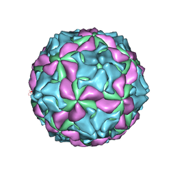 | | cryo-EM structure of human Aichi virus | | 分子名称: | Genome polyprotein, capsid protein VP0, capsid protein VP1 | | 著者 | Zhu, L, Wang, X.X, Ren, J.S, Tuthill, T.J, Fry, E.E, Rao, Z.H, Stuart, D.I. | | 登録日 | 2016-07-04 | | 公開日 | 2016-09-21 | | 最終更新日 | 2024-03-27 | | 実験手法 | ELECTRON MICROSCOPY (3.7 Å) | | 主引用文献 | Structure of human Aichi virus and implications for receptor binding
Nat Microbiol, 1, 2016
|
|
4IKA
 | | Crystal structure of EV71 3Dpol-VPg | | 分子名称: | 3Dpol, NICKEL (II) ION, VPg | | 著者 | Chen, C, Wang, Y.X, Lou, Z.Y. | | 登録日 | 2012-12-25 | | 公開日 | 2013-09-04 | | 最終更新日 | 2023-11-08 | | 実験手法 | X-RAY DIFFRACTION (2.7 Å) | | 主引用文献 | Crystal structure of enterovirus 71 RNA-dependent RNA polymerase complexed with its protein primer VPg: implication for a trans mechanism of VPg uridylylation
J.Virol., 87, 2013
|
|
3RJX
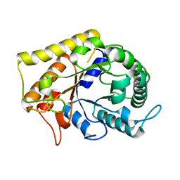 | | Crystal Structure of Hyperthermophilic Endo-Beta-1,4-glucanase | | 分子名称: | Endoglucanase FnCel5A | | 著者 | Zheng, B.S, Yang, W, Zhao, X.Y, Wang, Y.G, Lou, Z.Y, Rao, Z.H, Feng, Y. | | 登録日 | 2011-04-15 | | 公開日 | 2011-12-14 | | 最終更新日 | 2024-03-20 | | 実験手法 | X-RAY DIFFRACTION (2.4 Å) | | 主引用文献 | Crystal structure of hyperthermophilic endo-beta-1,4-glucanase: implications for catalytic mechanism and thermostability.
J.Biol.Chem., 287, 2012
|
|
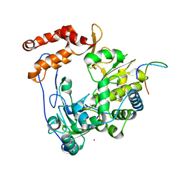
3S0Z
 | | Crystal structure of New Delhi Metallo-beta-lactamase (NDM-1) | | 分子名称: | Metallo-beta-lactamase, ZINC ION | | 著者 | Guo, Y, Wang, J, Niu, G.J, Shui, W.Q, Sun, Y.N, Lou, Z.Y, Rao, Z.H. | | 登録日 | 2011-05-13 | | 公開日 | 2011-06-01 | | 最終更新日 | 2023-11-01 | | 実験手法 | X-RAY DIFFRACTION (2.5 Å) | | 主引用文献 | A structural view of the antibiotic degradation enzyme NDM-1 from a superbug.
Protein Cell, 2011
|
|
4O7P
 | | Crystal structure of Mycobacterium tuberculosis maltose kinase MaK complexed with maltose | | 分子名称: | 2-[BIS-(2-HYDROXY-ETHYL)-AMINO]-2-HYDROXYMETHYL-PROPANE-1,3-DIOL, Maltokinase, SULFATE ION, ... | | 著者 | Li, J, Guan, X.T, Rao, Z.H. | | 登録日 | 2013-12-26 | | 公開日 | 2014-10-22 | | 最終更新日 | 2024-02-28 | | 実験手法 | X-RAY DIFFRACTION (2.9 Å) | | 主引用文献 | Homotypic dimerization of a maltose kinase for molecular scaffolding.
Sci Rep, 4, 2014
|
|
4RLW
 | | Crystal Structure of (3R)-hydroxyacyl-ACP dehydratase HadAB hetero-dimer from Mycobacterium tuberculosis complexed with Butein | | 分子名称: | (2E)-1-(2,4-dihydroxyphenyl)-3-(3,4-dihydroxyphenyl)prop-2-en-1-one, (3R)-hydroxyacyl-ACP dehydratase subunit HadA, (3R)-hydroxyacyl-ACP dehydratase subunit HadB, ... | | 著者 | Li, J, Dong, Y, Rao, Z.H. | | 登録日 | 2014-10-18 | | 公開日 | 2015-10-21 | | 最終更新日 | 2023-09-20 | | 実験手法 | X-RAY DIFFRACTION (2.196 Å) | | 主引用文献 | Molecular basis for the inhibition of beta-hydroxyacyl-ACP dehydratase HadAB complex from Mycobacterium tuberculosis by flavonoid inhibitors.
Protein Cell, 6, 2015
|
|
4RLU
 | | Crystal Structure of (3R)-hydroxyacyl-ACP dehydratase HadAB hetero-dimer from Mycobacterium tuberculosis complexed with 2',4,4'-trihydroxychalcone | | 分子名称: | (3R)-hydroxyacyl-ACP dehydratase subunit HadA, (3R)-hydroxyacyl-ACP dehydratase subunit HadB, 2',4,4'-TRIHYDROXYCHALCONE, ... | | 著者 | Li, J, Dong, Y, Rao, Z.H. | | 登録日 | 2014-10-18 | | 公開日 | 2015-10-21 | | 最終更新日 | 2023-09-20 | | 実験手法 | X-RAY DIFFRACTION (2.198 Å) | | 主引用文献 | Molecular basis for the inhibition of beta-hydroxyacyl-ACP dehydratase HadAB complex from Mycobacterium tuberculosis by flavonoid inhibitors.
Protein Cell, 6, 2015
|
|


