3X33
 
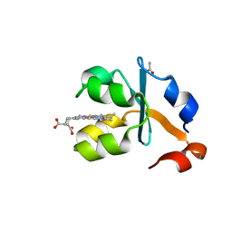 | | Crystal structure of the oxidized form of the solubilized domain of porcine cytochrome b5 in form 2 crystal | | 分子名称: | ACETATE ION, Cytochrome b5, PROTOPORPHYRIN IX CONTAINING FE | | 著者 | Hirano, Y, Kimura, S, Tamada, T. | | 登録日 | 2015-01-14 | | 公開日 | 2015-07-15 | | 最終更新日 | 2023-11-08 | | 実験手法 | X-RAY DIFFRACTION (0.93 Å) | | 主引用文献 | High-resolution crystal structures of the solubilized domain of porcine cytochrome b5.
Acta Crystallogr.,Sect.D, 71, 2015
|
|
3X34
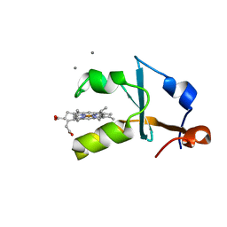 | | Crystal structure of the reduced form of the solubilized domain of porcine cytochrome b5 in form 1 crystal | | 分子名称: | CALCIUM ION, Cytochrome b5, PROTOPORPHYRIN IX CONTAINING FE | | 著者 | Hirano, Y, Kimura, S, Tamada, T. | | 登録日 | 2015-01-14 | | 公開日 | 2015-07-15 | | 最終更新日 | 2023-11-08 | | 実験手法 | X-RAY DIFFRACTION (0.76 Å) | | 主引用文献 | High-resolution crystal structures of the solubilized domain of porcine cytochrome b5.
Acta Crystallogr.,Sect.D, 71, 2015
|
|
3X32
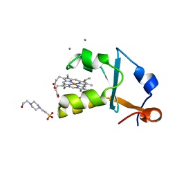 | | Crystal structure of the oxidized form of the solubilized domain of porcine cytochrome b5 in form 1 crystal | | 分子名称: | 4-(2-HYDROXYETHYL)-1-PIPERAZINE ETHANESULFONIC ACID, CALCIUM ION, Cytochrome b5, ... | | 著者 | Hirano, Y, Kimura, S, Tamada, T. | | 登録日 | 2015-01-14 | | 公開日 | 2015-07-15 | | 最終更新日 | 2023-11-08 | | 実験手法 | X-RAY DIFFRACTION (0.83 Å) | | 主引用文献 | High-resolution crystal structures of the solubilized domain of porcine cytochrome b5.
Acta Crystallogr.,Sect.D, 71, 2015
|
|
5Y6T
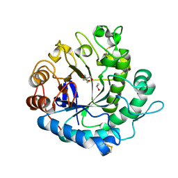 | | Crystal structure of endo-1,4-beta-mannanase from Eisenia fetida | | 分子名称: | 2-AMINO-2-HYDROXYMETHYL-PROPANE-1,3-DIOL, ISOPROPYL ALCOHOL, endo-1,4-beta-mannanase | | 著者 | Hirano, Y, Ueda, M, Tamada, T. | | 登録日 | 2017-08-15 | | 公開日 | 2018-06-27 | | 最終更新日 | 2023-11-22 | | 実験手法 | X-RAY DIFFRACTION (1.7 Å) | | 主引用文献 | Gene cloning, expression, and X-ray crystallographic analysis of a beta-mannanase from Eisenia fetida.
Enzyme.Microb.Technol., 117, 2018
|
|
5N6O
 | | Wild type human Rac1-GDP | | 分子名称: | GUANOSINE-5'-DIPHOSPHATE, MAGNESIUM ION, Ras-related C3 botulinum toxin substrate 1 | | 著者 | Cherfils, J, Ferrandez, Y. | | 登録日 | 2017-02-15 | | 公開日 | 2017-12-27 | | 最終更新日 | 2024-05-08 | | 実験手法 | X-RAY DIFFRACTION (2.59 Å) | | 主引用文献 | Allosteric inhibition of the guanine nucleotide exchange factor DOCK5 by a small molecule.
Sci Rep, 7, 2017
|
|
6H7E
 | | GEF regulatory domain | | 分子名称: | ADENOSINE-3',5'-CYCLIC-MONOPHOSPHATE, SULFATE ION, cDNA FLJ56134, ... | | 著者 | Ferrandez, Y, Cherfils, J, Peurois, F. | | 登録日 | 2018-07-31 | | 公開日 | 2020-02-19 | | 最終更新日 | 2023-12-20 | | 実験手法 | X-RAY DIFFRACTION (2.3 Å) | | 主引用文献 | Membranes prime the RapGEF EPAC1 to transduce cAMP signaling.
Nat Commun, 14, 2023
|
|
6IQE
 | | Human prohibitin 2 | | 分子名称: | Prohibitin-2 | | 著者 | Hirano, Y, Koshiba, T, Tamada, T. | | 登録日 | 2018-11-07 | | 公開日 | 2019-09-25 | | 最終更新日 | 2023-11-22 | | 実験手法 | X-RAY DIFFRACTION (1.701 Å) | | 主引用文献 | Structural Basis of Mitochondrial Scaffolds by Prohibitin Complexes: Insight into a Role of the Coiled-Coil Region.
Iscience, 19, 2019
|
|
6L0V
 | | Structure of RLD2 BRX domain bound to LZY3 CCL motif | | 分子名称: | 1,2-ETHANEDIOL, DI(HYDROXYETHYL)ETHER, NGR2, ... | | 著者 | Hirano, Y, Futrutani, M, Nishimura, T, Taniguchi, M, Morita, M.T, Hakoshima, T. | | 登録日 | 2019-09-27 | | 公開日 | 2020-02-05 | | 最終更新日 | 2024-03-27 | | 実験手法 | X-RAY DIFFRACTION (1.347 Å) | | 主引用文献 | Polar recruitment of RLD by LAZY1-like protein during gravity signaling in root branch angle control.
Nat Commun, 11, 2020
|
|
6L0W
 | | Structure of RLD2 BRX domain bound to LZY3 CCL motif | | 分子名称: | 1,2-ETHANEDIOL, CITRATE ANION, NGR2, ... | | 著者 | Hirano, Y, Futrutani, M, Nishimura, T, Taniguchi, M, Morita, M.T, Hakoshima, T. | | 登録日 | 2019-09-27 | | 公開日 | 2020-02-05 | | 実験手法 | X-RAY DIFFRACTION (1.591 Å) | | 主引用文献 | Polar recruitment of RLD by LAZY1-like protein during gravity signaling in root branch angle control.
Nat Commun, 11, 2020
|
|
5WQR
 | | High resolution structure of high-potential iron-sulfur protein in the reduced state | | 分子名称: | GLYCEROL, High-potential iron-sulfur protein, IRON/SULFUR CLUSTER, ... | | 著者 | Ohno, H, Takeda, K, Niwa, S, Tsujinaka, T, Hanazono, Y, Hirano, Y, Miki, K. | | 登録日 | 2016-11-28 | | 公開日 | 2017-06-07 | | 最終更新日 | 2023-11-08 | | 実験手法 | X-RAY DIFFRACTION (0.8 Å) | | 主引用文献 | Crystallographic characterization of the high-potential iron-sulfur protein in the oxidized state at 0.8 angstrom resolution
PLoS ONE, 12, 2017
|
|
1YGJ
 | | Crystal Structure of Pyridoxal Kinase in Complex with Roscovitine and Derivatives | | 分子名称: | (2R)-2-({6-[BENZYL(METHYL)AMINO]-9-ISOPROPYL-9H-PURIN-2-YL}AMINO)BUTAN-1-OL, Pyridoxal kinase | | 著者 | Tang, L, Li, M.-H, Cao, P, Wang, F, Chang, W.-R, Bach, S, Reinhardt, J, Ferandin, Y, Koken, M, Galons, H, Wan, Y, Gray, N, Meijer, L, Jiang, T, Liang, D.-C. | | 登録日 | 2005-01-05 | | 公開日 | 2005-07-05 | | 最終更新日 | 2024-03-13 | | 実験手法 | X-RAY DIFFRACTION (2.7 Å) | | 主引用文献 | Crystal structure of pyridoxal kinase in complex with roscovitine and derivatives
J.Biol.Chem., 280, 2005
|
|
1YHJ
 | | Crystal Structure of Pyridoxal Kinase in Complex with Roscovitine and Derivatives | | 分子名称: | (2R)-2-{[6-(BENZYLOXY)-9-ISOPROPYL-9H-PURIN-2-YL]AMINO}BUTAN-1-OL, Pyridoxal Kinase | | 著者 | Tang, L, Li, M.-H, Cao, P, Wang, F, Chang, W.-R, Bach, S, Reinhardt, J, Ferandin, Y, Koken, M, Galons, H, Wan, Y, Gray, N, Meijer, L, Jiang, T, Liang, D.-C. | | 登録日 | 2005-01-09 | | 公開日 | 2005-07-05 | | 最終更新日 | 2024-03-13 | | 実験手法 | X-RAY DIFFRACTION (2.8 Å) | | 主引用文献 | Crystal structure of pyridoxal kinase in complex with roscovitine and derivatives.
J.Biol.Chem., 280, 2005
|
|
1YGK
 | | Crystal Structure of Pyridoxal Kinase in Complex with Roscovitine and Derivatives | | 分子名称: | Pyridoxal kinase, R-ROSCOVITINE | | 著者 | Tang, L, Li, M.-H, Cao, P, Wang, F, Chang, W.-R, Bach, S, Reinhardt, J, Ferandin, Y, Koken, M, Galons, H, Wan, Y, Gray, N, Meijer, L, Jiang, T, Liang, D.-C. | | 登録日 | 2005-01-05 | | 公開日 | 2005-07-05 | | 最終更新日 | 2024-03-13 | | 実験手法 | X-RAY DIFFRACTION (2.6 Å) | | 主引用文献 | Crystal structure of pyridoxal kinase in complex with roscovitine and derivatives
J.Biol.Chem., 280, 2005
|
|
6CQF
 | | Crystal structure of HPK1 in complex an inhibitor G1858 | | 分子名称: | Mitogen-activated protein kinase kinase kinase kinase 1, N-{2-(3,3-difluoropyrrolidin-1-yl)-6-[(3R)-pyrrolidin-3-yl]pyrimidin-4-yl}-1-(propan-2-yl)-1H-pyrazolo[4,3-c]pyridin-6-amine | | 著者 | Wu, P, Lehoux, I, Mortara, K, Franke, Y, Chan, B.K, Wang, W. | | 登録日 | 2018-03-15 | | 公開日 | 2018-12-19 | | 最終更新日 | 2024-03-13 | | 実験手法 | X-RAY DIFFRACTION (2.246 Å) | | 主引用文献 | Hematopoietic Progenitor Kinase-1 Structure in a Domain-Swapped Dimer.
Structure, 27, 2019
|
|
5YGE
 | | ArgA complexed with AceCoA and glutamate | | 分子名称: | ACETYL COENZYME *A, Amino-acid acetyltransferase, CACODYLIC ACID, ... | | 著者 | Yang, X, Wu, L, Ran, Y, Xu, A, Zhang, B, Yang, X, Zhang, R, Rao, Z, Li, J. | | 登録日 | 2017-09-22 | | 公開日 | 2017-10-11 | | 最終更新日 | 2024-03-27 | | 実験手法 | X-RAY DIFFRACTION (2.039 Å) | | 主引用文献 | Crystal structure of l-glutamate N-acetyltransferase ArgA from Mycobacterium tuberculosis
Biochim. Biophys. Acta, 1865, 2017
|
|
5W96
 | | Solution structure of phage derived peptide inhibitor of frizzled 7 receptor | | 分子名称: | Fz7 binding peptide | | 著者 | Nile, A.H, de Sousa e Melo, F, Mukund, S, Piskol, R, Hansen, S, Zhou, L, Zhang, Y, Fu, Y, Gogol, E.B, Komuves, L.G, Modrusan, Z, Angers, S, Franke, Y, Koth, C, Fairbrother, W.J, Wang, W, de Sauvage, F.J, Hannoush, R.N. | | 登録日 | 2017-06-22 | | 公開日 | 2018-04-18 | | 最終更新日 | 2023-06-14 | | 実験手法 | SOLUTION NMR | | 主引用文献 | A selective peptide inhibitor of Frizzled 7 receptors disrupts intestinal stem cells.
Nat. Chem. Biol., 14, 2018
|
|
5WQQ
 | | High resolution structure of high-potential iron-sulfur protein in the oxidized state | | 分子名称: | GLYCEROL, High-potential iron-sulfur protein, IRON/SULFUR CLUSTER, ... | | 著者 | Ohno, H, Takeda, K, Niwa, S, Tsujinaka, T, Hanazono, Y, Hirano, Y, Miki, K. | | 登録日 | 2016-11-28 | | 公開日 | 2017-06-07 | | 最終更新日 | 2023-11-08 | | 実験手法 | X-RAY DIFFRACTION (0.8 Å) | | 主引用文献 | Crystallographic characterization of the high-potential iron-sulfur protein in the oxidized state at 0.8 angstrom resolution
PLoS ONE, 12, 2017
|
|
8RIJ
 | | Discovery of the first orally bioavailable ADAMTS7 inhibitor BAY-9835 | | 分子名称: | CALCIUM ION, CHLORIDE ION, DI(HYDROXYETHYL)ETHER, ... | | 著者 | Schafer, M, Meibom, D, Wasnaire, P, Beyer, K, Broehl, A, Cancho-Grande, Y, Elowe, N, Henninger, K, Johannes, S, Jungmann, N, Krainz, T, Lindner, N, Maassen, S, MacDonald, B, Menshykau, D, Mittendorf, J, Sanchez, G, Stefan, E, Torge, A, Xing, Y, Zubov, D. | | 登録日 | 2023-12-18 | | 公開日 | 2024-02-21 | | 最終更新日 | 2024-03-06 | | 実験手法 | X-RAY DIFFRACTION (1.96 Å) | | 主引用文献 | BAY-9835: Discovery of the First Orally Bioavailable ADAMTS7 Inhibitor.
J.Med.Chem., 67, 2024
|
|
3AAC
 | | Small heat shock protein hsp14.0 with the mutations of I120F and I122F in the form II crystal | | 分子名称: | Putative uncharacterized protein ST1653 | | 著者 | Takeda, K, Hayashi, T, Abe, T, Hirano, Y, Hanazono, Y, Yohda, M, Miki, K. | | 登録日 | 2009-11-13 | | 公開日 | 2010-11-17 | | 最終更新日 | 2024-03-13 | | 実験手法 | X-RAY DIFFRACTION (2.4 Å) | | 主引用文献 | Dimer structure and conformational variability in the N-terminal region of an archaeal small heat shock protein, StHsp14.0
J.Struct.Biol., 174, 2011
|
|
3AAB
 | | Small heat shock protein hsp14.0 with the mutations of I120F and I122F in the form I crystal | | 分子名称: | GLYCEROL, ISOPROPYL ALCOHOL, Putative uncharacterized protein ST1653 | | 著者 | Takeda, K, Hayashi, T, Abe, T, Hirano, Y, Hanazono, Y, Yohda, M, Miki, K. | | 登録日 | 2009-11-13 | | 公開日 | 2010-11-17 | | 最終更新日 | 2024-03-13 | | 実験手法 | X-RAY DIFFRACTION (1.851 Å) | | 主引用文献 | Dimer structure and conformational variability in the N-terminal region of an archaeal small heat shock protein, StHsp14.0
J.Struct.Biol., 174, 2011
|
|
3A39
 | | Crystal Structure of High-Potential Iron-Sulfur Protein from Thermochromatium tepidum at 0.72 angstrom resolution | | 分子名称: | GLYCEROL, High-potential iron-sulfur protein, IRON/SULFUR CLUSTER, ... | | 著者 | Takeda, K, Kusumoto, K, Hirano, Y, Miki, K. | | 登録日 | 2009-06-11 | | 公開日 | 2009-10-27 | | 最終更新日 | 2023-11-01 | | 実験手法 | X-RAY DIFFRACTION (0.72 Å) | | 主引用文献 | Detailed assessment of X-ray induced structural perturbation in a crystalline state protein.
J.Struct.Biol., 169, 2010
|
|
7MDP
 | | KRas G12C in complex with G-2897 | | 分子名称: | 1,2-ETHANEDIOL, 2,5,8,11,14,17-HEXAOXANONADECAN-19-OL, 4-(trifluoromethyl)-1,3-benzothiazol-2-amine, ... | | 著者 | Oh, A, Frank, Y, Wang, W. | | 登録日 | 2021-04-05 | | 公開日 | 2022-03-16 | | 最終更新日 | 2023-10-18 | | 実験手法 | X-RAY DIFFRACTION (1.96 Å) | | 主引用文献 | Conformation-locking antibodies for the discovery and characterization of KRAS inhibitors.
Nat.Biotechnol., 40, 2022
|
|
3BHT
 | | Structure of phosphorylated Thr160 CDK2/cyclin A in complex with the inhibitor meriolin 3 | | 分子名称: | 4-(4-methoxy-1H-pyrrolo[2,3-b]pyridin-3-yl)pyrimidin-2-amine, Cell division protein kinase 2, Cyclin-A2, ... | | 著者 | Echalier, A, Bettayeb, K, Ferandin, Y, Lozach, O, Clement, M, Valette, A, Liger, F, Marquet, B, Morris, J.C, Endicott, J.A, Joseph, B, Meijer, L. | | 登録日 | 2007-11-29 | | 公開日 | 2008-02-12 | | 最終更新日 | 2011-07-13 | | 実験手法 | X-RAY DIFFRACTION (2 Å) | | 主引用文献 | Meriolins, a new class of cell death inducing kinase inhibitors with enhanced selectivity for cyclin-dependent kinases
Cancer Res., 67, 2007
|
|
3BHV
 | | Structure of phosphorylated Thr160 CDK2/cyclin A in complex with the inhibitor variolin B | | 分子名称: | 9-amino-5-(2-aminopyrimidin-4-yl)pyrido[3',2':4,5]pyrrolo[1,2-c]pyrimidin-4-ol, Cell division protein kinase 2, Cyclin-A2, ... | | 著者 | Echalier, A, Bettayeb, K, Ferandin, Y, Lozach, O, Clement, M, Valette, A, Liger, F, Marquet, B, Morris, J.C, Endicott, J.A, Joseph, B, Meijer, L. | | 登録日 | 2007-11-29 | | 公開日 | 2008-02-12 | | 最終更新日 | 2011-07-13 | | 実験手法 | X-RAY DIFFRACTION (2.1 Å) | | 主引用文献 | Meriolins, a new class of cell death inducing kinase inhibitors with enhanced selectivity for cyclin-dependent kinases
Cancer Res., 67, 2007
|
|
2Z5C
 | | Crystal Structure of a Novel Chaperone Complex for Yeast 20S Proteasome Assembly | | 分子名称: | Proteasome component PUP2, Protein YPL144W, Uncharacterized protein YLR021W | | 著者 | Yashiroda, H, Mizushima, T, Okamoto, K, Kameyama, T, Hayashi, H, Kishimoto, T, Kasahara, M, Kurimoto, E, Sakata, E, Suzuki, A, Hirano, Y, Murata, S, Kato, K, Yamane, T, Tanaka, K. | | 登録日 | 2007-07-03 | | 公開日 | 2008-01-22 | | 最終更新日 | 2023-11-01 | | 実験手法 | X-RAY DIFFRACTION (2.9 Å) | | 主引用文献 | Crystal structure of a chaperone complex that contributes to the assembly of yeast 20S proteasomes
Nat.Struct.Mol.Biol., 15, 2008
|
|


