Loading
PDBj
メニューPDBj@FacebookPDBj@TwitterPDBj@YouTubewwPDB FoundationwwPDB
RCSB PDBPDBeBMRBAdv. SearchSearch help
Search by PDB author
1VKB
DownloadVisualize
BU of 1vkb by Molmil
Crystal structure of an aig2-like protein (a2ld1, ggact, mgc7867) from mus musculus at 1.90 A resolution
分子名称: FORMIC ACID, hypothetical protein
著者Joint Center for Structural Genomics (JCSG)
登録日2004-05-07
公開日2004-05-18
最終更新日2023-01-25
実験手法X-RAY DIFFRACTION (1.9 Å)
主引用文献Crystal structure of a conserved hypothetical protein (gi: 13879369) from Mouse at 1.90 A resolution reveals a new fold.
Proteins, 61, 2005
1VKY
DownloadVisualize
BU of 1vky by Molmil
Crystal structure of S-adenosylmethionine tRNA ribosyltransferase (TM0574) from Thermotoga maritima at 2.00 A resolution
分子名称: S-adenosylmethionine:tRNA ribosyltransferase-isomerase, UNKNOWN LIGAND
著者Joint Center for Structural Genomics (JCSG)
登録日2004-06-28
公開日2004-08-17
最終更新日2024-05-22
実験手法X-RAY DIFFRACTION (2 Å)
主引用文献Crystal structure of S-adenosylmethionine:tRNA ribosyltransferase-isomerase (QueA) from Thermotoga maritima at 2.0 A resolution reveals a new fold.
Proteins, 59, 2005
1VPZ
DownloadVisualize
BU of 1vpz by Molmil
Crystal structure of a putative carbon storage regulator protein (csra, pa0905) from pseudomonas aeruginosa at 2.05 A resolution
分子名称: Carbon storage regulator homolog
著者Joint Center for Structural Genomics (JCSG)
登録日2004-11-24
公開日2004-12-14
最終更新日2023-01-25
実験手法X-RAY DIFFRACTION (2.05 Å)
主引用文献Crystal structure of the global regulatory protein CsrA from Pseudomonas putida at 2.05 A resolution reveals a new fold.
Proteins, 61, 2005
1VL4
DownloadVisualize
BU of 1vl4 by Molmil
CRYSTAL STRUCTURE OF A PUTATIVE MODULATOR OF A DNA GYRASE (TM0727) FROM THERMOTOGA MARITIMA MSB8 AT 1.95 A RESOLUTION
分子名称: pmbA-related protein
著者Joint Center for Structural Genomics (JCSG)
登録日2004-07-09
公開日2004-09-21
最終更新日2024-05-22
実験手法X-RAY DIFFRACTION (1.95 Å)
主引用文献Crystal structure of a putative modulator of DNA gyrase (pmbA) from Thermotoga maritima at 1.95 A resolution reveals a new fold.
Proteins, 61, 2005
1VLR
DownloadVisualize
BU of 1vlr by Molmil
Crystal structure of mRNA decapping enzyme (DcpS) from Mus musculus at 1.83 A resolution
分子名称: 1,2-ETHANEDIOL, mRNA decapping enzyme
著者Joint Center for Structural Genomics (JCSG)
登録日2004-08-10
公開日2004-08-24
最終更新日2023-09-20
実験手法X-RAY DIFFRACTION (1.83 Å)
主引用文献Crystal structure of an Apo mRNA decapping enzyme (DcpS) from Mouse at 1.83 A resolution.
Proteins, 60, 2005
1Z9F
DownloadVisualize
BU of 1z9f by Molmil
Crystal structure of single stranded DNA-binding protein (TM0604) from Thermotoga maritima at 2.60 A resolution
分子名称: Single-strand binding protein
著者Joint Center for Structural Genomics (JCSG)
登録日2005-04-01
公開日2005-04-12
最終更新日2023-09-20
実験手法X-RAY DIFFRACTION (2.3 Å)
主引用文献Crystal structure of a single-stranded DNA-binding protein (TM0604) from Thermotoga maritima at 2.60 A resolution.
Proteins, 63, 2006
7MT4
DownloadVisualize
BU of 7mt4 by Molmil
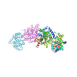
Crystal structure of tryptophan Synthase in complex with F9, NH4+, pH7.8 - alpha aminoacrylate form - E(A-A)
分子名称: 2-({[4-(TRIFLUOROMETHOXY)PHENYL]SULFONYL}AMINO)ETHYL DIHYDROGEN PHOSPHATE, 2-{[(E)-{3-hydroxy-2-methyl-5-[(phosphonooxy)methyl]pyridin-4-yl}methylidene]amino}prop-2-enoic acid, AMMONIUM ION, ...
著者Drago, V, Hilario, E, Dunn, M.F, Mueser, T.C, Mueller, L.J.
登録日2021-05-12
公開日2021-12-29
最終更新日2023-10-18
実験手法X-RAY DIFFRACTION (1.4 Å)
主引用文献Imaging active site chemistry and protonation states: NMR crystallography of the tryptophan synthase alpha-aminoacrylate intermediate.
Proc.Natl.Acad.Sci.USA, 119, 2022
7MT5
DownloadVisualize
BU of 7mt5 by Molmil
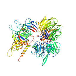
Crystal structure of tryptophan synthase in complex with F9, Cs+, pH7.8 - alpha aminoacrylate form - E(A-A)
分子名称: 2-({[4-(TRIFLUOROMETHOXY)PHENYL]SULFONYL}AMINO)ETHYL DIHYDROGEN PHOSPHATE, 2-{[(E)-{3-hydroxy-2-methyl-5-[(phosphonooxy)methyl]pyridin-4-yl}methylidene]amino}prop-2-enoic acid, CESIUM ION, ...
著者Drago, V, Hilario, E, Dunn, M.F, Mueser, T.C, Mueller, L.J.
登録日2021-05-12
公開日2021-12-29
最終更新日2023-10-18
実験手法X-RAY DIFFRACTION (1.5 Å)
主引用文献Imaging active site chemistry and protonation states: NMR crystallography of the tryptophan synthase alpha-aminoacrylate intermediate.
Proc.Natl.Acad.Sci.USA, 119, 2022
3H50
DownloadVisualize
BU of 3h50 by Molmil
CRYSTAL STRUCTURE OF A TETRACENOMYCIN POLYKETIDE SYNTHESIS PROTEIN (TCMJ) FROM XANTHOMONAS CAMPESTRIS PV. CAMPESTRIS AT 1.60 A RESOLUTION
分子名称: ACETATE ION, Tetracenomycin polyketide synthesis protein, ZINC ION
著者Joint Center for Structural Genomics (JCSG)
登録日2009-04-21
公開日2009-05-05
最終更新日2023-02-01
実験手法X-RAY DIFFRACTION (1.6 Å)
主引用文献Conformational changes associated with the binding of zinc acetate at the putative active site of XcTcmJ, a cupin from Xanthomonas campestris pv. campestris.
Acta Crystallogr.,Sect.F, 66, 2010
1VR3
DownloadVisualize
BU of 1vr3 by Molmil
Crystal structure of Acireductone dioxygenase (13543033) from Mus musculus at 2.06 A resolution
分子名称: Acireductone dioxygenase, ISOPROPYL ALCOHOL, NICKEL (II) ION, ...
著者Joint Center for Structural Genomics (JCSG)
登録日2005-01-28
公開日2005-02-15
最終更新日2023-01-25
実験手法X-RAY DIFFRACTION (2.06 Å)
主引用文献Crystal structure of acireductone dioxygenase (ARD) from Mus musculus at 2.06 angstrom resolution.
Proteins, 64, 2006
7MT6
DownloadVisualize
BU of 7mt6 by Molmil
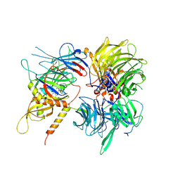
Crystal structure of tryptophan synthase in complex with F9, Cs+, benzimidazole, pH7.8 - alpha aminoacrylate form - E(A-A)(BZI)
分子名称: 2-({[4-(TRIFLUOROMETHOXY)PHENYL]SULFONYL}AMINO)ETHYL DIHYDROGEN PHOSPHATE, 2-{[(E)-{3-hydroxy-2-methyl-5-[(phosphonooxy)methyl]pyridin-4-yl}methylidene]amino}prop-2-enoic acid, BENZIMIDAZOLE, ...
著者Drago, V, Hilario, E, Dunn, M.F, Mueser, T.C, Mueller, L.J.
登録日2021-05-12
公開日2021-12-29
最終更新日2023-10-18
実験手法X-RAY DIFFRACTION (1.7 Å)
主引用文献Imaging active site chemistry and protonation states: NMR crystallography of the tryptophan synthase alpha-aminoacrylate intermediate.
Proc.Natl.Acad.Sci.USA, 119, 2022
1VQR
DownloadVisualize
BU of 1vqr by Molmil
Crystal structure of a virulence factor (cj0248) from campylobacter jejuni subsp. jejuni at 2.25 A resolution
分子名称: hypothetical protein Cj0248
著者Joint Center for Structural Genomics (JCSG)
登録日2004-12-17
公開日2004-12-28
最終更新日2023-01-25
実験手法X-RAY DIFFRACTION (2.25 Å)
主引用文献Crystal structure of virulence factor CJ0248 from Campylobacter jejuni at 2.25 A resolution reveals a new fold.
Proteins, 62, 2006
1VR8
DownloadVisualize
BU of 1vr8 by Molmil
Crystal structure of GTP binding regulator (TM1622) from Thermotoga Maritima at 1.75 A resolution
分子名称: AZIDE ION, GLYCEROL, GTP binding regulator
著者Joint Center for Structural Genomics (JCSG)
登録日2005-02-16
公開日2005-03-15
最終更新日2023-01-25
実験手法X-RAY DIFFRACTION (1.75 Å)
主引用文献Crystal structure of an ORFan protein (TM1622) from Thermotoga maritima at 1.75 A resolution reveals a fold similar to the Ran-binding protein Mog1p.
Proteins, 65, 2006
1VK3
DownloadVisualize
BU of 1vk3 by Molmil
Crystal structure of Phosphoribosylformylglycinamidine synthase II (TM1246) from Thermotoga maritima at 2.15 A resolution
分子名称: CHLORIDE ION, Phosphoribosylformylglycinamidine synthase II
著者Joint Center for Structural Genomics (JCSG)
登録日2004-04-23
公開日2004-05-11
最終更新日2023-01-25
実験手法X-RAY DIFFRACTION (2.15 Å)
主引用文献Crystal structure of phosphoribosylformylglycinamidine synthase II (smPurL) from Thermotoga maritima at 2.15 A resolution.
Proteins, 63, 2006
1ZKG
DownloadVisualize
BU of 1zkg by Molmil
Crystal structure of Transcriptional regulator, TETR family (tm1030) from Thermotoga maritima at 2.30 A resolution
分子名称: UNKNOWN LIGAND, transcriptional regulator, TetR family
著者Joint Center for Structural Genomics (JCSG)
登録日2005-05-02
公開日2005-07-26
最終更新日2023-01-25
実験手法X-RAY DIFFRACTION (2.3 Å)
主引用文献Crystal structure of TM1030 from Thermotoga maritima at 2.3 A resolution reveals molecular details of its transcription repressor function.
Proteins, 68, 2007
1ZCZ
DownloadVisualize
BU of 1zcz by Molmil
Crystal structure of Phosphoribosylaminoimidazolecarboxamide formyltransferase / IMP cyclohydrolase (TM1249) from THERMOTOGA MARITIMA at 1.88 A resolution
分子名称: Bifunctional purine biosynthesis protein purH, POTASSIUM ION, TETRAETHYLENE GLYCOL
著者Joint Center for Structural Genomics (JCSG)
登録日2005-04-13
公開日2005-04-26
最終更新日2023-09-20
実験手法X-RAY DIFFRACTION (1.88 Å)
主引用文献Crystal structure of AICAR transformylase IMP cyclohydrolase (TM1249) from Thermotoga maritima at 1.88 A resolution.
Proteins, 71, 2008
6UD7
DownloadVisualize
BU of 6ud7 by Molmil
Crystal structure of full-length human DCAF15-DDB1(deltaBPB)-DDA1-RBM39 in complex with indisulam
分子名称: DDB1- and CUL4-associated factor 15, DET1- and DDB1-associated protein 1, DNA damage-binding protein 1,DNA damage-binding protein 1, ...
著者Bussiere, D.E, Shu, W, Xie, L, Knapp, M.
登録日2019-09-18
公開日2019-12-18
最終更新日2020-01-22
実験手法X-RAY DIFFRACTION (2.3 Å)
主引用文献Structural basis of indisulam-mediated RBM39 recruitment to DCAF15 E3 ligase complex.
Nat.Chem.Biol., 16, 2020
6UE5
DownloadVisualize
BU of 6ue5 by Molmil
Crystal structure of full-length human DCAF15-DDB1-deltaPBP-DDA1-RBM39 in complex with 4-(aminomethyl)-N-(3-cyano-4-methyl-1H-indol-7-yl)benzenesulfonamide
分子名称: 4-(aminomethyl)-N-(3-cyano-4-methyl-1H-indol-7-yl)benzene-1-sulfonamide, DDB1- and CUL4-associated factor 15, DET1- and DDB1-associated protein 1, ...
著者Knapp, M.S, Shu, W, Xie, L, Bussiere, D.E.
登録日2019-09-20
公開日2019-12-18
最終更新日2023-10-11
実験手法X-RAY DIFFRACTION (2.61 Å)
主引用文献Structural basis of indisulam-mediated RBM39 recruitment to DCAF15 E3 ligase complex.
Nat.Chem.Biol., 16, 2020
1VR0
DownloadVisualize
BU of 1vr0 by Molmil
Crystal structure of putative 2-phosphosulfolactate phosphatase (15026306) from Clostridium acetobutylicum at 2.6 A resolution
分子名称: (2R)-3-SULFOLACTIC ACID, MAGNESIUM ION, Probable 2-phosphosulfolactate phosphatase
著者Joint Center for Structural Genomics (JCSG)
登録日2005-01-26
公開日2005-02-15
最終更新日2023-01-25
実験手法X-RAY DIFFRACTION (2.49 Å)
主引用文献Crystal structure of 2-phosphosulfolactate phosphatase (ComB) from Clostridium acetobutylicum at 2.6 A resolution reveals a new fold with a novel active site.
Proteins, 65, 2006
1VK9
DownloadVisualize
BU of 1vk9 by Molmil
CRYSTAL STRUCTURE OF A DUF1893 family protein (TM1506) FROM THERMOTOGA MARITIMA AT 2.70 A RESOLUTION
分子名称: UNKNOWN LIGAND, ZINC ION, conserved hypothetical protein TM1506
著者Joint Center for Structural Genomics (JCSG)
登録日2004-05-06
公開日2004-05-18
最終更新日2023-01-25
実験手法X-RAY DIFFRACTION (2.7 Å)
主引用文献Crystal structure of an ADP-ribosylated protein with a cytidine deaminase-like fold, but unknown function (TM1506), from Thermotoga maritima at 2.70 A resolution.
Proteins, 71, 2008
1VQ3
DownloadVisualize
BU of 1vq3 by Molmil
Crystal structure of Phosphoribosylformylglycinamidine synthase, purS subunit (EC 6.3.5.3) (TM1244) from Thermotoga maritima at 1.90 A resolution
分子名称: Phosphoribosylformylglycinamidine synthase, purS subunit
著者Joint Center for Structural Genomics (JCSG)
登録日2004-12-15
公開日2004-12-28
最終更新日2023-09-20
実験手法X-RAY DIFFRACTION (1.9 Å)
主引用文献Crystal structure of phosphoribosylformyl-glycinamidine synthase II, PurS subunit (TM1244) from Thermotoga maritima at 1.90 A resolution.
Proteins, 65, 2006
2A6A
DownloadVisualize
BU of 2a6a by Molmil
Crystal structure of Glycoprotein endopeptidase (tm0874) from THERMOTOGA MARITIMA at 2.50 A resolution
分子名称: UNKNOWN LIGAND, hypothetical protein TM0874
著者Joint Center for Structural Genomics (JCSG)
登録日2005-07-02
公開日2005-07-19
最終更新日2023-01-25
実験手法X-RAY DIFFRACTION (2.5 Å)
主引用文献Structure of an essential bacterial protein YeaZ (TM0874) from Thermotoga maritima at 2.5 A resolution.
Acta Crystallogr.,Sect.F, 66, 2010
3H0N
DownloadVisualize
BU of 3h0n by Molmil
Crystal structure of a duf1470 family protein (jann_2411) from jannaschia sp. ccs1 at 1.45 A resolution
分子名称: ACETATE ION, GLYCEROL, NICKEL (II) ION, ...
著者Joint Center for Structural Genomics (JCSG)
登録日2009-04-09
公開日2009-04-21
最終更新日2023-02-01
実験手法X-RAY DIFFRACTION (1.45 Å)
主引用文献The structure of Jann_2411 (DUF1470) from Jannaschia sp. at 1.45  resolution reveals a new fold (the ABATE domain) and suggests its possible role as a transcription regulator.
Acta Crystallogr.,Sect.F, 66, 2010
2FG0
DownloadVisualize
BU of 2fg0 by Molmil
Crystal structure of a putative gamma-d-glutamyl-l-diamino acid endopeptidase (npun_r0659) from nostoc punctiforme pcc 73102 at 1.79 A resolution
分子名称: COG0791: Cell wall-associated hydrolases (invasion-associated proteins), GLYCEROL
著者Joint Center for Structural Genomics (JCSG)
登録日2005-12-20
公開日2006-01-10
最終更新日2023-01-25
実験手法X-RAY DIFFRACTION (1.79 Å)
主引用文献Structural Basis of Murein Peptide Specificity of a gamma-D-Glutamyl-L-Diamino Acid Endopeptidase.
Structure, 17, 2009
2FNO
DownloadVisualize
BU of 2fno by Molmil
Crystal structure of a glutathione s-transferase (atu5508) from agrobacterium tumefaciens str. c58 at 2.00 A resolution
分子名称: AGR_pAT_752p, THIOCYANATE ION
著者Joint Center for Structural Genomics (JCSG)
登録日2006-01-11
公開日2006-02-14
最終更新日2023-01-25
実験手法X-RAY DIFFRACTION (2 Å)
主引用文献Comparative structural analysis of a novel glutathioneS-transferase (ATU5508) from Agrobacterium tumefaciens at 2.0 A resolution.
Proteins, 65, 2006

223166

件を2024-07-31に公開中

PDB statisticsPDBj update infoContact PDBjnumon