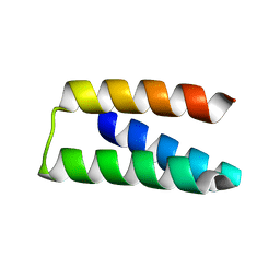
4G4Z
 
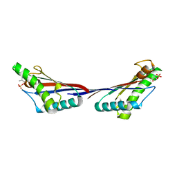 | |
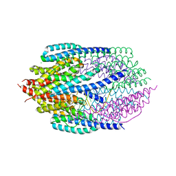
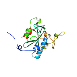
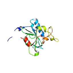
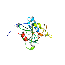
5I2Q
 | | Structure of EF-hand containing protein | | 分子名称: | CALCIUM ION, EF-hand domain-containing protein D2 | | 著者 | Park, K.R, Kwon, M.S, An, J.Y, Lee, J.G, Youn, H.S, Lee, Y, Kang, J.Y, Kim, T.G, Lim, J.J, Park, J.S, Lee, S.H, Song, W.K, Cheong, H, Jun, C, Eom, S.H. | | 登録日 | 2016-02-09 | | 公開日 | 2016-12-28 | | 最終更新日 | 2023-11-08 | | 実験手法 | X-RAY DIFFRACTION (1.935 Å) | | 主引用文献 | Structural implications of Ca(2+)-dependent actin-bundling function of human EFhd2/Swiprosin-1.
Sci Rep, 6, 2016
|
|
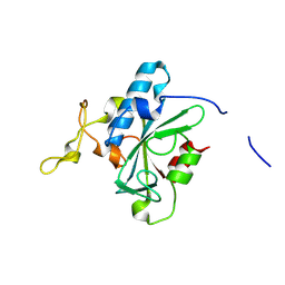
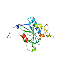
5I2O
 | | Structure of EF-hand containing protein | | 分子名称: | CALCIUM ION, EF-hand domain-containing protein D2 | | 著者 | Park, K.R, Kwon, M.S, An, J.Y, Lee, J.G, Youn, H.S, Lee, Y, Kang, J.Y, Kim, T.G, Lim, J.J, Park, J.S, Lee, S.H, Song, W.K, Cheong, H, Jun, C, Eom, S.H. | | 登録日 | 2016-02-09 | | 公開日 | 2016-12-28 | | 最終更新日 | 2023-11-08 | | 実験手法 | X-RAY DIFFRACTION (1.952 Å) | | 主引用文献 | Structural implications of Ca(2+)-dependent actin-bundling function of human EFhd2/Swiprosin-1.
Sci Rep, 6, 2016
|
|
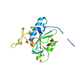
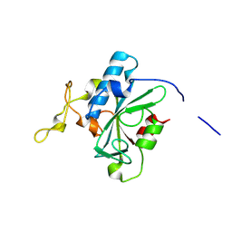
5I2L
 | | Structure of EF-hand containing protein | | 分子名称: | CALCIUM ION, EF-hand domain-containing protein D2 | | 著者 | Park, K.R, Kwon, M.S, An, J.Y, Lee, J.G, Youn, H.S, Lee, Y, Kang, J.Y, Kim, T.G, Lim, J.J, Park, J.S, Lee, S.H, Song, W.K, Cheong, H, Jun, C, Eom, S.H. | | 登録日 | 2016-02-09 | | 公開日 | 2016-12-28 | | 最終更新日 | 2024-03-20 | | 実験手法 | X-RAY DIFFRACTION (1.85 Å) | | 主引用文献 | Structural implications of Ca(2+)-dependent actin-bundling function of human EFhd2/Swiprosin-1.
Sci Rep, 6, 2016
|
|
5Y7D
 | | Crystal structure of human Endothelial-overexpressed LPS associated factor 1 | | 分子名称: | CHLORIDE ION, GLYCEROL, Protein CXorf40A, ... | | 著者 | Park, S.H, Kim, M.J, Park, J.S, Kim, H.J, Han, B.W. | | 登録日 | 2017-08-17 | | 公開日 | 2018-08-22 | | 最終更新日 | 2024-03-27 | | 実験手法 | X-RAY DIFFRACTION (1.71 Å) | | 主引用文献 | Crystal Structure of Human EOLA1 Implies Its Possibility of RNA Binding.
Molecules, 24, 2019
|
|
7E5Z
 | | Dehydrogenase holoenzyme | | 分子名称: | 2-AMINO-5,6-DIMERCAPTO-7-METHYL-3,7,8A,9-TETRAHYDRO-8-OXA-1,3,9,10-TETRAAZA-ANTHRACEN-4-ONE GUANOSINE DINUCLEOTIDE, FE2/S2 (INORGANIC) CLUSTER, FLAVIN MONONUCLEOTIDE, ... | | 著者 | Roh, S.H, Park, J.S, Heo, Y.Y. | | 登録日 | 2021-02-21 | | 公開日 | 2022-02-23 | | 最終更新日 | 2022-03-02 | | 実験手法 | ELECTRON MICROSCOPY (3.6 Å) | | 主引用文献 | Dehydrogenase holoenzyme
To Be Published
|
|
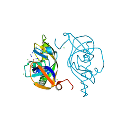
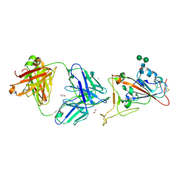
3ZC1
 | |
3ZC0
 | | Structure of AfC3PO - duplex RNA complex | | 分子名称: | 5'-R(*UP*UP*CP*GP*AP*CP*GP*CP*GP*UP*CP*GP*AP*AP*UP*U)-3', AFTRAX, CHLORIDE ION, ... | | 著者 | Parizotto, E.A, Lowe, E.D, Parker, J.S. | | 登録日 | 2012-11-14 | | 公開日 | 2013-01-23 | | 最終更新日 | 2023-12-20 | | 実験手法 | X-RAY DIFFRACTION (2.982 Å) | | 主引用文献 | Structural Basis for Duplex RNA Recognition and Cleavage by Archaeoglobus Fulgidus C3Po.
Nat.Struct.Mol.Biol., 20, 2013
|
|
1IJS
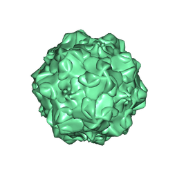 | | CPV (STRAIN D) mutant A300D, complex (VIRAL COAT/DNA), VP2, PH=7.5, T=4 DEGREES C | | 分子名称: | DNA (5'-D(*AP*C)-3'), DNA (5'-D(*CP*CP*AP*CP*CP*CP*CP*AP*A)-3'), PROTEIN (PARVOVIRUS COAT PROTEIN) | | 著者 | Llamas-Saiz, A.L, Agbandje-McKenna, M, Parker, J.S.L, Wahid, A.T.M, Parrish, C.R, Rossmann, M.G. | | 登録日 | 1996-09-12 | | 公開日 | 1996-12-23 | | 最終更新日 | 2024-04-03 | | 実験手法 | X-RAY DIFFRACTION (3.25 Å) | | 主引用文献 | Structural analysis of a mutation in canine parvovirus which controls antigenicity and host range.
Virology, 225, 1996
|
|
1DJM
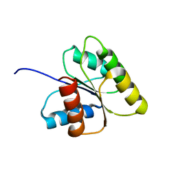 | | SOLUTION STRUCTURE OF BEF3-ACTIVATED CHEY FROM ESCHERICHIA COLI | | 分子名称: | CHEMOTAXIS PROTEIN Y | | 著者 | Cho, H.S, Lee, S.Y, Yan, D, Pan, X, Parkinson, J.S, Kustu, S, Wemmer, D.E, Pelton, J.G. | | 登録日 | 1999-12-03 | | 公開日 | 2000-04-05 | | 最終更新日 | 2024-05-22 | | 実験手法 | SOLUTION NMR | | 主引用文献 | NMR structure of activated CheY.
J.Mol.Biol., 297, 2000
|
|
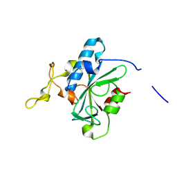
7OPB
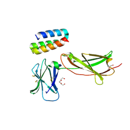 | | IL7R in complex with an antagonist | | 分子名称: | 1,2-ETHANEDIOL, DI(HYDROXYETHYL)ETHER, IL7R binder, ... | | 著者 | Markovic, I, Verschueren, K.H.G, Verstraete, K, Savvides, S.N. | | 登録日 | 2021-05-31 | | 公開日 | 2022-05-11 | | 最終更新日 | 2024-01-31 | | 実験手法 | X-RAY DIFFRACTION (2.144 Å) | | 主引用文献 | Design of protein-binding proteins from the target structure alone.
Nature, 605, 2022
|
|
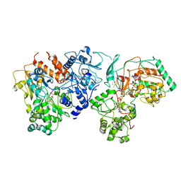
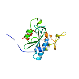
7S5B
 | |
7CM4
 | | Crystal Structure of COVID-19 virus spike receptor-binding domain complexed with a neutralizing antibody CT-P59 | | 分子名称: | 1,2-ETHANEDIOL, IgG heavy chain, IgG light chain, ... | | 著者 | Kim, Y.G, Jeong, J.H, Bae, J.S, Lee, J. | | 登録日 | 2020-07-24 | | 公開日 | 2021-01-20 | | 最終更新日 | 2023-11-29 | | 実験手法 | X-RAY DIFFRACTION (2.71 Å) | | 主引用文献 | A therapeutic neutralizing antibody targeting receptor binding domain of SARS-CoV-2 spike protein.
Nat Commun, 12, 2021
|
|
8JJI
 | | Crystal structure of QR-hNTAQ1 C28S | | 分子名称: | Protein N-terminal glutamine amidohydrolase | | 著者 | Kang, J.M, Han, B.W. | | 登録日 | 2023-05-30 | | 公開日 | 2024-06-26 | | 実験手法 | X-RAY DIFFRACTION (2.206 Å) | | 主引用文献 | Structural study for substrate recognition of human N-terminal glutamine amidohydrolase 1 in the arginine N-degron pathway.
Protein Sci., 33, 2024
|
|
8JJG
 | | Crystal structure of QW-hNTAQ1 C28S | | 分子名称: | Protein N-terminal glutamine amidohydrolase | | 著者 | Kang, J.M, Han, B.W. | | 登録日 | 2023-05-30 | | 公開日 | 2024-06-26 | | 実験手法 | X-RAY DIFFRACTION (1.45 Å) | | 主引用文献 | Structural study for substrate recognition of human N-terminal glutamine amidohydrolase 1 in the arginine N-degron pathway.
Protein Sci., 33, 2024
|
|
8JK1
 | | Crystal structure of QA-hNTAQ1 C28S | | 分子名称: | Protein N-terminal glutamine amidohydrolase | | 著者 | Kang, J.M, Han, B.W. | | 登録日 | 2023-05-31 | | 公開日 | 2024-06-26 | | 実験手法 | X-RAY DIFFRACTION (2.067 Å) | | 主引用文献 | Structural study for substrate recognition of human N-terminal glutamine amidohydrolase 1 in the arginine N-degron pathway.
Protein Sci., 33, 2024
|
|
8JJH
 | | Crystal structure of QH-hNTAQ1 C28S | | 分子名称: | Protein N-terminal glutamine amidohydrolase | | 著者 | Kang, J.M, Han, B.W. | | 登録日 | 2023-05-30 | | 公開日 | 2024-06-26 | | 実験手法 | X-RAY DIFFRACTION (1.61 Å) | | 主引用文献 | Structural study for substrate recognition of human N-terminal glutamine amidohydrolase 1 in the arginine N-degron pathway.
Protein Sci., 33, 2024
|
|
8JK0
 | | Crystal structure of QL-hNTAQ1 C28S | | 分子名称: | Protein N-terminal glutamine amidohydrolase | | 著者 | Kang, J.M, Han, B.W. | | 登録日 | 2023-05-31 | | 公開日 | 2024-06-26 | | 実験手法 | X-RAY DIFFRACTION (1.45 Å) | | 主引用文献 | Structural study for substrate recognition of human N-terminal glutamine amidohydrolase 1 in the arginine N-degron pathway.
Protein Sci., 33, 2024
|
|
8JJU
 | | Crystal structure of QD-hNTAQ1 C28S | | 分子名称: | Protein N-terminal glutamine amidohydrolase | | 著者 | Kang, J.M, Han, B.W. | | 登録日 | 2023-05-31 | | 公開日 | 2024-06-26 | | 実験手法 | X-RAY DIFFRACTION (1.46 Å) | | 主引用文献 | Structural study for substrate recognition of human N-terminal glutamine amidohydrolase 1 in the arginine N-degron pathway.
Protein Sci., 33, 2024
|
|
8JJY
 | | Crystal structure of QN-hNTAQ1 C28S | | 分子名称: | Protein N-terminal glutamine amidohydrolase | | 著者 | Kang, J.M, Han, B.W. | | 登録日 | 2023-05-31 | | 公開日 | 2024-06-26 | | 実験手法 | X-RAY DIFFRACTION (1.69 Å) | | 主引用文献 | Structural study for substrate recognition of human N-terminal glutamine amidohydrolase 1 in the arginine N-degron pathway.
Protein Sci., 33, 2024
|
|
8JJW
 | | Crystal structure of QG-hNTAQ1 C28S | | 分子名称: | MAGNESIUM ION, Protein N-terminal glutamine amidohydrolase | | 著者 | Kang, J.M, Han, B.W. | | 登録日 | 2023-05-31 | | 公開日 | 2024-06-26 | | 実験手法 | X-RAY DIFFRACTION (1.4 Å) | | 主引用文献 | Structural study for substrate recognition of human N-terminal glutamine amidohydrolase 1 in the arginine N-degron pathway.
Protein Sci., 33, 2024
|
|
8JJZ
 | | Crystal structure of QQ-hNTAQ1 C28S | | 分子名称: | Protein N-terminal glutamine amidohydrolase | | 著者 | Kang, J.M, Han, B.W. | | 登録日 | 2023-05-31 | | 公開日 | 2024-06-26 | | 実験手法 | X-RAY DIFFRACTION (2.03 Å) | | 主引用文献 | Structural study for substrate recognition of human N-terminal glutamine amidohydrolase 1 in the arginine N-degron pathway.
Protein Sci., 33, 2024
|
|
8JJF
 | | Crystal structure of QE-hNTAQ1 C28S | | 分子名称: | Protein N-terminal glutamine amidohydrolase | | 著者 | Kang, J.M, Han, B.W. | | 登録日 | 2023-05-30 | | 公開日 | 2024-06-26 | | 実験手法 | X-RAY DIFFRACTION (1.51 Å) | | 主引用文献 | Structural study for substrate recognition of human N-terminal glutamine amidohydrolase 1 in the arginine N-degron pathway.
Protein Sci., 33, 2024
|
|
8JK2
 | | Crystal structure of QF-hNTAQ1 C28S | | 分子名称: | Protein N-terminal glutamine amidohydrolase | | 著者 | Kang, J.M, Han, B.W. | | 登録日 | 2023-05-31 | | 公開日 | 2024-06-26 | | 実験手法 | X-RAY DIFFRACTION (1.742 Å) | | 主引用文献 | Structural study for substrate recognition of human N-terminal glutamine amidohydrolase 1 in the arginine N-degron pathway.
Protein Sci., 33, 2024
|
|
8JJX
 | | Crystal structure of QS-hNTAQ1 C28S | | 分子名称: | Protein N-terminal glutamine amidohydrolase | | 著者 | Kang, J.M, Han, B.W. | | 登録日 | 2023-05-31 | | 公開日 | 2024-06-26 | | 実験手法 | X-RAY DIFFRACTION (1.7 Å) | | 主引用文献 | Structural study for substrate recognition of human N-terminal glutamine amidohydrolase 1 in the arginine N-degron pathway.
Protein Sci., 33, 2024
|
|


