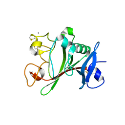
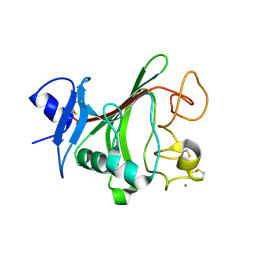
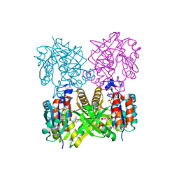
6P7S
 
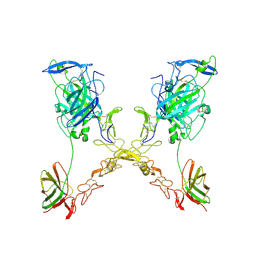
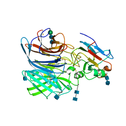 | | Crystal Structure of the Cedar henipavirus Attachment G Glycoprotein globular domain in complex with the receptor ephrin-B1 | | 分子名称: | 2-acetamido-2-deoxy-beta-D-glucopyranose, 2-acetamido-2-deoxy-beta-D-glucopyranose-(1-4)-2-acetamido-2-deoxy-beta-D-glucopyranose, Attachment glycoprotein, ... | | 著者 | Xu, K, Nikolov, D.B, Xu, Y. | | 登録日 | 2019-06-06 | | 公開日 | 2019-09-25 | | 最終更新日 | 2024-10-23 | | 実験手法 | X-RAY DIFFRACTION (3.49 Å) | | 主引用文献 | Structural and functional analyses reveal promiscuous and species specific use of ephrin receptors by Cedar virus.
Proc.Natl.Acad.Sci.USA, 116, 2019
|
|
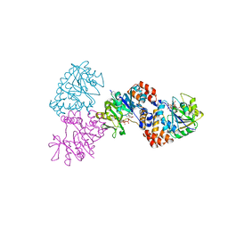
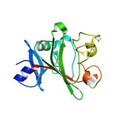
6P7Y
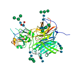 | | Crystal Structure of the Cedar henipavirus Attachment G Glycoprotein globular domain in complex with the receptor ephrin-B2 | | 分子名称: | 2-acetamido-2-deoxy-beta-D-glucopyranose, 2-acetamido-2-deoxy-beta-D-glucopyranose-(1-4)-[alpha-L-fucopyranose-(1-6)]2-acetamido-2-deoxy-beta-D-glucopyranose, Attachment glycoprotein, ... | | 著者 | Xu, K, Nikolov, D.B, Xu, Y. | | 登録日 | 2019-06-06 | | 公開日 | 2019-09-25 | | 最終更新日 | 2024-11-20 | | 実験手法 | X-RAY DIFFRACTION (2.844 Å) | | 主引用文献 | Structural and functional analyses reveal promiscuous and species specific use of ephrin receptors by Cedar virus.
Proc.Natl.Acad.Sci.USA, 116, 2019
|
|
1NUK
 | |
1Z3U
 | |
1Z3S
 | |
1MP4
 | | W224H VARIANT OF S. ENTERICA RmlA | | 分子名称: | URIDINE-5'-DIPHOSPHATE-GLUCOSE, W224H Variant of S. Enterica RmlA Bound to UDP-Glucose | | 著者 | Barton, W.A, Biggins, J.B, Jiang, J, Thorson, J.S, Nikolov, D.B. | | 登録日 | 2002-09-11 | | 公開日 | 2002-10-09 | | 最終更新日 | 2024-02-14 | | 実験手法 | X-RAY DIFFRACTION (2.2 Å) | | 主引用文献 | Expanding pyrimidine diphosphosugar libraries via structure-based nucleotidylyltransferase engineering
Proc.Natl.Acad.Sci.USA, 99, 2002
|
|
1MP5
 | | Y177F VARIANT OF S. ENTERICA RmlA | | 分子名称: | URIDINE-5'-DIPHOSPHATE-GLUCOSE, Y177F VARIANT OF S. ENTERICA RmlA BOUND TO UDP-GLUCOSE | | 著者 | Barton, W.A, Biggins, J.B, Jiang, J, Thorson, J.S, Nikolov, D.B. | | 登録日 | 2002-09-11 | | 公開日 | 2002-10-09 | | 最終更新日 | 2024-02-14 | | 実験手法 | X-RAY DIFFRACTION (2.75 Å) | | 主引用文献 | Expanding pyrimidine diphosphosugar libraries via structure-based nucleotidylyltransferase engineering
Proc.Natl.Acad.Sci.USA, 99, 2002
|
|
1P8T
 | | Crystal structure of Nogo-66 Receptor | | 分子名称: | 2-acetamido-2-deoxy-alpha-D-glucopyranose, 2-acetamido-2-deoxy-beta-D-glucopyranose, Reticulon 4 receptor | | 著者 | Barton, W.A, Liu, B.P, Tzvetkova, D, Jeffrey, P.D, Fournier, A.E, Sah, D, Cate, R, Strittmatter, S.M, Nikolov, D.B. | | 登録日 | 2003-05-07 | | 公開日 | 2003-05-20 | | 最終更新日 | 2024-11-13 | | 実験手法 | X-RAY DIFFRACTION (3.2 Å) | | 主引用文献 | Structure and axon outgrowth inhibitor binding of the Nogo-66 receptor and related proteins
Embo J., 22, 2003
|
|
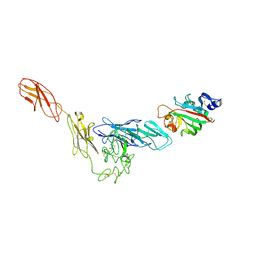
4JYO
 | | Structural basis for angiopoietin-1 mediated signaling initiation | | 分子名称: | Angiopoietin-1, CALCIUM ION | | 著者 | Yu, X, Seegar, T.C.M, Dalton, A.C, Tzvetkova-Robev, D, Goldgur, Y, Nikolov, D.B, Barton, W.A. | | 登録日 | 2013-03-31 | | 公開日 | 2013-05-08 | | 最終更新日 | 2024-11-06 | | 実験手法 | X-RAY DIFFRACTION (2.5 Å) | | 主引用文献 | Structural basis for angiopoietin-1-mediated signaling initiation.
Proc.Natl.Acad.Sci.USA, 110, 2013
|
|
4JZC
 | | Angiopoietin-2 fibrinogen domain TAG mutant | | 分子名称: | Angiopoietin-2 | | 著者 | Yu, X, Seegar, T.C.M, Dalton, A.C, Tzvetkova-Robev, D, Goldgur, Y, Nikolov, D.B, Barton, W.A. | | 登録日 | 2013-04-02 | | 公開日 | 2013-05-08 | | 最終更新日 | 2024-11-20 | | 実験手法 | X-RAY DIFFRACTION (1.9 Å) | | 主引用文献 | Structural basis for angiopoietin-1-mediated signaling initiation.
Proc.Natl.Acad.Sci.USA, 110, 2013
|
|
4K0V
 | | Structural basis for angiopoietin-1 mediated signaling initiation | | 分子名称: | Angiopoietin-1, TEK tyrosine kinase variant | | 著者 | Yu, X, Seegar, T.C.M, Dalton, A.C, Tzvetkova-Robev, D, Goldgur, Y, Nikolov, D.B, Barton, W.A. | | 登録日 | 2013-04-04 | | 公開日 | 2013-05-08 | | 最終更新日 | 2024-11-06 | | 実験手法 | X-RAY DIFFRACTION (4.51 Å) | | 主引用文献 | Structural basis for angiopoietin-1-mediated signaling initiation.
Proc.Natl.Acad.Sci.USA, 110, 2013
|
|
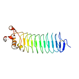
4PLM
 | | Crystal Structure of Chicken Netrin-1 (LN-LE3) | | 分子名称: | 2-acetamido-2-deoxy-beta-D-glucopyranose, CALCIUM ION, CHLORIDE ION, ... | | 著者 | Xu, K, Nikolov, D.B. | | 登録日 | 2014-05-18 | | 公開日 | 2014-06-18 | | 最終更新日 | 2024-10-16 | | 実験手法 | X-RAY DIFFRACTION (2.801 Å) | | 主引用文献 | Neural migration. Structures of netrin-1 bound to two receptors provide insight into its axon guidance mechanism.
Science, 344, 2014
|
|
4PLN
 | |
4P8S
 | | Crystal structure of Nogo-receptor-2 | | 分子名称: | 2-acetamido-2-deoxy-alpha-D-glucopyranose-(1-4)-2-acetamido-2-deoxy-beta-D-glucopyranose, 2-acetamido-2-deoxy-beta-D-glucopyranose, 2-acetamido-2-deoxy-beta-D-glucopyranose-(1-4)-2-acetamido-2-deoxy-beta-D-glucopyranose, ... | | 著者 | Semavina, M, Saha, N, Kolev, M.V, Giger, R.J, Himanen, J.P, Nikolov, D.B. | | 登録日 | 2014-04-01 | | 公開日 | 2014-04-09 | | 最終更新日 | 2024-10-16 | | 実験手法 | X-RAY DIFFRACTION (1.8 Å) | | 主引用文献 | Crystal structure of the Nogo-receptor-2.
Protein Sci., 2011
|
|
1IIN
 | | thymidylyltransferase complexed with UDP-glucose | | 分子名称: | URIDINE-5'-DIPHOSPHATE-GLUCOSE, glucose-1-phosphate thymidylyltransferase | | 著者 | Barton, W.A, Lesniak, J, Biggins, J.B, Jeffrey, P.D, Jiang, J, Rajashankar, K.R, Thorson, J.S, Nikolov, D.B. | | 登録日 | 2001-04-23 | | 公開日 | 2001-05-09 | | 最終更新日 | 2024-02-07 | | 実験手法 | X-RAY DIFFRACTION (2.1 Å) | | 主引用文献 | Structure, mechanism and engineering of a nucleotidylyltransferase as a first step toward glycorandomization.
Nat.Struct.Biol., 8, 2001
|
|
4PLO
 | |
1FVI
 | | CRYSTAL STRUCTURE OF CHLORELLA VIRUS DNA LIGASE-ADENYLATE | | 分子名称: | ADENOSINE MONOPHOSPHATE, CHLORELLA VIRUS DNA LIGASE-ADENYLATE, SULFATE ION | | 著者 | Odell, M, Sriskanda, V, Shuman, S, Nikolov, D.B. | | 登録日 | 2000-09-20 | | 公開日 | 2000-11-22 | | 最終更新日 | 2024-11-20 | | 実験手法 | X-RAY DIFFRACTION (2 Å) | | 主引用文献 | Crystal structure of eukaryotic DNA ligase-adenylate illuminates the mechanism of nick sensing and strand joining.
Mol.Cell, 6, 2000
|
|
1IIM
 | | thymidylyltransferase complexed with TTP | | 分子名称: | THYMIDINE-5'-TRIPHOSPHATE, glucose-1-phosphate thymidylyltransferase | | 著者 | Barton, W.A, Lesniak, J, Biggins, J.B, Jeffrey, P.D, Jiang, J, Rajashankar, K.R, Thorson, J.S, Nikolov, D.B. | | 登録日 | 2001-04-23 | | 公開日 | 2001-05-09 | | 最終更新日 | 2024-02-07 | | 実験手法 | X-RAY DIFFRACTION (2.1 Å) | | 主引用文献 | Structure, mechanism and engineering of a nucleotidylyltransferase as a first step toward glycorandomization.
Nat.Struct.Biol., 8, 2001
|
|
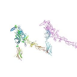
1KGY
 | | Crystal Structure of the EphB2-ephrinB2 complex | | 分子名称: | EPHRIN TYPE-B RECEPTOR 2, EPHRIN-B2 | | 著者 | Himanen, J.P, Rajashankar, K.R, Lackmann, M, Cowan, C.A, Henkemeyer, M, Nikolov, D.B. | | 登録日 | 2001-11-28 | | 公開日 | 2002-05-28 | | 最終更新日 | 2024-10-30 | | 実験手法 | X-RAY DIFFRACTION (2.7 Å) | | 主引用文献 | Crystal structure of an Eph receptor-ephrin complex.
Nature, 414, 2001
|
|
1QWI
 | |
3FL7
 | | Crystal structure of the human ephrin A2 ectodomain | | 分子名称: | 2-acetamido-2-deoxy-beta-D-glucopyranose, CHLORIDE ION, Ephrin receptor, ... | | 著者 | Walker, J.R, Yermekbayeva, L, Seitova, A, Butler-Cole, C, Bountra, C, Weigelt, J, Arrowsmith, C.H, Edwards, A.M, Bochkarev, A, Dhe-Paganon, S, Structural Genomics Consortium (SGC) | | 登録日 | 2008-12-18 | | 公開日 | 2009-01-27 | | 最終更新日 | 2024-10-30 | | 実験手法 | X-RAY DIFFRACTION (2.5 Å) | | 主引用文献 | Architecture of Eph receptor clusters.
Proc.Natl.Acad.Sci.USA, 107, 2010
|
|
4YN0
 | |
4LI1
 | |
3ETP
 | | The crystal structure of the ligand-binding domain of the EphB2 receptor at 2.0 A resolution | | 分子名称: | Ephrin type-B receptor 2 | | 著者 | Goldgur, Y, Paavilainen, S, Nikolov, D.B, Himanen, J.P. | | 登録日 | 2008-10-08 | | 公開日 | 2008-10-21 | | 最終更新日 | 2024-11-06 | | 実験手法 | X-RAY DIFFRACTION (2 Å) | | 主引用文献 | Structure of the ligand-binding domain of the EphB2 receptor at 2 A resolution.
Acta Crystallogr.,Sect.F, 65, 2009
|
|
6CMI
 | |


