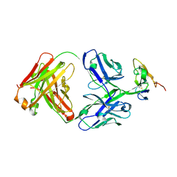
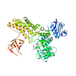
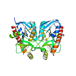
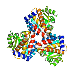
7FC5
 
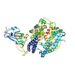 | | Crystal structure of SARS-CoV-2 RBD and horse ACE2 | | 分子名称: | 2-acetamido-2-deoxy-beta-D-glucopyranose, Angiotensin-converting enzyme, Spike protein S1 | | 著者 | Wang, X.Q, Lan, J, Ge, J.W. | | 登録日 | 2021-07-13 | | 公開日 | 2022-06-22 | | 最終更新日 | 2023-11-29 | | 実験手法 | X-RAY DIFFRACTION (2.894 Å) | | 主引用文献 | Structural insights into the binding of SARS-CoV-2, SARS-CoV, and hCoV-NL63 spike receptor-binding domain to horse ACE2.
Structure, 30, 2022
|
|
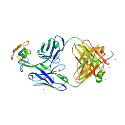
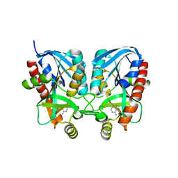
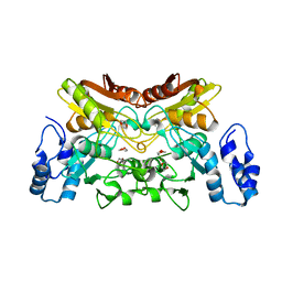
7FC6
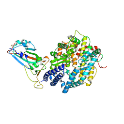 | | Crystal structure of SARS-CoV RBD and horse ACE2 | | 分子名称: | 2-acetamido-2-deoxy-beta-D-glucopyranose, 2-acetamido-2-deoxy-beta-D-glucopyranose-(1-4)-2-acetamido-2-deoxy-beta-D-glucopyranose, Angiotensin-converting enzyme, ... | | 著者 | Wang, X.Q, Lan, J, Ge, J.W. | | 登録日 | 2021-07-13 | | 公開日 | 2022-07-13 | | 最終更新日 | 2023-11-29 | | 実験手法 | X-RAY DIFFRACTION (2.655 Å) | | 主引用文献 | Structural insights into the binding of SARS-CoV-2, SARS-CoV, and hCoV-NL63 spike receptor-binding domain to horse ACE2.
Structure, 30, 2022
|
|
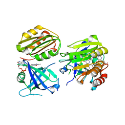
8D9Y
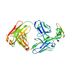 | | Crystal structure of Taipan alpha-neurotoxin in complex with Centi-3FTX-D09 antibody | | 分子名称: | 1-(2-METHOXY-ETHOXY)-2-{2-[2-(2-METHOXY-ETHOXY]-ETHOXY}-ETHANE, 4-(2-HYDROXYETHYL)-1-PIPERAZINE ETHANESULFONIC ACID, Centi-3FTX-D09 Fab heavy chain, ... | | 著者 | Pletnev, S, Verardi, R, Tully, E.S, Glanville, J, Kwong, P.D. | | 登録日 | 2022-06-12 | | 公開日 | 2023-06-14 | | 実験手法 | X-RAY DIFFRACTION (2.2 Å) | | 主引用文献 | Venom protection by antibody from a snakebite hyperimmune subject
To Be Published
|
|
8D9Z
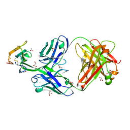 | | Crystal structure of Cobra alpha-neurotoxin in complex with Centi-3FTX-D09 antibody | | 分子名称: | ACETATE ION, Centi-3FTX-D09 Fab heavy chain, Centi-3FTX-D09 Fab light chain, ... | | 著者 | Pletnev, S, Verardi, R, Tully, E.S, Glanville, J, Kwong, P.D. | | 登録日 | 2022-06-12 | | 公開日 | 2023-06-14 | | 実験手法 | X-RAY DIFFRACTION (1.8 Å) | | 主引用文献 | Venom protection by antibody from a snakebite hyperimmune subject
To Be Published
|
|
8DA1
 | | Crystal structure of Krait alpha-neurotoxin in complex with Centi-3FTX-D09 antibody | | 分子名称: | Alpha-bungarotoxin, Centi-3FTX-D09 Fab heavy chain, Centi-3FTX-D09 Fab light chain | | 著者 | Pletnev, S, Verardi, R, Tully, E.S, Glanville, J, Kwong, P.D. | | 登録日 | 2022-06-12 | | 公開日 | 2023-06-14 | | 実験手法 | X-RAY DIFFRACTION (2.67 Å) | | 主引用文献 | Venom protection by antibody from a snakebite hyperimmune subject
To Be Published
|
|
8DA0
 | | Crystal structure of Mamba alpha-neurotoxin in complex with Centi-3FTX-D09 antibody | | 分子名称: | ACETATE ION, Alpha-elapitoxin-Dpp2d, Centi-3FTX-D09 Fab heavy chain, ... | | 著者 | Pletnev, S, Verardi, R, Tully, E.S, Glanville, J, Kwong, P.D. | | 登録日 | 2022-06-12 | | 公開日 | 2023-06-14 | | 実験手法 | X-RAY DIFFRACTION (2.2 Å) | | 主引用文献 | Venom protection by antibody from a snakebite hyperimmune subject
To Be Published
|
|
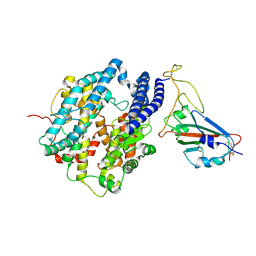
4KPB
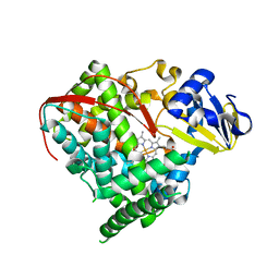 | | Crystal structure of cytochrome P450 BM-3 R47E mutant | | 分子名称: | Cytochrome P450 BM-3, PROTOPORPHYRIN IX CONTAINING FE | | 著者 | Sadre-Bazzaz, K, Catalano, J, McDermott, A.E, Tong, L. | | 登録日 | 2013-05-13 | | 公開日 | 2013-07-24 | | 最終更新日 | 2024-02-28 | | 実験手法 | X-RAY DIFFRACTION (2.1 Å) | | 主引用文献 | Structural Evidence: A Single Charged Residue Affects Substrate Binding in Cytochrome P450 BM-3.
Biochemistry, 52, 2013
|
|
4KPA
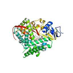 | | Crystal structure of cytochrome P450 BM-3 in complex with N-palmitoylglycine (NPG) | | 分子名称: | Cytochrome P450 BM-3, N-PALMITOYLGLYCINE, PROTOPORPHYRIN IX CONTAINING FE | | 著者 | Amadeo, G.A, Catalano, J, McDermott, A.E, Tong, L. | | 登録日 | 2013-05-13 | | 公開日 | 2013-07-24 | | 最終更新日 | 2024-02-28 | | 実験手法 | X-RAY DIFFRACTION (2 Å) | | 主引用文献 | Structural Evidence: A Single Charged Residue Affects Substrate Binding in Cytochrome P450 BM-3.
Biochemistry, 52, 2013
|
|
4UR9
 | | Structure of ligand bound glycosylhydrolase | | 分子名称: | 4-ethoxyquinazoline, CALCIUM ION, O-(2-ACETAMIDO-2-DEOXY D-GLUCOPYRANOSYLIDENE) AMINO-N-PHENYLCARBAMATE, ... | | 著者 | Darby, J.F, Landstroem, J, Roth, C, He, Y, Schultz, M, Davies, G.J, Hubbard, R.E. | | 登録日 | 2014-06-27 | | 公開日 | 2015-02-25 | | 最終更新日 | 2024-05-08 | | 実験手法 | X-RAY DIFFRACTION (2.2 Å) | | 主引用文献 | Discovery of Selective Small-Molecule Activators of a Bacterial Glycoside Hydrolase.
Angew.Chem.Int.Ed.Engl., 53, 2014
|
|
6IF8
 | | Aeromonas hydrophila MtaN-2 complexed with adenine | | 分子名称: | 5'-methylthioadenosine/S-adenosylhomocysteine nucleosidase, ADENINE | | 著者 | Chen, J, Liu, W, Wang, L, Shang, F, Lan, J, Chen, Y, Xu, Y. | | 登録日 | 2018-09-18 | | 公開日 | 2018-10-03 | | 最終更新日 | 2023-11-22 | | 実験手法 | X-RAY DIFFRACTION (2 Å) | | 主引用文献 | Aeromonas hydrophila MtaN-2 complexed with adenine
To Be Published
|
|
3ZUE
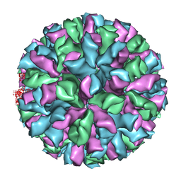 | | Rabbit Hemorrhagic Disease Virus (RHDV)capsid protein | | 分子名称: | CAPSID STRUCTURAL PROTEIN VP60 | | 著者 | Luque, D, Gonzalez, J.M, Gomez-Blanco, J, Marabini, R, Chichon, J, Mena, I, Angulo, I, Carrascosa, J.L, Verdaguer, N, Trus, B.L, Barcena, J, Caston, J.R. | | 登録日 | 2011-07-18 | | 公開日 | 2012-05-23 | | 最終更新日 | 2024-05-08 | | 実験手法 | ELECTRON MICROSCOPY (10.3 Å) | | 主引用文献 | Epitope Insertion at the N-Terminal Molecular Switch of the Rabbit Hemorrhagic Disease Virus T=3 Capsid Protein Leads to Larger T=4 Capsids.
J.Virol., 86, 2012
|
|
8I29
 | | Crystal structure of butanol dehydrogenase A (YqdH) in complex with NADH from Fusobacterium nucleatum | | 分子名称: | CHLORIDE ION, COBALT (II) ION, NADH-dependent butanol dehydrogenase A, ... | | 著者 | Bai, X, Lan, J, Bu, T.T, Wang, L.L, He, S.R, Zhang, J, Xu, Y.B. | | 登録日 | 2023-01-14 | | 公開日 | 2023-02-01 | | 最終更新日 | 2024-05-29 | | 実験手法 | X-RAY DIFFRACTION (2.72 Å) | | 主引用文献 | Crystal structure of butanol dehydrogenase A (YqdH) in complex with NADH from Fusobacterium nucleatum
To Be Published
|
|
1JS7
 | | Solution Structure of dAAUAA DNA Bulge | | 分子名称: | 5'-D(*CP*GP*TP*AP*GP*CP*CP*GP*AP*TP*GP*C)-3', 5'-D(*GP*CP*AP*TP*CP*GP*AP*AP*UP*AP*AP*GP*CP*TP*AP*CP*G)-3' | | 著者 | Gollmick, F.A, Lorenz, M, Dornberger, U, von Langen, J, Diekmann, S, Fritzsche, H. | | 登録日 | 2001-08-16 | | 公開日 | 2002-06-19 | | 最終更新日 | 2024-05-22 | | 実験手法 | SOLUTION NMR | | 主引用文献 | Solution structure of dAATAA and dAAUAA DNA bulges.
Nucleic Acids Res., 30, 2002
|
|
1JRV
 | | SOLUTION STRUCTURE OF DAATAA DNA BULGE | | 分子名称: | 5'-D(*CP*GP*TP*AP*GP*CP*CP*GP*AP*TP*GP*C)-3', 5'-D(*GP*CP*AP*TP*CP*GP*AP*AP*TP*AP*AP*GP*CP*TP*AP*CP*G)-3' | | 著者 | Gollmick, F.A, Lorenz, M, Dornberger, U, von Langen, J, Diekmann, S, Fritzsche, H. | | 登録日 | 2001-08-15 | | 公開日 | 2002-06-19 | | 最終更新日 | 2024-05-22 | | 実験手法 | SOLUTION NMR | | 主引用文献 | Solution structure of dAATAA and dAAUAA DNA bulges.
Nucleic Acids Res., 30, 2002
|
|
1JRW
 | | Solution Structure of dAATAA DNA Bulge | | 分子名称: | 5'-D(*CP*GP*TP*AP*GP*CP*CP*GP*AP*TP*GP*C)-3', 5'-D(*GP*CP*AP*TP*CP*GP*AP*AP*TP*AP*AP*GP*CP*TP*AP*CP*G)-3' | | 著者 | Gollmick, F.A, Lorenz, M, Dornberger, U, von Langen, J, Diekmann, S, Fritzsche, H. | | 登録日 | 2001-08-15 | | 公開日 | 2002-08-28 | | 最終更新日 | 2024-05-22 | | 実験手法 | SOLUTION NMR | | 主引用文献 | Solution structure of dAATAA and dAAUAA DNA bulges.
Nucleic Acids Res., 30, 2002
|
|
5FI4
 | | Discovery of imidazo[1,2-a]-pyridine inhibitors of pan-PI3 kinases that are efficacious in a mouse xenograft model | | 分子名称: | GLYCEROL, Phosphatidylinositol 3-kinase regulatory subunit alpha, Phosphatidylinositol 4,5-bisphosphate 3-kinase catalytic subunit alpha isoform, ... | | 著者 | Elling, R.A, Knapp, M.S, Han, W, Daniel, L.M, Xy, Y, Burger, M.T, Ni, Z, Smith, A, Lan, J, Williams, T, Verhagen, J, Huh, K, Merritt, H, Chan, J, Kaufman, S, Voliva, C.F, Pecchi, S. | | 登録日 | 2015-12-22 | | 公開日 | 2016-02-03 | | 最終更新日 | 2024-03-06 | | 実験手法 | X-RAY DIFFRACTION (2.5 Å) | | 主引用文献 | Discovery of imidazo[1,2-a]-pyridine inhibitors of pan-PI3 kinases that are efficacious in a mouse xenograft model.
Bioorg.Med.Chem.Lett., 26, 2016
|
|
6K2Q
 | | Aeromonas hydrophila MtaN-2 complexed with adenine | | 分子名称: | 5'-methylthioadenosine/S-adenosylhomocysteine nucleosidase, ADENINE | | 著者 | Chen, J, Liu, W, Wang, L, Shang, F, Lan, J, Chen, Y, Xu, Y. | | 登録日 | 2019-05-15 | | 公開日 | 2019-05-29 | | 最終更新日 | 2023-11-22 | | 実験手法 | X-RAY DIFFRACTION (2 Å) | | 主引用文献 | Crystal Structure of Aeromonas hydrophila Cytoplasmic 5'-Methylthioadenosine/S-Adenosylhomocysteine Nucleosidase.
Biochemistry, 58, 2019
|
|
6W77
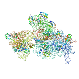 | | 30S-Inactivated-high-Mg2+ Class A | | 分子名称: | 30S ribosomal protein S10, 30S ribosomal protein S11, 30S ribosomal protein S12, ... | | 著者 | Jahagirdar, D, Jha, V, Basu, B, Gomez-Blanco, J, Vargas, J, Ortega, J. | | 登録日 | 2020-03-18 | | 公開日 | 2020-10-21 | | 最終更新日 | 2024-03-06 | | 実験手法 | ELECTRON MICROSCOPY (3.6 Å) | | 主引用文献 | Alternative conformations and motions adopted by 30S ribosomal subunits visualized by cryo-electron microscopy.
Rna, 26, 2020
|
|
6W7M
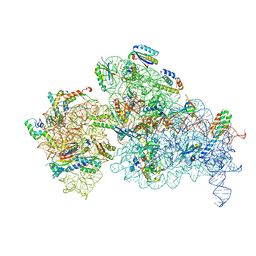 | | 30S-Inactive-high-Mg2+ + carbon layer | | 分子名称: | 16S rRNA, 30S ribosomal protein S10, 30S ribosomal protein S11, ... | | 著者 | Jahagirdar, D, Jha, V, Basu, B, Gomez-Blanco, J, Vargas, J, Ortega, J. | | 登録日 | 2020-03-19 | | 公開日 | 2020-10-21 | | 最終更新日 | 2024-03-06 | | 実験手法 | ELECTRON MICROSCOPY (3.8 Å) | | 主引用文献 | Alternative conformations and motions adopted by 30S ribosomal subunits visualized by cryo-electron microscopy.
Rna, 26, 2020
|
|
6K28
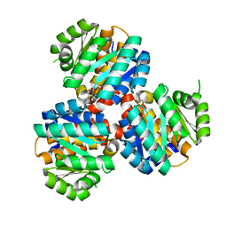 | | Crystal structure of the 5-(Hydroxyethyl)-methylthiazole Kinase ThiM from Klebsiella pneumonia in complex with TZE | | 分子名称: | 2-(4-METHYL-THIAZOL-5-YL)-ETHANOL, Hydroxyethylthiazole kinase, MAGNESIUM ION | | 著者 | Chen, Y, Wang, L, Shang, F, Lan, J, Liu, W, Xu, Y. | | 登録日 | 2019-05-13 | | 公開日 | 2019-07-10 | | 最終更新日 | 2024-03-27 | | 実験手法 | X-RAY DIFFRACTION (2.002 Å) | | 主引用文献 | Structural insight of the 5-(Hydroxyethyl)-methylthiazole kinase ThiM involving vitamin B1 biosynthetic pathway from the Klebsiella pneumoniae.
Biochem.Biophys.Res.Commun., 518, 2019
|
|
6JYY
 | | Crystal structure of the 5-(Hydroxyethyl)-methylthiazole Kinase ThiM from Klebsiella pneumonia | | 分子名称: | Hydroxyethylthiazole kinase | | 著者 | Chen, Y, Wang, L, Shang, F, Lan, J, Liu, W, Xu, Y. | | 登録日 | 2019-04-29 | | 公開日 | 2019-06-26 | | 最終更新日 | 2024-03-27 | | 実験手法 | X-RAY DIFFRACTION (2 Å) | | 主引用文献 | Structural insight of the 5-(Hydroxyethyl)-methylthiazole kinase ThiM involving vitamin B1 biosynthetic pathway from the Klebsiella pneumoniae.
Biochem.Biophys.Res.Commun., 518, 2019
|
|
6K63
 | | The crystal structure of cytidine deaminase from Klebsiella pneumoniae | | 分子名称: | 1,4-DIETHYLENE DIOXIDE, Cytidine deaminase, ZINC ION | | 著者 | Liu, W, Shang, F, Lan, J, Chen, Y, Wang, L, Xu, Y. | | 登録日 | 2019-06-01 | | 公開日 | 2019-07-31 | | 最終更新日 | 2023-11-22 | | 実験手法 | X-RAY DIFFRACTION (2.073 Å) | | 主引用文献 | Biochemical and structural analysis of the Klebsiella pneumoniae cytidine deaminase CDA.
Biochem.Biophys.Res.Commun., 519, 2019
|
|
6K2L
 | | Crystal structure of the Siderophore-interacting protein SipS from Aeromonas hydrophila | | 分子名称: | FLAVIN-ADENINE DINUCLEOTIDE, Siderophore-interacting protein | | 著者 | Shang, F, Lan, J, Liu, W, Xu, Y. | | 登録日 | 2019-05-14 | | 公開日 | 2019-06-12 | | 最終更新日 | 2024-03-27 | | 実験手法 | X-RAY DIFFRACTION (2.5 Å) | | 主引用文献 | Crystal structure of the Siderophore-interacting protein SIP from Aeromonas hydrophila.
Biochem.Biophys.Res.Commun., 519, 2019
|
|
7DQA
 | | Cryo-EM structure of SARS-CoV2 RBD-ACE2 complex | | 分子名称: | 2-acetamido-2-deoxy-beta-D-glucopyranose, Angiotensin-converting enzyme 2, CHLORIDE ION, ... | | 著者 | Wang, J, Lan, J, Wang, X.Q, Wang, H.W. | | 登録日 | 2020-12-22 | | 公開日 | 2021-12-29 | | 最終更新日 | 2022-07-13 | | 実験手法 | ELECTRON MICROSCOPY (2.8 Å) | | 主引用文献 | Reduced graphene oxide membrane as supporting film for high-resolution cryo-EM
Biophys Rep, 7, 2022
|
|
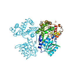
7E8M
 | | Crystal structure of SARS-CoV-2 antibody P2C-1F11 with mutated RBD | | 分子名称: | 2-acetamido-2-deoxy-beta-D-glucopyranose, Spike protein S1, antibody P2C-1F11 heavy chain, ... | | 著者 | Wang, X.Q, Zhang, L.Q, Ge, J.W, Wang, R.K, Lan, J. | | 登録日 | 2021-03-02 | | 公開日 | 2021-05-26 | | 最終更新日 | 2023-11-29 | | 実験手法 | X-RAY DIFFRACTION (2.09 Å) | | 主引用文献 | Analysis of SARS-CoV-2 variant mutations reveals neutralization escape mechanisms and the ability to use ACE2 receptors from additional species.
Immunity, 54, 2021
|
|


