6JPI
 
 | |
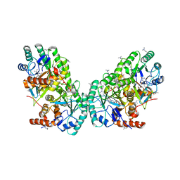
1C8J
 | | CRYSTAL STRUCTURE OF CYTOCHROME P450CAM MUTANT (F87W/Y96F) | | 分子名称: | CYTOCHROME P450-CAM, PROTOPORPHYRIN IX CONTAINING FE | | 著者 | Liu, Y, Jiang, F, Guo, Q, Chen, X, Jin, J, Sun, Y, Rao, Z. | | 登録日 | 2000-05-31 | | 公開日 | 2001-05-09 | | 最終更新日 | 2023-12-27 | | 実験手法 | X-RAY DIFFRACTION (2.1 Å) | | 主引用文献 | Crystal Structure of Cytochrome P450cam mutant (F87W/Y96F)
To be Published
|
|
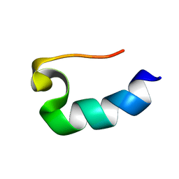
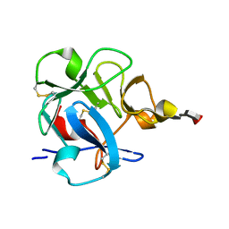
1RIM
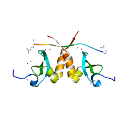
 | | E6-binding zinc finger (E6apc2) | | 分子名称: | E6apc2 peptide | | 著者 | Liu, Y, Liu, Z, Androphy, E, Chen, J, Baleja, J.D. | | 登録日 | 2003-11-17 | | 公開日 | 2004-08-03 | | 最終更新日 | 2024-05-22 | | 実験手法 | SOLUTION NMR | | 主引用文献 | Design and characterization of helical peptides that inhibit the E6 protein of papillomavirus.
Biochemistry, 43, 2004
|
|
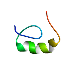
1RIJ
 | | E6-bind Trp-cage (E6apn1) | | 分子名称: | E6apn1 peptide | | 著者 | Liu, Y, Liu, Z, Androphy, E, Chen, J, Baleja, J.D. | | 登録日 | 2003-11-17 | | 公開日 | 2004-08-03 | | 最終更新日 | 2024-05-22 | | 実験手法 | SOLUTION NMR | | 主引用文献 | Design and characterization of helical peptides that inhibit the E6 protein of papillomavirus.
Biochemistry, 43, 2004
|
|
1RIK
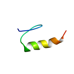 | | E6-binding zinc finger (E6apc1) | | 分子名称: | E6apc1 peptide | | 著者 | Liu, Y, Liu, Z, Androphy, E, Chen, J, Baleja, J.D. | | 登録日 | 2003-11-17 | | 公開日 | 2004-08-03 | | 最終更新日 | 2024-05-22 | | 実験手法 | SOLUTION NMR | | 主引用文献 | Design and characterization of helical peptides that inhibit the E6 protein of papillomavirus.
Biochemistry, 43, 2004
|
|
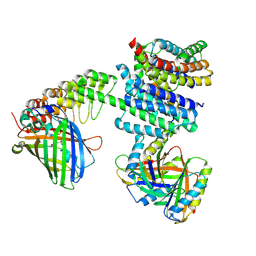
3A64
 | | Crystal structure of CcCel6C, a glycoside hydrolase family 6 enzyme, from Coprinopsis cinerea | | 分子名称: | Cellobiohydrolase, MAGNESIUM ION | | 著者 | Liu, Y, Yoshida, M, Kurakata, Y, Miyazaki, T, Nishikawa, A, Tonozuka, T. | | 登録日 | 2009-08-21 | | 公開日 | 2009-09-01 | | 最終更新日 | 2023-11-01 | | 実験手法 | X-RAY DIFFRACTION (1.6 Å) | | 主引用文献 | Crystal structure of a glycoside hydrolase family 6 enzyme, CcCel6C, a cellulase constitutively produced by Coprinopsis cinerea
Febs J., 277, 2010
|
|
5T1I
 | | CBX3 chromo shadow domain in complex with histone H3 peptide | | 分子名称: | Chromobox protein homolog 3, Histone H3.1, UNKNOWN ATOM OR ION | | 著者 | Liu, Y, Tempel, W, Bountra, C, Arrowsmith, C.H, Edwards, A.M, Min, J, Structural Genomics Consortium (SGC) | | 登録日 | 2016-08-19 | | 公開日 | 2016-09-14 | | 最終更新日 | 2023-10-04 | | 実験手法 | X-RAY DIFFRACTION (1.6 Å) | | 主引用文献 | Peptide recognition by heterochromatin protein 1 (HP1) chromoshadow domains revisited: Plasticity in the pseudosymmetric histone binding site of human HP1.
J. Biol. Chem., 292, 2017
|
|
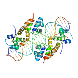
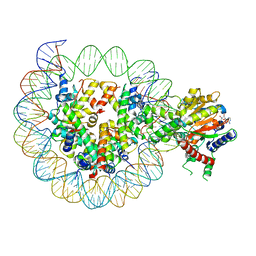
8WH5
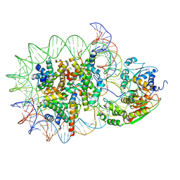 | | Structure of DDM1-nucleosome complex in the apo state | | 分子名称: | ATP-dependent DNA helicase DDM1, DNA (antisense strand), DNA (sense strand), ... | | 著者 | Liu, Y, Zhang, Z, Du, J. | | 登録日 | 2023-09-22 | | 公開日 | 2024-04-17 | | 実験手法 | ELECTRON MICROSCOPY (3.58 Å) | | 主引用文献 | Molecular basis of chromatin remodelling by DDM1 involved in plant DNA methylation.
Nat.Plants, 10, 2024
|
|
8WHB
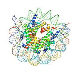 | | Structure of nucleosome core particle of Arabidopsis thaliana | | 分子名称: | DNA (antisense strand), DNA (sense strand), Histone H2A.6, ... | | 著者 | Liu, Y, Zhang, Z, Du, J. | | 登録日 | 2023-09-23 | | 公開日 | 2024-04-17 | | 実験手法 | ELECTRON MICROSCOPY (3.17 Å) | | 主引用文献 | Molecular basis of chromatin remodelling by DDM1 involved in plant DNA methylation.
Nat.Plants, 10, 2024
|
|
8WH8
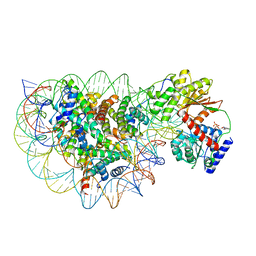 | | Structure of DDM1-nucleosome complex in ADP state | | 分子名称: | ADENOSINE-5'-DIPHOSPHATE, ATP-dependent DNA helicase DDM1, DNA (antisense strand), ... | | 著者 | Liu, Y, Zhang, Z, Du, J. | | 登録日 | 2023-09-22 | | 公開日 | 2024-04-17 | | 実験手法 | ELECTRON MICROSCOPY (3.6 Å) | | 主引用文献 | Molecular basis of chromatin remodelling by DDM1 involved in plant DNA methylation.
Nat.Plants, 10, 2024
|
|
8WH9
 | | Structure of DDM1-nucleosome complex in ADP-BeFx state | | 分子名称: | ADENOSINE-5'-DIPHOSPHATE, ATP-dependent DNA helicase DDM1, BERYLLIUM TRIFLUORIDE ION, ... | | 著者 | Liu, Y, Zhang, Z, Du, J. | | 登録日 | 2023-09-22 | | 公開日 | 2024-04-17 | | 実験手法 | ELECTRON MICROSCOPY (3.31 Å) | | 主引用文献 | Molecular basis of chromatin remodelling by DDM1 involved in plant DNA methylation.
Nat.Plants, 10, 2024
|
|
8WHA
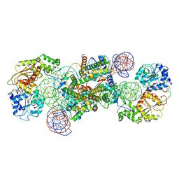 | | Structure of DDM1-nucleosome complex in the ADP-BeFx state with DDM1 bound to SHL2 and SHL-2 | | 分子名称: | ADENOSINE-5'-DIPHOSPHATE, ATP-dependent DNA helicase DDM1, BERYLLIUM TRIFLUORIDE ION, ... | | 著者 | Liu, Y, Zhang, Z, Du, J. | | 登録日 | 2023-09-22 | | 公開日 | 2024-04-17 | | 実験手法 | ELECTRON MICROSCOPY (4.05 Å) | | 主引用文献 | Molecular basis of chromatin remodelling by DDM1 involved in plant DNA methylation.
Nat.Plants, 10, 2024
|
|
7RDQ
 | |
1DQG
 | | CRYSTAL STRUCTURE OF THE CYSTEINE RICH DOMAIN OF MANNOSE RECEPTOR | | 分子名称: | MANNOSE RECEPTOR, SULFATE ION | | 著者 | Liu, Y, Chirino, A.J, Misulovin, Z, Leteux, C, Feizi, T, Nussenzweig, M.C, Bjorkman, P.J. | | 登録日 | 2000-01-04 | | 公開日 | 2000-05-10 | | 最終更新日 | 2011-07-13 | | 実験手法 | X-RAY DIFFRACTION (1.7 Å) | | 主引用文献 | Crystal structure of the cysteine-rich domain of mannose receptor complexed with a sulfated carbohydrate ligand.
J.Exp.Med., 191, 2000
|
|
1DQO
 | | Crystal structure of the cysteine rich domain of mannose receptor complexed with Acetylgalactosamine-4-sulfate | | 分子名称: | 2-acetamido-2-deoxy-4-O-sulfo-beta-D-galactopyranose, MANNOSE RECEPTOR | | 著者 | Liu, Y, Chirino, A.J, Misulovin, Z, Leteux, C, Feizi, T, Nussenzweig, M.C, Bjorkman, P.J. | | 登録日 | 2000-01-04 | | 公開日 | 2000-05-10 | | 最終更新日 | 2023-08-09 | | 実験手法 | X-RAY DIFFRACTION (2.2 Å) | | 主引用文献 | Crystal structure of the cysteine-rich domain of mannose receptor complexed with a sulfated carbohydrate ligand.
J.Exp.Med., 191, 2000
|
|
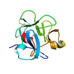
1A2W
 | | CRYSTAL STRUCTURE OF A 3D DOMAIN-SWAPPED DIMER OF BOVINE PANCREATIC RIBONUCLEASE A | | 分子名称: | CHLORIDE ION, RIBONUCLEASE A, SULFATE ION | | 著者 | Liu, Y, Hart, P.J, Schlunegger, M.P, Eisenberg, D.S. | | 登録日 | 1998-01-12 | | 公開日 | 1998-04-29 | | 最終更新日 | 2023-08-02 | | 実験手法 | X-RAY DIFFRACTION (2.1 Å) | | 主引用文献 | The crystal structure of a 3D domain-swapped dimer of RNase A at a 2.1-A resolution.
Proc.Natl.Acad.Sci.USA, 95, 1998
|
|
16VP
 | | CONSERVED CORE OF THE HERPES SIMPLEX VIRUS TRANSCRIPTIONAL REGULATORY PROTEIN VP16 | | 分子名称: | PROTEIN (VP16, VMW65, ATIF), ... | | 著者 | Liu, Y, Gong, W, Huang, C.C, Herr, W, Cheng, X. | | 登録日 | 1999-02-11 | | 公開日 | 1999-07-28 | | 最終更新日 | 2023-12-27 | | 実験手法 | X-RAY DIFFRACTION (2.1 Å) | | 主引用文献 | Crystal structure of the conserved core of the herpes simplex virus transcriptional regulatory protein VP16.
Genes Dev., 13, 1999
|
|
4BL6
 | | Bicaudal-D uses a parallel, homodimeric coiled coil with heterotypic registry to co-ordinate recruitment of cargos to dynein | | 分子名称: | ARGININE, PROTEIN BICAUDAL D | | 著者 | Liu, Y, Salter, H.K, Holding, A.N, Johnson, C.M, Stephens, E, Lukavsky, P.J, Walshaw, J, Bullock, S.L. | | 登録日 | 2013-05-02 | | 公開日 | 2013-06-12 | | 最終更新日 | 2017-07-12 | | 実験手法 | X-RAY DIFFRACTION (2.18 Å) | | 主引用文献 | Bicaudal-D Uses a Parallel, Homodimeric Coiled Coil with Heterotypic Registry to Coordinate Recruitment of Cargos to Dynein
Genes Dev., 27, 2013
|
|
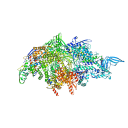
6MZI
 | | CryoEM structure of human enterovirus D68 expanded 1 particle (pH 6.5, 4 degrees Celsius, 3 min) | | 分子名称: | viral protein 1, viral protein 2, viral protein 3, ... | | 著者 | Liu, Y, Rossmann, M.G. | | 登録日 | 2018-11-05 | | 公開日 | 2018-12-19 | | 最終更新日 | 2024-03-13 | | 実験手法 | ELECTRON MICROSCOPY (3.46 Å) | | 主引用文献 | Molecular basis for the acid-initiated uncoating of human enterovirus D68.
Proc. Natl. Acad. Sci. U.S.A., 115, 2018
|
|
3A9B
 | | CcCel6C, a glycoside hydrolase family 6 enzyme, complexed with cellobiose | | 分子名称: | Cellobiohydrolase, MAGNESIUM ION, beta-D-glucopyranose, ... | | 著者 | Liu, Y, Yoshida, M, Kurakata, Y, Miyazaki, T, Nishikawa, A, Tonozuka, T. | | 登録日 | 2009-10-22 | | 公開日 | 2009-11-03 | | 最終更新日 | 2023-11-01 | | 実験手法 | X-RAY DIFFRACTION (1.2 Å) | | 主引用文献 | Crystal structure of a glycoside hydrolase family 6 enzyme, CcCel6C, a cellulase constitutively produced by Coprinopsis cinerea
Febs J., 277, 2010
|
|
3ABX
 | | CcCel6C, a glycoside hydrolase family 6 enzyme, complexed with p-nitrophenyl beta-D-cellotrioside | | 分子名称: | 4-nitrophenyl beta-D-glucopyranosyl-(1->4)-beta-D-glucopyranosyl-(1->4)-beta-D-glucopyranoside, Cellobiohydrolase, MAGNESIUM ION | | 著者 | Liu, Y, Yoshida, M, Kurakata, Y, Miyazaki, T, Nishikawa, A, Tonozuka, T. | | 登録日 | 2009-12-24 | | 公開日 | 2010-01-05 | | 最終更新日 | 2023-11-01 | | 実験手法 | X-RAY DIFFRACTION (1.4 Å) | | 主引用文献 | Crystal structure of a glycoside hydrolase family 6 enzyme, CcCel6C, a cellulase constitutively produced by Coprinopsis cinerea
Febs J., 277, 2010
|
|
3OIX
 | | Crystal structure of the putative dihydroorotate dehydrogenase from Streptococcus mutans | | 分子名称: | FLAVIN MONONUCLEOTIDE, GLYCEROL, Putative dihydroorotate dehydrogenase; dihydroorotate oxidase | | 著者 | Liu, Y, Gao, Z.Q, Liu, C.P, Dong, Y.H. | | 登録日 | 2010-08-20 | | 公開日 | 2010-09-08 | | 最終更新日 | 2023-11-01 | | 実験手法 | X-RAY DIFFRACTION (2.399 Å) | | 主引用文献 | Structure of the putative dihydroorotate dehydrogenase from Streptococcus mutans
Acta Crystallogr.,Sect.F, 67, 2011
|
|
6NHT
 | |
6NHV
 | |
1FWU
 | |


