Loading
PDBj
メニューPDBj@FacebookPDBj@TwitterPDBj@YouTubewwPDB FoundationwwPDB
RCSB PDBPDBeBMRBAdv. SearchSearch help
Search by PDB author
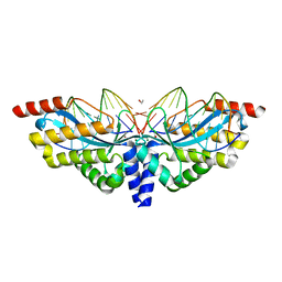
3E54
DownloadVisualize
BU of 3e54 by Molmil
Archaeal Intron-encoded Homing Endonuclease I-Vdi141I Complexed With DNA
分子名称: DNA (5'-D(*DCP*DTP*DGP*DAP*DCP*DTP*DCP*DTP*DCP*DTP*DTP*DAP*DA)-3'), DNA (5'-D(*DTP*DTP*DGP*DGP*DCP*DTP*DAP*DCP*DCP*DTP*DTP*DAP*DA)-3'), DNA (5'-D(P*DGP*DAP*DGP*DAP*DGP*DTP*DCP*DAP*DG)-3'), ...
著者Nomura, N, Nomura, Y, Sussman, D, Stoddard, B.L.
登録日2008-08-13
公開日2008-12-30
最終更新日2024-05-29
実験手法X-RAY DIFFRACTION (2.5 Å)
主引用文献Recognition of a common rDNA target site in archaea and eukarya by analogous LAGLIDADG and His-Cys box homing endonucleases
Nucleic Acids Res., 36, 2008
2GKO
DownloadVisualize
BU of 2gko by Molmil
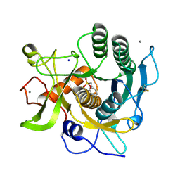
S41 Psychrophilic Protease
分子名称: CALCIUM ION, SODIUM ION, microbial serine proteinases; subtilisin, ...
著者Walter, R.L, Mekel, M.J, Grayling, R.A, Arnold, F.H, Wintrode, P.L, Almog, O.
登録日2006-04-03
公開日2007-05-01
最終更新日2024-10-16
実験手法X-RAY DIFFRACTION (1.4 Å)
主引用文献The crystal structures of the psychrophilic subtilisin S41 and the mesophilic subtilisin Sph reveal the same calcium-loaded state.
Proteins, 74, 2009
3D43
DownloadVisualize
BU of 3d43 by Molmil
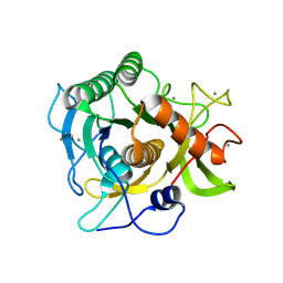
The crystal structure of Sph at 0.8A
分子名称: CALCIUM ION, Sphericase
著者Almog, O.
登録日2008-05-13
公開日2009-04-28
最終更新日2024-10-30
実験手法X-RAY DIFFRACTION (0.8 Å)
主引用文献The crystal structures of the psychrophilic subtilisin S41 and the mesophilic subtilisin Sph reveal the same calcium-loaded state.
Proteins, 74, 2009
5NW5
DownloadVisualize
BU of 5nw5 by Molmil
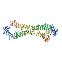
Crystal structure of the Rif1 N-terminal domain (RIF1-NTD) from Saccharomyces cerevisiae in complex with DNA
分子名称: DNA (30-MER), DNA (60-MER), Telomere length regulator protein RIF1
著者Bunker, R.D, Reinert, J.K, Shi, T, Thoma, N.H.
登録日2017-05-05
公開日2017-06-14
最終更新日2024-01-17
実験手法X-RAY DIFFRACTION (6.502 Å)
主引用文献Rif1 maintains telomeres and mediates DNA repair by encasing DNA ends.
Nat. Struct. Mol. Biol., 24, 2017
5NVR
DownloadVisualize
BU of 5nvr by Molmil
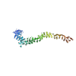
Crystal structure of the Rif1 N-terminal domain (RIF1-NTD) from Saccharomyces cerevisiae
分子名称: Telomere length regulator protein RIF1
著者Bunker, R.D, Shi, T, Thoma, N.H.
登録日2017-05-04
公開日2017-06-14
最終更新日2024-10-09
実験手法X-RAY DIFFRACTION (3.95 Å)
主引用文献Rif1 maintains telomeres and mediates DNA repair by encasing DNA ends.
Nat. Struct. Mol. Biol., 24, 2017
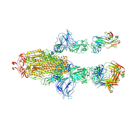
7MW6
DownloadVisualize
BU of 7mw6 by Molmil
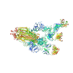
Structure of the SARS-CoV-2 Spike trimer with three RBDs up in complex with the Fab fragment of human neutralizing antibody clone 2
分子名称: 2-acetamido-2-deoxy-beta-D-glucopyranose, 2-acetamido-2-deoxy-beta-D-glucopyranose-(1-3)-2-acetamido-2-deoxy-beta-D-glucopyranose, 2-acetamido-2-deoxy-beta-D-glucopyranose-(1-4)-2-acetamido-2-deoxy-beta-D-glucopyranose, ...
著者Hu, Y, Xiong, Y.
登録日2021-05-15
公開日2022-03-16
最終更新日2024-10-09
実験手法ELECTRON MICROSCOPY (3.22 Å)
主引用文献Monospecific and bispecific monoclonal SARS-CoV-2 neutralizing antibodies that maintain potency against B.1.617.
Nat Commun, 13, 2022
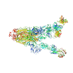
7MW4
DownloadVisualize
BU of 7mw4 by Molmil
Structure of the SARS-CoV-2 Spike trimer with one RBD down in complex with the Fab fragment of human neutralizing antibody clone 6
分子名称: 2-acetamido-2-deoxy-beta-D-glucopyranose, 2-acetamido-2-deoxy-beta-D-glucopyranose-(1-3)-2-acetamido-2-deoxy-beta-D-glucopyranose, 2-acetamido-2-deoxy-beta-D-glucopyranose-(1-4)-2-acetamido-2-deoxy-beta-D-glucopyranose, ...
著者Hu, Y, Xiong, Y.
登録日2021-05-15
公開日2022-03-16
最終更新日2023-11-01
実験手法ELECTRON MICROSCOPY (3.42 Å)
主引用文献Monospecific and bispecific monoclonal SARS-CoV-2 neutralizing antibodies that maintain potency against B.1.617.
Nat Commun, 13, 2022
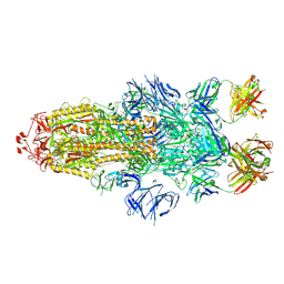
7MW2
DownloadVisualize
BU of 7mw2 by Molmil
Structure of the SARS-CoV-2 Spike trimer with all RBDs down in complex with the Fab fragment of human neutralizing antibody clone 6
分子名称: 2-acetamido-2-deoxy-beta-D-glucopyranose, 2-acetamido-2-deoxy-beta-D-glucopyranose-(1-4)-2-acetamido-2-deoxy-beta-D-glucopyranose, Fab of antibody clone 6, ...
著者Hu, Y, Xiong, Y.
登録日2021-05-15
公開日2022-03-16
最終更新日2024-10-23
実験手法ELECTRON MICROSCOPY (2.97 Å)
主引用文献Monospecific and bispecific monoclonal SARS-CoV-2 neutralizing antibodies that maintain potency against B.1.617.
Nat Commun, 13, 2022
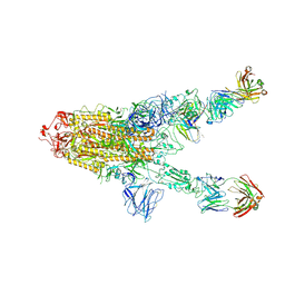
7MW5
DownloadVisualize
BU of 7mw5 by Molmil
Structure of the SARS-CoV-2 Spike trimer with one RBD down in complex with the Fab fragment of human neutralizing antibody clone 2
分子名称: 2-acetamido-2-deoxy-beta-D-glucopyranose, 2-acetamido-2-deoxy-beta-D-glucopyranose-(1-4)-2-acetamido-2-deoxy-beta-D-glucopyranose, Fab of antibody clone 2, ...
著者Hu, Y, Xiong, Y.
登録日2021-05-15
公開日2022-03-16
最終更新日2024-10-23
実験手法ELECTRON MICROSCOPY (3.42 Å)
主引用文献Monospecific and bispecific monoclonal SARS-CoV-2 neutralizing antibodies that maintain potency against B.1.617.
Nat Commun, 13, 2022
7MW3
DownloadVisualize
BU of 7mw3 by Molmil
Structure of the SARS-CoV-2 Spike trimer with two RBDs down in complex with the Fab fragment of human neutralizing antibody clone 6
分子名称: 2-acetamido-2-deoxy-beta-D-glucopyranose, 2-acetamido-2-deoxy-beta-D-glucopyranose-(1-3)-2-acetamido-2-deoxy-beta-D-glucopyranose, 2-acetamido-2-deoxy-beta-D-glucopyranose-(1-4)-2-acetamido-2-deoxy-beta-D-glucopyranose, ...
著者Hu, Y, Xiong, Y.
登録日2021-05-15
公開日2022-03-16
最終更新日2023-11-01
実験手法ELECTRON MICROSCOPY (3.15 Å)
主引用文献Monospecific and bispecific monoclonal SARS-CoV-2 neutralizing antibodies that maintain potency against B.1.617.
Nat Commun, 13, 2022
4Y68
DownloadVisualize
BU of 4y68 by Molmil
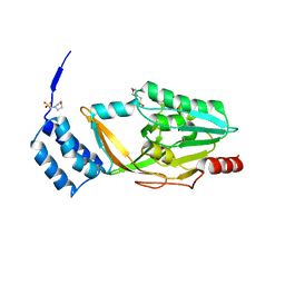
Structure of a lipoprotein from Streptococcus agalactiae
分子名称: 2-(N-MORPHOLINO)-ETHANESULFONIC ACID, Putative nisin-resistance protein
著者Khosa, S, Hoeppner, A, Smits, S.H.
登録日2015-02-12
公開日2016-01-20
最終更新日2024-05-08
実験手法X-RAY DIFFRACTION (2.21 Å)
主引用文献Structural basis of lantibiotic recognition by the nisin resistance protein from Streptococcus agalactiae.
Sci Rep, 6, 2016
5DCL
DownloadVisualize
BU of 5dcl by Molmil
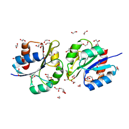
Structure of a lantibiotic response regulator: N terminal domain of the nisin resistance regulator NsrR
分子名称: 1,2-ETHANEDIOL, PhoB family transcriptional regulator
著者Khosa, S, Kleinschrodt, D, Hoeppner, A, Smits, S.H.
登録日2015-08-24
公開日2016-03-16
最終更新日2024-05-08
実験手法X-RAY DIFFRACTION (1.41 Å)
主引用文献Structure of the Response Regulator NsrR from Streptococcus agalactiae, Which Is Involved in Lantibiotic Resistance.
Plos One, 11, 2016
5DCM
DownloadVisualize
BU of 5dcm by Molmil
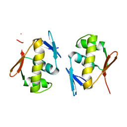
Structure of a lantibiotic response regulator: C-terminal domain of the nisin resistance regulator NsrR
分子名称: PhoB family transcriptional regulator
著者Khosa, S, Kleinschrodt, D, Hoeppner, A, Smits, S.H.J.
登録日2015-08-24
公開日2016-07-06
最終更新日2024-05-08
実験手法X-RAY DIFFRACTION (1.6 Å)
主引用文献Structure of the Response Regulator NsrR from Streptococcus agalactiae, Which Is Involved in Lantibiotic Resistance.
Plos One, 11, 2016
<12


227111

件を2024-11-06に公開中

PDB statisticsPDBj update infoContact PDBjnumon