Loading
PDBj
メニューPDBj@FacebookPDBj@TwitterPDBj@YouTubewwPDB FoundationwwPDB
RCSB PDBPDBeBMRBAdv. SearchSearch help
Search by PDB author
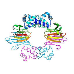
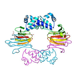
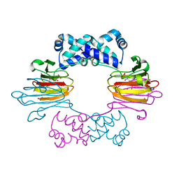
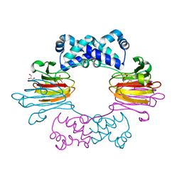
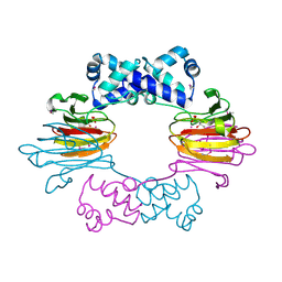
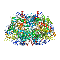
5JSO
DownloadVisualize
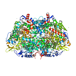
BU of 5jso by Molmil
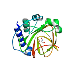
Structures of DddQ from Ruegeria lac. Reveal Key Residues for Metal Binding and Catalysis - TRIS bound
分子名称: 2-AMINO-2-HYDROXYMETHYL-PROPANE-1,3-DIOL, CHLORIDE ION, Dimethlysulfonioproprionate lyase DddQ, ...
著者Brummett, A.E, Dey, M.
登録日2016-05-09
公開日2017-02-22
最終更新日2023-09-27
実験手法X-RAY DIFFRACTION (2 Å)
主引用文献New Mechanistic Insight from Substrate- and Product-Bound Structures of the Metal-Dependent Dimethylsulfoniopropionate Lyase DddQ.
Biochemistry, 55, 2016
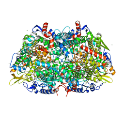
3M30
DownloadVisualize
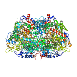
BU of 3m30 by Molmil
Structural Insight into Methyl-Coenzyme M Reductase Chemistry using Coenzyme B Analogues
分子名称: 1,2-ETHANEDIOL, 1-THIOETHANESULFONIC ACID, ACETATE ION, ...
著者Cedervall, P.E, Dey, M, Ragsdale, S.W, Wilmot, C.M.
登録日2010-03-08
公開日2010-09-15
最終更新日2017-11-08
実験手法X-RAY DIFFRACTION (1.45 Å)
主引用文献Structural insight into methyl-coenzyme M reductase chemistry using coenzyme B analogues.
Biochemistry, 49, 2010
3M2R
DownloadVisualize
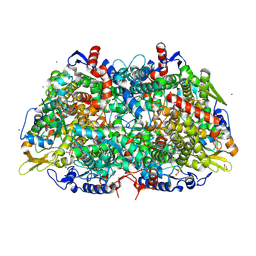
BU of 3m2r by Molmil
Structural Insight into Methyl-Coenzyme M Reductase Chemistry using Coenzyme B Analogues
分子名称: 1,2-ETHANEDIOL, 1-THIOETHANESULFONIC ACID, Coenzyme B, ...
著者Cedervall, P.E, Dey, M, Ragsdale, S.W, Wilmot, C.M.
登録日2010-03-08
公開日2010-09-15
最終更新日2017-11-08
実験手法X-RAY DIFFRACTION (1.3 Å)
主引用文献Structural insight into methyl-coenzyme M reductase chemistry using coenzyme B analogues.
Biochemistry, 49, 2010
3M1V
DownloadVisualize
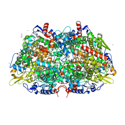
BU of 3m1v by Molmil
Structural Insight into Methyl-Coenzyme M Reductase Chemistry using Coenzyme B Analogues
分子名称: 1,2-ETHANEDIOL, 1-THIOETHANESULFONIC ACID, ACETATE ION, ...
著者Cedervall, P.E, Dey, M, Ragsdale, S.W, Wilmot, C.M.
登録日2010-03-05
公開日2010-09-15
最終更新日2017-11-08
実験手法X-RAY DIFFRACTION (1.45 Å)
主引用文献Structural insight into methyl-coenzyme M reductase chemistry using coenzyme B analogues.
Biochemistry, 49, 2010
3M2U
DownloadVisualize
BU of 3m2u by Molmil
Structural Insight into Methyl-Coenzyme M Reductase Chemistry using Coenzyme B Analogues
分子名称: 1,2-ETHANEDIOL, 1-THIOETHANESULFONIC ACID, ACETATE ION, ...
著者Cedervall, P.E, Dey, M, Ragsdale, S.W, Wilmot, C.M.
登録日2010-03-08
公開日2010-09-15
最終更新日2017-03-29
実験手法X-RAY DIFFRACTION (1.4 Å)
主引用文献Structural insight into methyl-coenzyme M reductase chemistry using coenzyme B analogues.
Biochemistry, 49, 2010
3M32
DownloadVisualize
BU of 3m32 by Molmil
Structural Insight into Methyl-Coenzyme M Reductase Chemistry using Coenzyme B Analogues
分子名称: 1,2-ETHANEDIOL, 1-THIOETHANESULFONIC ACID, ACETATE ION, ...
著者Cedervall, P.E, Dey, M, Ragsdale, S.W, Wilmot, C.M.
登録日2010-03-08
公開日2010-09-15
最終更新日2017-11-08
実験手法X-RAY DIFFRACTION (1.35 Å)
主引用文献Structural insight into methyl-coenzyme M reductase chemistry using coenzyme B analogues.
Biochemistry, 49, 2010
4J1X
DownloadVisualize
BU of 4j1x by Molmil
Crystal Structure of Fe(II)-HppE with alternative substrate (S)-1-HPP
分子名称: Epoxidase, FE (II) ION, GLYCEROL, ...
著者Drennan, C.L, Dey, M.
登録日2013-02-03
公開日2013-04-03
最終更新日2023-09-20
実験手法X-RAY DIFFRACTION (2.8 Å)
主引用文献Mechanistic studies of an unprecedented enzyme-catalysed 1,2-phosphono-migration reaction.
Nature, 496, 2013
4J1W
DownloadVisualize
BU of 4j1w by Molmil
Crystal Structure of Fe(II)-HppE with alternative substrate (R)-1-HPP
分子名称: Epoxidase, FE (II) ION, GLYCEROL, ...
著者Drennan, C.L, Dey, M.
登録日2013-02-03
公開日2013-04-03
最終更新日2024-02-28
実験手法X-RAY DIFFRACTION (2.72 Å)
主引用文献Mechanistic studies of an unprecedented enzyme-catalysed 1,2-phosphono-migration reaction.
Nature, 496, 2013
3POT
DownloadVisualize
BU of 3pot by Molmil
Structural analysis of a Ni(III)-methyl species in methyl-coenzyme M reductase from Methanothermobacter marburgensis
分子名称: 1,2-ETHANEDIOL, 1-THIOETHANESULFONIC ACID, Coenzyme B, ...
著者Cedervall, P.E, Wilmot, C.M.
登録日2010-11-23
公開日2011-04-13
最終更新日2023-09-06
実験手法X-RAY DIFFRACTION (1.2 Å)
主引用文献Structural Analysis of a Ni-Methyl Species in Methyl-Coenzyme M Reductase from Methanothermobacter marburgensis.
J.Am.Chem.Soc., 133, 2011
3SCG
DownloadVisualize
BU of 3scg by Molmil
Fe(II)-HppE with R-HPP
分子名称: Epoxidase, FE (II) ION, [(2R)-2-hydroxypropyl]phosphonic acid
著者Drennan, C.L.
登録日2011-06-07
公開日2011-07-06
最終更新日2023-09-13
実験手法X-RAY DIFFRACTION (3 Å)
主引用文献Structural basis of regiospecificity of a mononuclear iron enzyme in antibiotic fosfomycin biosynthesis.
J.Am.Chem.Soc., 133, 2011
3SCF
DownloadVisualize
BU of 3scf by Molmil
Fe(II)-HppE with S-HPP and NO
分子名称: (S)-2-HYDROXYPROPYLPHOSPHONIC ACID, Epoxidase, FE (II) ION, ...
著者Drennan, C.L.
登録日2011-06-07
公開日2011-07-06
最終更新日2023-09-13
実験手法X-RAY DIFFRACTION (2.85 Å)
主引用文献Structural basis of regiospecificity of a mononuclear iron enzyme in antibiotic fosfomycin biosynthesis.
J.Am.Chem.Soc., 133, 2011
3SCH
DownloadVisualize
BU of 3sch by Molmil
Co(II)-HppE with R-HPP
分子名称: COBALT (II) ION, Epoxidase, [(2R)-2-hydroxypropyl]phosphonic acid
著者Drennan, C.L.
登録日2011-06-07
公開日2011-07-06
最終更新日2023-12-06
実験手法X-RAY DIFFRACTION (2.1 Å)
主引用文献Structural basis of regiospecificity of a mononuclear iron enzyme in antibiotic fosfomycin biosynthesis.
J.Am.Chem.Soc., 133, 2011
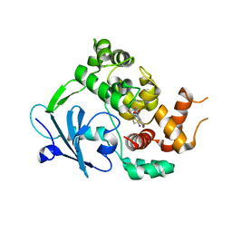
6RLW
DownloadVisualize
BU of 6rlw by Molmil
Structure of the human 8-oxoguanine DNA Glycosylase hOGG1 in complex with inhibitor TH5487
分子名称: 4-(4-bromanyl-2-oxidanylidene-3~{H}-benzimidazol-1-yl)-~{N}-(4-iodophenyl)piperidine-1-carboxamide, N-glycosylase/DNA lyase
著者Masuyer, G, Stenmark, P.
登録日2019-05-03
公開日2020-07-22
最終更新日2024-01-24
実験手法X-RAY DIFFRACTION (2 Å)
主引用文献Targeting OGG1 arrests cancer cell proliferation by inducing replication stress.
Nucleic Acids Res., 48, 2020
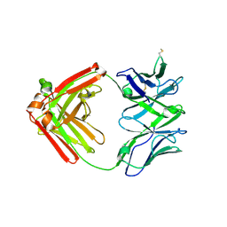
6XRJ
DownloadVisualize
BU of 6xrj by Molmil
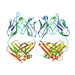
Crystal structure of the disulfide linked DH717.1 Fab dimer, derived from a macaque HIV-1 vaccine-induced Env glycan-reactive neutralizing antibody B cell lineage
分子名称: DH717.1 heavy chain Fab fragment, DH717.1 light chain Fab fragment
著者Manne, K, Nicely, N.I, Acharya, P.
登録日2020-07-13
公開日2020-12-30
最終更新日2023-10-18
実験手法X-RAY DIFFRACTION (3.15 Å)
主引用文献Fab-dimerized glycan-reactive antibodies are a structural category of natural antibodies.
Cell, 184, 2021
7THT
DownloadVisualize
BU of 7tht by Molmil
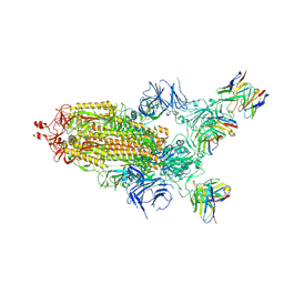
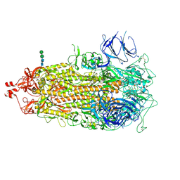
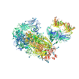
CryoEM structure of SARS-CoV-2 S protein in complex with Receptor Binding Domain antibody DH1042
分子名称: 2-acetamido-2-deoxy-beta-D-glucopyranose, 2-acetamido-2-deoxy-beta-D-glucopyranose-(1-4)-2-acetamido-2-deoxy-beta-D-glucopyranose, DH1042 heavy chain, ...
著者Manne, K, May, A, Acharya, P.
登録日2022-01-12
公開日2022-02-16
最終更新日2023-04-12
実験手法ELECTRON MICROSCOPY (3.42 Å)
主引用文献Structural diversity of the SARS-CoV-2 Omicron spike.
Mol.Cell, 82, 2022
7TL9
DownloadVisualize
BU of 7tl9 by Molmil
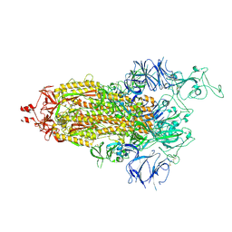
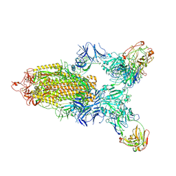
SARS-CoV-2 Omicron 1-RBD up Spike Protein Trimer without the P986-P987 stabilizing mutations (S-GSAS-Omicron)
分子名称: 2-acetamido-2-deoxy-beta-D-glucopyranose, 2-acetamido-2-deoxy-beta-D-glucopyranose-(1-4)-2-acetamido-2-deoxy-beta-D-glucopyranose, Spike glycoprotein, ...
著者Stalls, V, Acharya, P.
登録日2022-01-18
公開日2022-02-23
最終更新日2023-04-12
実験手法ELECTRON MICROSCOPY (3.5 Å)
主引用文献Structural diversity of the SARS-CoV-2 Omicron spike.
Mol.Cell, 82, 2022
7TEI
DownloadVisualize
BU of 7tei by Molmil
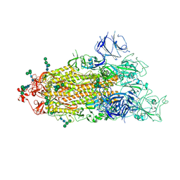
SARS-CoV-2 Omicron 1-RBD up Spike Protein Trimer without the P986-P987 stabilizing mutations (S-GSAS-Omicron)
分子名称: 2-acetamido-2-deoxy-beta-D-glucopyranose, 2-acetamido-2-deoxy-beta-D-glucopyranose-(1-4)-2-acetamido-2-deoxy-beta-D-glucopyranose, Spike glycoprotein, ...
著者Stalls, V, Acharya, P.
登録日2022-01-05
公開日2022-02-16
最終更新日2023-04-12
実験手法ELECTRON MICROSCOPY (3.4 Å)
主引用文献Structural diversity of the SARS-CoV-2 Omicron spike.
Mol.Cell, 82, 2022
7TF8
DownloadVisualize
BU of 7tf8 by Molmil
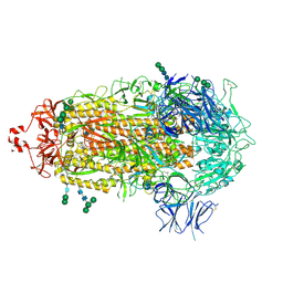
SARS-CoV-2 Omicron 3-RBD down Spike Protein Trimer without the P986-P987 stabilizing mutations (S-GSAS-Omicron)
分子名称: 2-acetamido-2-deoxy-beta-D-glucopyranose, 2-acetamido-2-deoxy-beta-D-glucopyranose-(1-4)-2-acetamido-2-deoxy-beta-D-glucopyranose, Spike glycoprotein, ...
著者Stalls, V, Acharya, P.
登録日2022-01-06
公開日2022-02-16
最終更新日2023-04-12
実験手法ELECTRON MICROSCOPY (3.36 Å)
主引用文献Structural diversity of the SARS-CoV-2 Omicron spike.
Mol.Cell, 82, 2022
7TL1
DownloadVisualize
BU of 7tl1 by Molmil
SARS-CoV-2 Omicron 3-RBD down Spike Protein Trimer without the P986-P987 stabilizing mutations (S-GSAS-Omicron)
分子名称: 2-acetamido-2-deoxy-beta-D-glucopyranose, 2-acetamido-2-deoxy-beta-D-glucopyranose-(1-4)-2-acetamido-2-deoxy-beta-D-glucopyranose, Spike glycoprotein, ...
著者Stalls, V, Acharya, P.
登録日2022-01-18
公開日2022-02-23
最終更新日2023-04-12
実験手法ELECTRON MICROSCOPY (3.5 Å)
主引用文献Structural diversity of the SARS-CoV-2 Omicron spike.
Mol.Cell, 82, 2022
7L06
DownloadVisualize
BU of 7l06 by Molmil
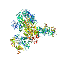
Cryo-EM structure of SARS-CoV-2 2P S ectodomain bound to two copies of domain-swapped antibody 2G12
分子名称: 2-acetamido-2-deoxy-beta-D-glucopyranose, 2-acetamido-2-deoxy-beta-D-glucopyranose-(1-4)-2-acetamido-2-deoxy-beta-D-glucopyranose, 2G12 heavy chain, ...
著者Manne, K, Henderson, R, Acharya, P.
登録日2020-12-11
公開日2020-12-30
最終更新日2021-06-09
実験手法ELECTRON MICROSCOPY (3.3 Å)
主引用文献Fab-dimerized glycan-reactive antibodies are a structural category of natural antibodies.
Cell, 184, 2021
7L6M
DownloadVisualize
BU of 7l6m by Molmil
Cryo-EM structure of DH898.1 Fab-dimer from local refinement of the Fab-dimer bound near the CD4 binding site of HIV-1 Env CH848 SOSIP trimer
分子名称: DH898.1 Fab heavy chain, DH898.1 Fab light chain
著者Manne, K, Edwards, R.J, Acharya, P.
登録日2020-12-23
公開日2021-02-10
最終更新日2021-06-09
実験手法ELECTRON MICROSCOPY (4.7 Å)
主引用文献Fab-dimerized glycan-reactive antibodies are a structural category of natural antibodies.
Cell, 184, 2021
7L02
DownloadVisualize
BU of 7l02 by Molmil
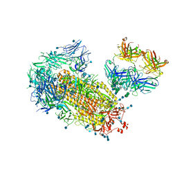
Cryo-EM structure of SARS-CoV-2 2P S ectodomain bound to one copy of domain-swapped antibody 2G12
分子名称: 2-acetamido-2-deoxy-beta-D-glucopyranose, 2-acetamido-2-deoxy-beta-D-glucopyranose-(1-4)-2-acetamido-2-deoxy-beta-D-glucopyranose, 2G12 heavy chain, ...
著者Manne, K, Henderson, R, Acharya, P.
登録日2020-12-10
公開日2020-12-30
最終更新日2021-06-09
実験手法ELECTRON MICROSCOPY (3.2 Å)
主引用文献Fab-dimerized glycan-reactive antibodies are a structural category of natural antibodies.
Cell, 184, 2021
7L09
DownloadVisualize
BU of 7l09 by Molmil
Cryo-EM structure of SARS-CoV-2 2P S ectodomain bound domain-swapped antibody 2G12 from masked 3D refinement
分子名称: 2-acetamido-2-deoxy-beta-D-glucopyranose, 2-acetamido-2-deoxy-beta-D-glucopyranose-(1-4)-2-acetamido-2-deoxy-beta-D-glucopyranose, 2G12 heavy chain, ...
著者Manne, K, Henderson, R, Acharya, P.
登録日2020-12-11
公開日2020-12-30
最終更新日2021-06-09
実験手法ELECTRON MICROSCOPY (3.1 Å)
主引用文献Fab-dimerized glycan-reactive antibodies are a structural category of natural antibodies.
Cell, 184, 2021
7LD1
DownloadVisualize
BU of 7ld1 by Molmil
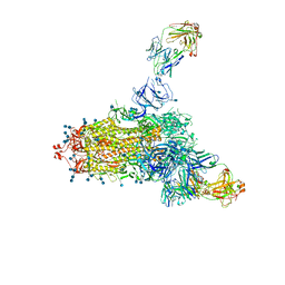
Structure of SARS-CoV-2 S protein in complex with Receptor Binding Domain antibody DH1047
分子名称: 2-acetamido-2-deoxy-beta-D-glucopyranose, 2-acetamido-2-deoxy-beta-D-glucopyranose-(1-4)-2-acetamido-2-deoxy-beta-D-glucopyranose, DH1047 heavy chain, ...
著者Manne, K, Acharya, P.
登録日2021-01-12
公開日2021-01-27
最終更新日2021-08-18
実験手法ELECTRON MICROSCOPY (3.4 Å)
主引用文献In vitro and in vivo functions of SARS-CoV-2 infection-enhancing and neutralizing antibodies.
Cell, 184, 2021
7LCN
DownloadVisualize
BU of 7lcn by Molmil
Structure of SARS-CoV-2 S protein in complex with N-terminal domain antibody DH1050.1
分子名称: 2-acetamido-2-deoxy-beta-D-glucopyranose, 2-acetamido-2-deoxy-beta-D-glucopyranose-(1-4)-2-acetamido-2-deoxy-beta-D-glucopyranose, DH1050.1 heavy chain, ...
著者Manne, K, Acharya, P.
登録日2021-01-11
公開日2021-01-27
最終更新日2021-08-18
実験手法ELECTRON MICROSCOPY (3.35 Å)
主引用文献In vitro and in vivo functions of SARS-CoV-2 infection-enhancing and neutralizing antibodies.
Cell, 184, 2021

222624

件を2024-07-17に公開中

PDB statisticsPDBj update infoContact PDBjnumon