9J91
 
 | |
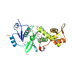
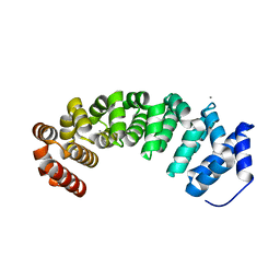
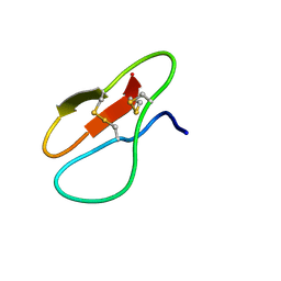
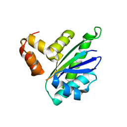
5H65
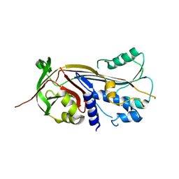
 | | Crystal structure of human POT1 and TPP1 | | 分子名称: | Adrenocortical dysplasia protein homolog, Protection of telomeres protein 1, ZINC ION | | 著者 | Chen, C, Wu, J, Lei, M. | | 登録日 | 2016-11-10 | | 公開日 | 2017-05-31 | | 最終更新日 | 2024-03-20 | | 実験手法 | X-RAY DIFFRACTION (2.1 Å) | | 主引用文献 | Structural insights into POT1-TPP1 interaction and POT1 C-terminal mutations in human cancer.
Nat Commun, 8, 2017
|
|
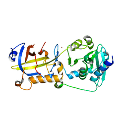
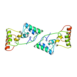
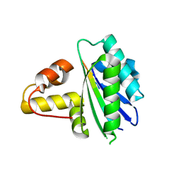
4D49
 | | Crystal structure of computationally designed armadillo repeat proteins for modular peptide recognition. | | 分子名称: | ARGININE, ARMADILLO REPEAT PROTEIN ARM00027, POLY ARG DECAPEPTIDE | | 著者 | Reichen, C, Forzani, C, Zhou, T, Parmeggiani, F, Fleishman, S.J, Mittl, P.R.E, Madhurantakam, C, Honegger, A, Ewald, C, Zerbe, O, Baker, D, Caflisch, A, Pluckthun, A. | | 登録日 | 2014-10-27 | | 公開日 | 2016-01-13 | | 最終更新日 | 2024-05-01 | | 実験手法 | X-RAY DIFFRACTION (2.09 Å) | | 主引用文献 | Computationally Designed Armadillo Repeat Proteins for Modular Peptide Recognition.
J.Mol.Biol., 428, 2016
|
|
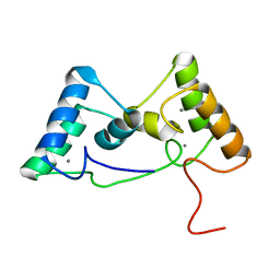
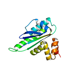
4D4E
 | | Crystal structure of computationally designed armadillo repeat proteins for modular peptide recognition. | | 分子名称: | ARMADILLO REPEAT PROTEIN ARM00016, GLYCEROL | | 著者 | Reichen, C, Forzani, C, Zhou, T, Parmeggiani, F, Fleishman, S.J, Mittl, P.R.E, Madhurantakam, C, Honegger, A, Ewald, C, Zerbe, O, Baker, D, Caflisch, A, Pluckthun, A. | | 登録日 | 2014-10-28 | | 公開日 | 2016-01-13 | | 最終更新日 | 2024-05-01 | | 実験手法 | X-RAY DIFFRACTION (2 Å) | | 主引用文献 | Computationally Designed Armadillo Repeat Proteins for Modular Peptide Recognition.
J.Mol.Biol., 428, 2016
|
|
8W13
 | | Crystal structure of MYST acetyltransferase domain in complex with N-(1-(5-bromo-2-methoxyphenyl)-1H-1,2,3-triazol-4-yl)-2-methoxybenzenesulfonamide | | 分子名称: | 1,2-ETHANEDIOL, CHLORIDE ION, Histone acetyltransferase KAT8, ... | | 著者 | Chen, C, Dou, Y, Wang, M, Xu, C, Buesking, A. | | 登録日 | 2024-02-15 | | 公開日 | 2024-09-11 | | 最終更新日 | 2024-10-23 | | 実験手法 | X-RAY DIFFRACTION (1.81 Å) | | 主引用文献 | Identification of triazolyl KAT6 inhibitors via a templated fragment approach.
Bioorg.Med.Chem.Lett., 113, 2024
|
|
8X39
 | |
8X3A
 | |
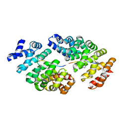
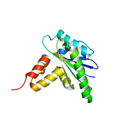
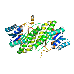
4V3Q
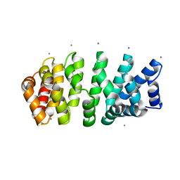 | | Designed armadillo repeat protein with 4 internal repeats, 2nd generation C-cap and 3rd generation N-cap. | | 分子名称: | CALCIUM ION, GLYCEROL, YIII_M4_AII | | 著者 | Reichen, C, Madhurantakam, C, Pluckthun, A, Mittl, P. | | 登録日 | 2014-10-20 | | 公開日 | 2016-01-13 | | 最終更新日 | 2024-01-10 | | 実験手法 | X-RAY DIFFRACTION (1.8 Å) | | 主引用文献 | Structures of designed armadillo-repeat proteins show propagation of inter-repeat interface effects.
Acta Crystallogr D Struct Biol, 72, 2016
|
|
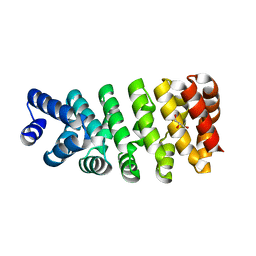
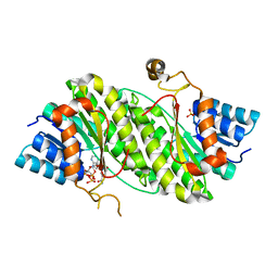
4V3O
 | | Designed armadillo repeat protein with 5 internal repeats, 2nd generation C-cap and 3rd generation N-cap. | | 分子名称: | ACETATE ION, CALCIUM ION, YIII_M5_AII | | 著者 | Reichen, C, Madhurantakam, C, Pluckthun, A, Mittl, P. | | 登録日 | 2014-10-20 | | 公開日 | 2016-01-13 | | 最終更新日 | 2024-01-10 | | 実験手法 | X-RAY DIFFRACTION (2 Å) | | 主引用文献 | Structures of Designed Armadillo-Repeat Proteins Show Propagation of Inter-Repeat Interface Effects
Acta Crystallogr.,Sect.D, 72, 2016
|
|
4V3R
 | | Designed armadillo repeat protein with 5 internal repeats, 2nd generation C-cap and 3rd generation N-cap. | | 分子名称: | MAGNESIUM ION, YIII_M5_AII | | 著者 | Reichen, C, Madhurantakam, C, Pluckthun, A, Mittl, P. | | 登録日 | 2014-10-20 | | 公開日 | 2016-01-13 | | 最終更新日 | 2024-01-10 | | 実験手法 | X-RAY DIFFRACTION (1.95 Å) | | 主引用文献 | Structures of Designed Armadillo-Repeat Proteins Show Propagation of Inter-Repeat Interface Effects
Acta Crystallogr.,Sect.D, 72, 2016
|
|
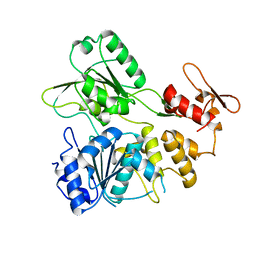
3HMY
 | | Crystal structure of HCR/T complexed with GT2 | | 分子名称: | GLYCEROL, N-acetyl-alpha-neuraminic acid-(2-8)-N-acetyl-alpha-neuraminic acid-(2-8)-N-acetyl-alpha-neuraminic acid, SULFATE ION, ... | | 著者 | Chen, C, Fu, Z, Kim, J.-J.P, Barbieri, J.T, Baldwin, M.R. | | 登録日 | 2009-05-29 | | 公開日 | 2009-07-14 | | 最終更新日 | 2024-10-16 | | 実験手法 | X-RAY DIFFRACTION (2 Å) | | 主引用文献 | Gangliosides as high affinity receptors for tetanus neurotoxin.
J.Biol.Chem., 284, 2009
|
|
7YHH
 | |
7YHG
 | |
7YHF
 | |
7YHI
 | |
8H61
 | | Ketoreductase CpKR mutant - M2 | | 分子名称: | Mutant M2 of ketoreductase CpKR, NADPH DIHYDRO-NICOTINAMIDE-ADENINE-DINUCLEOTIDE PHOSPHATE | | 著者 | Chen, C, Pan, J, Xu, J.H. | | 登録日 | 2022-10-14 | | 公開日 | 2023-10-18 | | 実験手法 | X-RAY DIFFRACTION (1.9 Å) | | 主引用文献 | Computational redesign of a robust ketoreductase for asymmetric synthesis of enantiopure diltiazem precursor.
To Be Published
|
|
3HN1
 | | Crystal structure of HCR/T complexed with GT2 and lactose | | 分子名称: | N-acetyl-alpha-neuraminic acid-(2-8)-N-acetyl-alpha-neuraminic acid, SULFATE ION, Tetanus toxin, ... | | 著者 | Chen, C, Fu, Z, Kim, J.-J.P, Barbieri, J.T, Baldwin, M.R. | | 登録日 | 2009-05-29 | | 公開日 | 2009-07-14 | | 最終更新日 | 2024-10-16 | | 実験手法 | X-RAY DIFFRACTION (2.1 Å) | | 主引用文献 | Gangliosides as high affinity receptors for tetanus neurotoxin.
J.Biol.Chem., 284, 2009
|
|
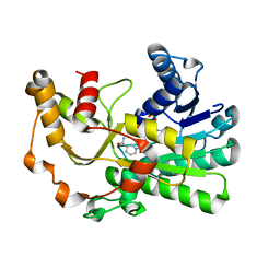
2F43
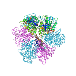 | | Rat liver F1-ATPase | | 分子名称: | ADENOSINE-5'-DIPHOSPHATE, ADENOSINE-5'-TRIPHOSPHATE, ATP synthase alpha chain, ... | | 著者 | Chen, C, Saxena, A.K, Simcoke, W.N, Garboczi, D.N, Pedersen, P.L, Ko, Y.H. | | 登録日 | 2005-11-22 | | 公開日 | 2006-03-07 | | 最終更新日 | 2024-12-25 | | 実験手法 | X-RAY DIFFRACTION (3 Å) | | 主引用文献 | Mitochondrial ATP synthase: Crystal structure of the catalytic F1 unit in a vanadate-induced transition-like state and implications for mechanism.
J.Biol.Chem., 281, 2006
|
|
8HDJ
 | |
8HER
 | |
8HEP
 | |
8HEQ
 | |
4DML
 | |
4DMM
 | |
8YNJ
 | |


