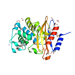
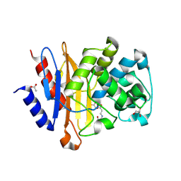
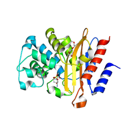
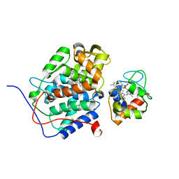
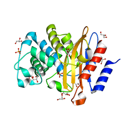
6H2A
 
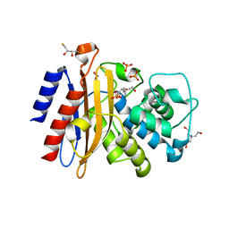 | | Structure of S70A BlaC from Mycobacterium tuberculosis obtained from crystals produced in the presence of DTT | | 分子名称: | 2,3-DIHYDROXY-1,4-DITHIOBUTANE, Beta-lactamase, GLYCEROL, ... | | 著者 | Tassoni, R, Pannu, N.S, Ubbink, M. | | 登録日 | 2018-07-13 | | 公開日 | 2019-01-23 | | 最終更新日 | 2024-01-17 | | 実験手法 | X-RAY DIFFRACTION (2.54 Å) | | 主引用文献 | New Conformations of Acylation Adducts of Inhibitors of beta-Lactamase from Mycobacterium tuberculosis.
Biochemistry, 58, 2019
|
|
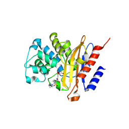
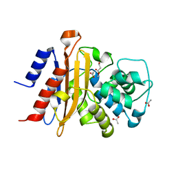
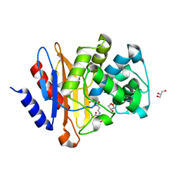
6H27
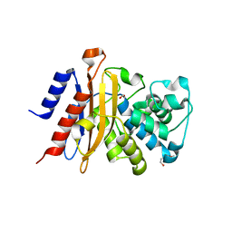 | | Structure of S70C BlaC from Mycobacterium tuberculosis | | 分子名称: | 1,2-ETHANEDIOL, 2-(2-METHOXYETHOXY)ETHANOL, BlaC | | 著者 | Tassoni, R, Pannu, N.S, Ubbink, M. | | 登録日 | 2018-07-13 | | 公開日 | 2019-01-23 | | 最終更新日 | 2024-01-17 | | 実験手法 | X-RAY DIFFRACTION (1.63 Å) | | 主引用文献 | New Conformations of Acylation Adducts of Inhibitors of beta-Lactamase from Mycobacterium tuberculosis.
Biochemistry, 58, 2019
|
|
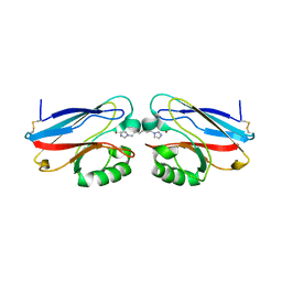
2OJ1
 | | Disulfide-linked dimer of azurin N42C/M64E double mutant | | 分子名称: | Azurin, COPPER (II) ION | | 著者 | de Jongh, T.E, Hoffmann, M, Einsle, O, Cavazzini, D, Rossi, G.L, Ubbink, M, Canters, G.W. | | 登録日 | 2007-01-12 | | 公開日 | 2007-11-27 | | 最終更新日 | 2024-04-03 | | 実験手法 | X-RAY DIFFRACTION (2.3 Å) | | 主引用文献 | Inter- and intramolecular electron transfer in modified azurin dimers
Eur.J.Inorg.Chem., 2007, 2007
|
|
7A5U
 | |
7A74
 | | Structure of G132N BlaC from Mycobacterium tuberculosis | | 分子名称: | 1,2-ETHANEDIOL, Beta-lactamase, GLYCEROL, ... | | 著者 | Chikunova, A, Ahmad, M.U, Perrakis, A, Ubbink, M. | | 登録日 | 2020-08-27 | | 公開日 | 2021-05-19 | | 最終更新日 | 2024-01-31 | | 実験手法 | X-RAY DIFFRACTION (1.6 Å) | | 主引用文献 | Two beta-Lactamase Variants with Reduced Clavulanic Acid Inhibition Display Different Millisecond Dynamics.
Antimicrob.Agents Chemother., 65, 2021
|
|
2M56
 | | The structure of the complex of cytochrome P450cam and its electron donor putidaredoxin determined by paramagnetic NMR spectroscopy | | 分子名称: | CAMPHOR, Camphor 5-monooxygenase, FE2/S2 (INORGANIC) CLUSTER, ... | | 著者 | Hiruma, Y, Hass, M.A.S, Ubbink, M. | | 登録日 | 2013-02-14 | | 公開日 | 2013-08-21 | | 最終更新日 | 2024-05-15 | | 実験手法 | SOLUTION NMR | | 主引用文献 | The structure of the cytochrome p450cam-putidaredoxin complex determined by paramagnetic NMR spectroscopy and crystallography.
J.Mol.Biol., 425, 2013
|
|
2BZC
 | |
2BZ7
 | |
2P80
 | |
2IWE
 | | Structure of a cavity mutant (H117G) of Pseudomonas aeruginosa azurin | | 分子名称: | 1,1'-HEXANE-1,6-DIYLBIS(1H-IMIDAZOLE), AZURIN, ZINC ION | | 著者 | De Jongh, T.E, Van Roon, A.M.M, Prudencio, M, Ubbink, M, Canters, G.W. | | 登録日 | 2006-06-29 | | 公開日 | 2007-06-12 | | 最終更新日 | 2023-12-13 | | 実験手法 | X-RAY DIFFRACTION (2.83 Å) | | 主引用文献 | Click-Chemistry with an Active Site Variant of Azurin
Eur.J.Inorg.Chem., 2006, 2006
|
|
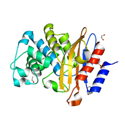
6H2C
 | | Structure of BlaC from Mycobacterium tuberculosis bound to the trans-enamine adduct derived from clavulanic acid. | | 分子名称: | (2E)-3-[(4-hydroxy-2-oxobutyl)amino]prop-2-enal, ACETATE ION, Beta-lactamase, ... | | 著者 | Tassoni, R, Pannu, N.S, Ubbink, M. | | 登録日 | 2018-07-13 | | 公開日 | 2019-01-23 | | 最終更新日 | 2024-01-17 | | 実験手法 | X-RAY DIFFRACTION (1.93 Å) | | 主引用文献 | New Conformations of Acylation Adducts of Inhibitors of beta-Lactamase from Mycobacterium tuberculosis.
Biochemistry, 58, 2019
|
|
6H2I
 | |
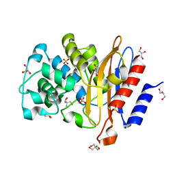
6H2K
 | | Structure of BlaC from Mycobacterium tuberculosis bound to the trans-enamine adduct of sulbactam. | | 分子名称: | ACETATE ION, Beta-lactamase, DI(HYDROXYETHYL)ETHER, ... | | 著者 | Tassoni, R, Pannu, N.S, Ubbink, M. | | 登録日 | 2018-07-13 | | 公開日 | 2019-01-23 | | 最終更新日 | 2024-01-17 | | 実験手法 | X-RAY DIFFRACTION (1.9 Å) | | 主引用文献 | New Conformations of Acylation Adducts of Inhibitors of beta-Lactamase from Mycobacterium tuberculosis.
Biochemistry, 58, 2019
|
|
6H2H
 | | Structure of BlaC from Mycobacterium tuberculosis covalently bound to avibactam. | | 分子名称: | (2S,5R)-1-formyl-5-[(sulfooxy)amino]piperidine-2-carboxamide, Beta-lactamase, POLYETHYLENE GLYCOL (N=34) | | 著者 | Tassoni, R, Pannu, N.S, Ubbink, M. | | 登録日 | 2018-07-13 | | 公開日 | 2019-01-23 | | 最終更新日 | 2024-01-17 | | 実験手法 | X-RAY DIFFRACTION (1.62 Å) | | 主引用文献 | New Conformations of Acylation Adducts of Inhibitors of beta-Lactamase from Mycobacterium tuberculosis.
Biochemistry, 58, 2019
|
|
6H2G
 | |
2KAJ
 | |
2JXM
 | | Ensemble of twenty structures of the Prochlorothrix hollandica plastocyanin- cytochrome f complex | | 分子名称: | COPPER (II) ION, Cytochrome f, HEME C, ... | | 著者 | Hulsker, R, Baranova, M, Bullerjahn, G, Ubbink, M. | | 登録日 | 2007-11-22 | | 公開日 | 2008-02-12 | | 最終更新日 | 2024-05-29 | | 実験手法 | SOLUTION NMR | | 主引用文献 | Dynamics in the transient complex of plastocyanin-cytochrome f from Prochlorothrix hollandica.
J.Am.Chem.Soc., 130, 2008
|
|
2GB8
 | | Solution structure of the complex between yeast iso-1-cytochrome c and yeast cytochrome c peroxidase | | 分子名称: | Cytochrome c iso-1, Cytochrome c peroxidase, HEME C, ... | | 著者 | Volkov, A.N, Worrall, J.A.R, Ubbink, M. | | 登録日 | 2006-03-10 | | 公開日 | 2006-11-21 | | 最終更新日 | 2022-03-09 | | 実験手法 | SOLUTION NMR | | 主引用文献 | Solution structure and dynamics of the complex between cytochrome c and cytochrome c peroxidase determined by paramagnetic NMR.
Proc.Natl.Acad.Sci.Usa, 103, 2006
|
|
7A72
 | | Structure of G132S BlaC from Mycobacterium tuberculosis bound to the trans-enamine adduct of sulbactam | | 分子名称: | ACETATE ION, Beta-lactamase, GLYCEROL, ... | | 著者 | Chikunova, A, Ahmad, M.U, Perrakis, A, Ubbink, M. | | 登録日 | 2020-08-27 | | 公開日 | 2021-07-07 | | 最終更新日 | 2024-01-31 | | 実験手法 | X-RAY DIFFRACTION (1.3 Å) | | 主引用文献 | The G132S Mutation Enhances the Resistance of Mycobacterium tuberculosis beta-Lactamase against Sulbactam.
Biochemistry, 60, 2021
|
|
7A5W
 | | Structure of D172N BlaC from Mycobacterium tuberculosis | | 分子名称: | 1,2-ETHANEDIOL, Beta-lactamase | | 著者 | Chikunova, A, Ahmad, M.U, Perrakis, A, Ubbink, M. | | 登録日 | 2020-08-24 | | 公開日 | 2021-07-07 | | 最終更新日 | 2024-01-31 | | 実験手法 | X-RAY DIFFRACTION (1.4 Å) | | 主引用文献 | The G132S Mutation Enhances the Resistance of Mycobacterium tuberculosis beta-Lactamase against Sulbactam.
Biochemistry, 60, 2021
|
|
7A5T
 | | Crystal structure of A55E mutant of BlaC from Mycobacterium tuberculosis | | 分子名称: | Beta-lactamase, GLYCEROL, PHOSPHATE ION | | 著者 | Chikunova, A, Ahmad, M.U, Perrakis, A, Ubbink, M. | | 登録日 | 2020-08-21 | | 公開日 | 2021-07-07 | | 最終更新日 | 2024-01-31 | | 実験手法 | X-RAY DIFFRACTION (1.4 Å) | | 主引用文献 | The G132S Mutation Enhances the Resistance of Mycobacterium tuberculosis beta-Lactamase against Sulbactam.
Biochemistry, 60, 2021
|
|
7A71
 | | Structure of G132S BlaC from Mycobacterium tuberculosis | | 分子名称: | 2-AMINO-2-HYDROXYMETHYL-PROPANE-1,3-DIOL, Beta-lactamase, GLYCEROL, ... | | 著者 | Chikunova, A, Ahmad, M.U, Perrakis, A, Ubbink, M. | | 登録日 | 2020-08-27 | | 公開日 | 2021-07-07 | | 最終更新日 | 2024-01-31 | | 実験手法 | X-RAY DIFFRACTION (1.4 Å) | | 主引用文献 | The G132S Mutation Enhances the Resistance of Mycobacterium tuberculosis beta-Lactamase against Sulbactam.
Biochemistry, 60, 2021
|
|
7A6Z
 | | Structure of P226G BlaC from Mycobacterium tuberculosis | | 分子名称: | 1,2-ETHANEDIOL, Beta-lactamase, GLYCEROL, ... | | 著者 | Chikunova, A, Ahmad, M.U, Perrakis, A, Ubbink, M. | | 登録日 | 2020-08-27 | | 公開日 | 2021-10-06 | | 最終更新日 | 2024-04-10 | | 実験手法 | X-RAY DIFFRACTION (1.3 Å) | | 主引用文献 | Conserved proline residues prevent dimerization and aggregation in the beta-lactamase BlaC.
Protein Sci., 33, 2024
|
|
2BH5
 | | X-ray structure of the M100K variant of ferric cyt c-550 from Paracoccus versutus determined at 295 K. | | 分子名称: | CYTOCHROME C-550, HEME C | | 著者 | Worrall, J.A.R, van Roon, A.-M.M, Ubbink, M, Canters, G.W. | | 登録日 | 2005-01-07 | | 公開日 | 2005-05-11 | | 最終更新日 | 2023-12-13 | | 実験手法 | X-RAY DIFFRACTION (1.95 Å) | | 主引用文献 | The Effect of Replacing the Axial Methionine Ligand with a Lysine Residue in Cytochrome C-550 from Paracoccus Versutus Assessed by X-Ray Crystallography and Unfolding.
FEBS J., 272, 2005
|
|
2BGV
 | | X-ray structure of ferric cytochrome c-550 from Paracoccus versutus | | 分子名称: | CYTOCHROME C-550, HEME C | | 著者 | Worrall, J.A.R, Van Roon, A.-M.M, Ubbink, M, Canters, G.W. | | 登録日 | 2005-01-05 | | 公開日 | 2005-05-11 | | 最終更新日 | 2024-05-01 | | 実験手法 | X-RAY DIFFRACTION (1.9 Å) | | 主引用文献 | The Effect of Replacing the Axial Methionine Ligand with a Lysine Residue in Cytochrome C-550 from Paracoccus Versutus Assessed by X-Ray Crystallography and Unfolding.
FEBS J., 272, 2005
|
|


