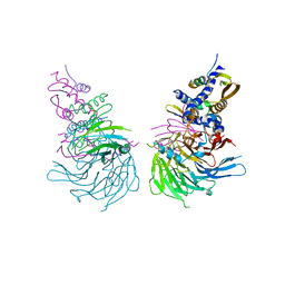
4MNX
 
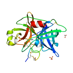
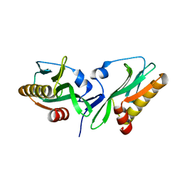
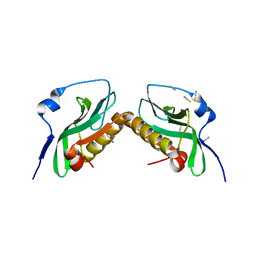
 | | Crystal structure of urokinase-type plasminogen activator (uPA) complexed with bicyclic peptide UK811 | | 分子名称: | 1,1',1''-(1,3,5-triazinane-1,3,5-triyl)tripropan-1-one, GLYCEROL, SULFATE ION, ... | | 著者 | Chen, S, Pojer, F, Heinis, C. | | 登録日 | 2013-09-11 | | 公開日 | 2014-02-05 | | 最終更新日 | 2017-11-15 | | 実験手法 | X-RAY DIFFRACTION (1.85 Å) | | 主引用文献 | Peptide ligands stabilized by small molecules.
Angew.Chem.Int.Ed.Engl., 53, 2014
|
|
4OS7
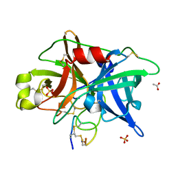
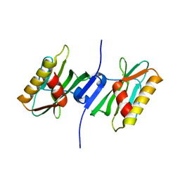
 | | Crystal structure of urokinase-type plasminogen activator (uPA) complexed with bicyclic peptide UK607 (bicyclic) | | 分子名称: | ACETATE ION, GLYCEROL, SULFATE ION, ... | | 著者 | Chen, S, Pojer, F, Heinis, C. | | 登録日 | 2014-02-12 | | 公開日 | 2014-09-24 | | 最終更新日 | 2021-06-02 | | 実験手法 | X-RAY DIFFRACTION (2 Å) | | 主引用文献 | Dithiol amino acids can structurally shape and enhance the ligand-binding properties of polypeptides.
Nat Chem, 6, 2014
|
|
4OS2
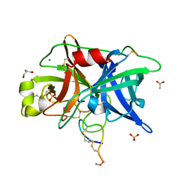
 | | Crystal structure of urokinase-type plasminogen activator (uPA) complexed with bicyclic peptide UK602 (bicyclic 1) | | 分子名称: | ACETATE ION, SULFATE ION, Urokinase-type plasminogen activator, ... | | 著者 | Chen, S, Pojer, F, Heinis, C. | | 登録日 | 2014-02-12 | | 公開日 | 2014-09-24 | | 最終更新日 | 2021-06-02 | | 実験手法 | X-RAY DIFFRACTION (1.79 Å) | | 主引用文献 | Dithiol amino acids can structurally shape and enhance the ligand-binding properties of polypeptides.
Nat Chem, 6, 2014
|
|
3U9G
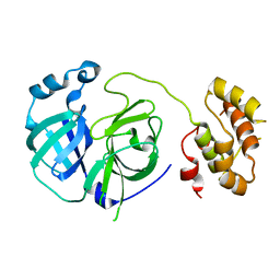
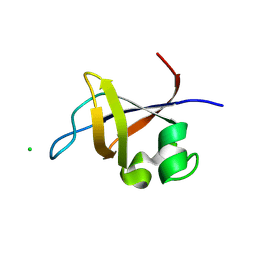
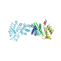
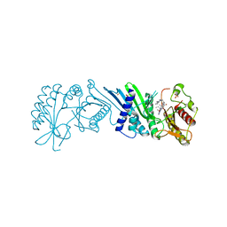
 | | Crystal structure of the Zinc finger antiviral protein | | 分子名称: | ZINC ION, Zinc finger CCCH-type antiviral protein 1 | | 著者 | Chen, S, Xu, Y, Zhang, K, Wang, X, Sun, J, Gao, G, Liu, Y. | | 登録日 | 2011-10-18 | | 公開日 | 2012-03-14 | | 最終更新日 | 2024-03-20 | | 実験手法 | X-RAY DIFFRACTION (1.801 Å) | | 主引用文献 | Structure of N-terminal domain of ZAP indicates how a zinc-finger protein recognizes complex RNA.
Nat.Struct.Mol.Biol., 19, 2012
|
|
4OS5
 | |
4OS1
 | | Crystal structure of urokinase-type plasminogen activator (uPA) complexed with bicyclic peptide UK601 (bicyclic 1) | | 分子名称: | ACETATE ION, SULFATE ION, Urokinase-type plasminogen activator, ... | | 著者 | Chen, S, Pojer, F, Heinis, C. | | 登録日 | 2014-02-12 | | 公開日 | 2014-09-24 | | 最終更新日 | 2021-06-02 | | 実験手法 | X-RAY DIFFRACTION (2.2 Å) | | 主引用文献 | Dithiol amino acids can structurally shape and enhance the ligand-binding properties of polypeptides.
Nat Chem, 6, 2014
|
|
4OS6
 | | Crystal structure of urokinase-type plasminogen activator (uPA) complexed with bicyclic peptide UK604 (bicyclic 2) | | 分子名称: | ACETATE ION, SULFATE ION, Urokinase-type plasminogen activator, ... | | 著者 | Chen, S, Pojer, F, Heinis, C. | | 登録日 | 2014-02-12 | | 公開日 | 2014-09-24 | | 最終更新日 | 2021-06-02 | | 実験手法 | X-RAY DIFFRACTION (1.75 Å) | | 主引用文献 | Dithiol amino acids can structurally shape and enhance the ligand-binding properties of polypeptides.
Nat Chem, 6, 2014
|
|
4OS4
 | | Crystal structure of urokinase-type plasminogen activator (uPA) complexed with bicyclic peptide UK603 (bicyclic 1) | | 分子名称: | ACETATE ION, CHLORIDE ION, GLYCEROL, ... | | 著者 | Chen, S, Pojer, F, Heinis, C. | | 登録日 | 2014-02-12 | | 公開日 | 2014-09-24 | | 最終更新日 | 2021-06-02 | | 実験手法 | X-RAY DIFFRACTION (2 Å) | | 主引用文献 | Dithiol amino acids can structurally shape and enhance the ligand-binding properties of polypeptides.
Nat Chem, 6, 2014
|
|
2PWX
 | |
8EZB
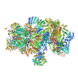
 | | NHEJ Long-range complex with ATP | | 分子名称: | ADENOSINE-5'-TRIPHOSPHATE, DNA (30-MER), DNA (31-MER), ... | | 著者 | Chen, S, He, Y. | | 登録日 | 2022-10-31 | | 公開日 | 2023-06-14 | | 最終更新日 | 2024-06-19 | | 実験手法 | ELECTRON MICROSCOPY (8.9 Å) | | 主引用文献 | Cryo-EM visualization of DNA-PKcs structural intermediates in NHEJ.
Sci Adv, 9, 2023
|
|
8EZ9
 | | Dimeric complex of DNA-PKcs | | 分子名称: | DNA-dependent protein kinase catalytic subunit, unknown region of DNA-PKcs | | 著者 | Chen, S, He, Y. | | 登録日 | 2022-10-31 | | 公開日 | 2023-06-14 | | 最終更新日 | 2024-06-19 | | 実験手法 | ELECTRON MICROSCOPY (5.67 Å) | | 主引用文献 | Cryo-EM visualization of DNA-PKcs structural intermediates in NHEJ.
Sci Adv, 9, 2023
|
|
8EZA
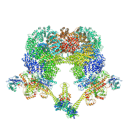 | | NHEJ Long-range complex with PAXX | | 分子名称: | ADENOSINE-5'-TRIPHOSPHATE, DNA (30-MER), DNA (31-MER), ... | | 著者 | Chen, S, He, Y. | | 登録日 | 2022-10-31 | | 公開日 | 2023-06-14 | | 最終更新日 | 2024-06-19 | | 実験手法 | ELECTRON MICROSCOPY (4.39 Å) | | 主引用文献 | Cryo-EM visualization of DNA-PKcs structural intermediates in NHEJ.
Sci Adv, 9, 2023
|
|
6NQ3
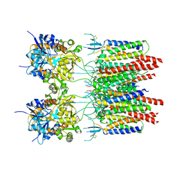
 | | Crystal Structure of a SUZ12-RBBP4-PHF19-JARID2 Heterotetrameric Complex | | 分子名称: | Histone-binding protein RBBP4, PHD finger protein 19, Polycomb protein SUZ12, ... | | 著者 | Chen, S, Jiao, L, Liu, X. | | 登録日 | 2019-01-19 | | 公開日 | 2020-01-29 | | 最終更新日 | 2024-03-13 | | 実験手法 | X-RAY DIFFRACTION (2.89 Å) | | 主引用文献 | A Dimeric Structural Scaffold for PRC2-PCL Targeting to CpG Island Chromatin.
Mol.Cell, 77, 2020
|
|
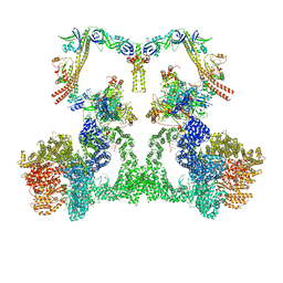
5T0G
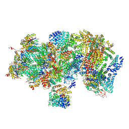 | | Structural basis for dynamic regulation of the human 26S proteasome | | 分子名称: | 26S protease regulatory subunit 10B, 26S protease regulatory subunit 4, 26S protease regulatory subunit 6A, ... | | 著者 | Chen, S, Wu, J, Lu, Y, Ma, Y.B, Lee, B.H, Yu, Z, Ouyang, Q, Finley, D, Kirschner, M.W, Mao, Y. | | 登録日 | 2016-08-16 | | 公開日 | 2016-10-19 | | 最終更新日 | 2016-11-30 | | 実験手法 | ELECTRON MICROSCOPY (4.4 Å) | | 主引用文献 | Structural basis for dynamic regulation of the human 26S proteasome.
Proc.Natl.Acad.Sci.USA, 113, 2016
|
|
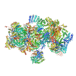
5T0H
 | | Structural basis for dynamic regulation of the human 26S proteasome | | 分子名称: | 26S protease regulatory subunit 10B, 26S protease regulatory subunit 4, 26S protease regulatory subunit 6A, ... | | 著者 | Chen, S, Wu, J, Lu, Y, Ma, Y.B, Lee, B.H, Yu, Z, Ouyang, Q, Finley, D, Kirschner, M.W, Mao, Y. | | 登録日 | 2016-08-16 | | 公開日 | 2016-10-19 | | 最終更新日 | 2016-11-30 | | 実験手法 | ELECTRON MICROSCOPY (6.8 Å) | | 主引用文献 | Structural basis for dynamic regulation of the human 26S proteasome.
Proc.Natl.Acad.Sci.USA, 113, 2016
|
|
5T0I
 | | Structural basis for dynamic regulation of the human 26S proteasome | | 分子名称: | 26S protease regulatory subunit 10B, 26S protease regulatory subunit 4, 26S protease regulatory subunit 6A, ... | | 著者 | Chen, S, Wu, J, Lu, Y, Ma, Y.B, Lee, B.H, Yu, Z, Ouyang, Q, Finley, D, Kirschner, M.W, Mao, Y. | | 登録日 | 2016-08-16 | | 公開日 | 2016-10-19 | | 最終更新日 | 2016-11-30 | | 実験手法 | ELECTRON MICROSCOPY (8 Å) | | 主引用文献 | Structural basis for dynamic regulation of the human 26S proteasome.
Proc.Natl.Acad.Sci.USA, 113, 2016
|
|
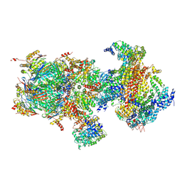
5T0C
 | | Structural basis for dynamic regulation of the human 26S proteasome | | 分子名称: | 26S protease regulatory subunit 10B, 26S protease regulatory subunit 4, 26S protease regulatory subunit 6A, ... | | 著者 | Chen, S, Wu, J, Lu, Y, Ma, Y.B, Lee, B.H, Yu, Z, Ouyang, Q, Finley, D, Kirschner, M.W, Mao, Y. | | 登録日 | 2016-08-15 | | 公開日 | 2016-10-19 | | 最終更新日 | 2018-07-18 | | 実験手法 | ELECTRON MICROSCOPY (3.8 Å) | | 主引用文献 | Structural basis for dynamic regulation of the human 26S proteasome.
Proc.Natl.Acad.Sci.USA, 113, 2016
|
|
5T0J
 | | Structural basis for dynamic regulation of the human 26S proteasome | | 分子名称: | 26S protease regulatory subunit 10B, 26S protease regulatory subunit 4, 26S protease regulatory subunit 6A, ... | | 著者 | Chen, S, Wu, J, Lu, Y, Ma, Y.B, Lee, B.H, Yu, Z, Ouyang, Q, Finley, D, Kirschner, M.W, Mao, Y. | | 登録日 | 2016-08-16 | | 公開日 | 2016-10-19 | | 最終更新日 | 2016-11-30 | | 実験手法 | ELECTRON MICROSCOPY (8 Å) | | 主引用文献 | Structural basis for dynamic regulation of the human 26S proteasome.
Proc.Natl.Acad.Sci.USA, 113, 2016
|
|
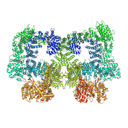
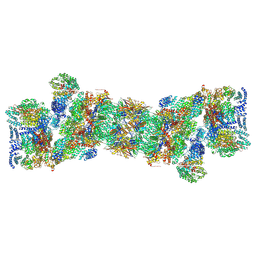
5VOU
 | | Structure of AMPA receptor-TARP complex | | 分子名称: | Glutamate receptor 2, Voltage-dependent calcium channel gamma-2 subunit | | 著者 | Chen, S, Zhao, Y, Wang, Y.S, Shekhar, M, Tajkhorshid, E, Gouaux, E. | | 登録日 | 2017-05-03 | | 公開日 | 2017-07-12 | | 最終更新日 | 2019-12-18 | | 実験手法 | ELECTRON MICROSCOPY (6.4 Å) | | 主引用文献 | Activation and Desensitization Mechanism of AMPA Receptor-TARP Complex by Cryo-EM.
Cell, 170, 2017
|
|
3O5Z
 | | Crystal structure of the SH3 domain from p85beta subunit of phosphoinositide 3-kinase (PI3K) | | 分子名称: | (4S)-2-METHYL-2,4-PENTANEDIOL, CHLORIDE ION, Phosphatidylinositol 3-kinase regulatory subunit beta | | 著者 | Chen, S, Xiao, Y, Ponnusamy, R, Tan, J, Lei, J, Hilgenfeld, R. | | 登録日 | 2010-07-28 | | 公開日 | 2011-08-10 | | 最終更新日 | 2014-09-10 | | 実験手法 | X-RAY DIFFRACTION (2.01 Å) | | 主引用文献 | X-ray structure of the SH3 domain of the phosphoinositide 3-kinase p85 beta subunit
Acta Crystallogr.,Sect.F, 67, 2011
|
|
3P38
 | |
3P31
 | |
3P39
 | |
7N6Z
 | | Crystal Structure of PI5P4KIIAlpha | | 分子名称: | Phosphatidylinositol 5-phosphate 4-kinase type-2 alpha, SULFATE ION | | 著者 | Chen, S, Ha, Y. | | 登録日 | 2021-06-09 | | 公開日 | 2021-06-30 | | 最終更新日 | 2023-10-18 | | 実験手法 | X-RAY DIFFRACTION (2.2 Å) | | 主引用文献 | Pharmacological inhibition of PI5P4K alpha / beta disrupts cell energy metabolism and selectively kills p53-null tumor cells.
Proc.Natl.Acad.Sci.USA, 118, 2021
|
|
7N7N
 | | Crystal Structure of PI5P4KIIAlpha complex with Volasertib | | 分子名称: | N-{trans-4-[4-(cyclopropylmethyl)piperazin-1-yl]cyclohexyl}-4-{[(7R)-7-ethyl-5-methyl-8-(1-methylethyl)-6-oxo-5,6,7,8-tetrahydropteridin-2-yl]amino}-3-methoxybenzamide, Phosphatidylinositol 5-phosphate 4-kinase type-2 alpha, SULFATE ION | | 著者 | Chen, S, Ha, Y. | | 登録日 | 2021-06-10 | | 公開日 | 2021-06-30 | | 最終更新日 | 2023-10-18 | | 実験手法 | X-RAY DIFFRACTION (2.3 Å) | | 主引用文献 | Pharmacological inhibition of PI5P4K alpha / beta disrupts cell energy metabolism and selectively kills p53-null tumor cells.
Proc.Natl.Acad.Sci.USA, 118, 2021
|
|


