4DWJ
 
 | | Crystal structure of Thymidylate Kinase from Staphylococcus aureus in complex with Thymidine Monophosphate | | 分子名称: | THYMIDINE-5'-PHOSPHATE, Thymidylate kinase | | 著者 | Filippova, E.V, Minasov, G, Shuvalova, L, Kiryukhina, O, Jedrzejczak, R, Babnigg, G, Rubin, E, Sacchettini, J, Joachimiak, A, Anderson, W.F, Midwest Center for Structural Genomics (MCSG), Structures of Mtb Proteins Conferring Susceptibility to Known Mtb Inhibitors (MTBI) | | 登録日 | 2012-02-24 | | 公開日 | 2012-03-07 | | 最終更新日 | 2023-09-13 | | 実験手法 | X-RAY DIFFRACTION (2.74 Å) | | 主引用文献 | Crystal structure of Thymidylate Kinase from Staphylococcus aureus in complex with Thymidine Monophosphate
To be Published
|
|
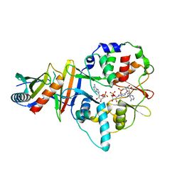
4DY6
 | | Crystal structure of NAD kinase 1 from Listeria monocytogenes in complex with 2'-phosphate bis(adenosine)-5'-diphosphate | | 分子名称: | CITRIC ACID, [(2R,3S,4R,5R)-5-(6-AMINO-9H-PURIN-9-YL)-3,4-DIHYDROXYTETRAHYDROFURAN-2-YL]METHYL [(2R,3R,4R,5R)-5-(6-AMINO-9H-PURIN-9-YL)-3-HYDROXY-4-(PHOSPHONOOXY)TETRAHYDROFURAN-2-YL]METHYL DIHYDROGEN DIPHOSPHATE, inorganic polyphosphate/ATP-NAD kinase 1 | | 著者 | Poncet-Montange, G, Assairi, L, Arold, S, Pochet, S, Labesse, G. | | 登録日 | 2012-02-28 | | 公開日 | 2012-03-07 | | 最終更新日 | 2023-09-13 | | 実験手法 | X-RAY DIFFRACTION (2.2 Å) | | 主引用文献 | NAD kinases use substrate-assisted catalysis for specific recognition of NAD.
J.Biol.Chem., 282, 2007
|
|
4DYQ
 | |
4DZJ
 | |
8JI8
 | | Crystal Structure of Prophenoloxidase PPO6 chimeric mutant (F215EASNRAIVD224 to G215DGPDSVVR223) from Aedes aegypti | | 分子名称: | 1,2-ETHANEDIOL, 2-AMINO-2-HYDROXYMETHYL-PROPANE-1,3-DIOL, COPPER (II) ION, ... | | 著者 | Zhu, X, Zhang, L, Yang, X, Bao, P, Ren, D, Han, Q. | | 登録日 | 2023-05-26 | | 公開日 | 2023-11-01 | | 実験手法 | X-RAY DIFFRACTION (2.65 Å) | | 主引用文献 | Mosquitoes have evolved two types of prophenoloxidases
To Be Published
|
|
8JI5
 | | Crystal structure of AetD in complex with 5-bromo-L-tryptophan and two Fe2+ | | 分子名称: | 5-bromo-L-tryptophan, AetD, FE (II) ION, ... | | 著者 | Li, H, Dai, L, Zheng, H.B, Chen, C.-C, Guo, R.-T. | | 登録日 | 2023-05-26 | | 公開日 | 2023-11-15 | | 最終更新日 | 2023-11-29 | | 実験手法 | X-RAY DIFFRACTION (2.01 Å) | | 主引用文献 | The structural and functional investigation into an unusual nitrile synthase.
Nat Commun, 14, 2023
|
|
8JI4
 | | Crystal structure of AetD in complex with 5-bromo-L-tryptophan | | 分子名称: | 2-AMINO-2-HYDROXYMETHYL-PROPANE-1,3-DIOL, 5-bromo-L-tryptophan, AetD, ... | | 著者 | Li, H, Dai, L, Zheng, H.B, Chen, C.-C, Guo, R.-T. | | 登録日 | 2023-05-26 | | 公開日 | 2023-11-15 | | 最終更新日 | 2023-11-29 | | 実験手法 | X-RAY DIFFRACTION (1.67 Å) | | 主引用文献 | The structural and functional investigation into an unusual nitrile synthase.
Nat Commun, 14, 2023
|
|
4E2P
 | | Crystal Structure of a Post-tailoring Hydroxylase (HmtN) Involved in the Himastatin Biosynthesis | | 分子名称: | Cytochrome P450 107B1 (P450CVIIB1), MAGNESIUM ION, PROTOPORPHYRIN IX CONTAINING FE | | 著者 | Zhang, H.D, Chen, J, Wang, H, Huang, L, Zhang, H.J. | | 登録日 | 2012-03-09 | | 公開日 | 2013-03-13 | | 最終更新日 | 2023-11-08 | | 実験手法 | X-RAY DIFFRACTION (2.36 Å) | | 主引用文献 | Crystal Structure of a Post-tailoring Hydroxylase (HmtN) Involved in the Himastatin Biosynthesis
To be Published
|
|
8JI3
 | | Crystal structure of AetD in complex with 5,7-dibromo-L-tryptophan and two Fe2+ | | 分子名称: | (2S)-2-azanyl-3-[5,7-bis(bromanyl)-1H-indol-3-yl]propanoic acid, AetD, FE (II) ION, ... | | 著者 | Li, H, Dai, L, Zheng, H.B, Chen, C.-C, Guo, R.-T. | | 登録日 | 2023-05-26 | | 公開日 | 2023-11-15 | | 最終更新日 | 2023-11-29 | | 実験手法 | X-RAY DIFFRACTION (1.78 Å) | | 主引用文献 | The structural and functional investigation into an unusual nitrile synthase.
Nat Commun, 14, 2023
|
|
8JI6
 | | Crystal structure of AetD in complex with L-tryptophan | | 分子名称: | 2-AMINO-2-HYDROXYMETHYL-PROPANE-1,3-DIOL, AetD, FE (II) ION, ... | | 著者 | Li, H, Dai, L, Zheng, H.B, Chen, C.-C, Guo, R.-T. | | 登録日 | 2023-05-26 | | 公開日 | 2023-11-15 | | 最終更新日 | 2023-11-29 | | 実験手法 | X-RAY DIFFRACTION (1.9 Å) | | 主引用文献 | The structural and functional investigation into an unusual nitrile synthase.
Nat Commun, 14, 2023
|
|
8JI2
 | | Crystal structure of AetD in complex with 5,7-dibromo-L-tryptophan | | 分子名称: | (2S)-2-azanyl-3-[5,7-bis(bromanyl)-1H-indol-3-yl]propanoic acid, 2-AMINO-2-HYDROXYMETHYL-PROPANE-1,3-DIOL, AetD, ... | | 著者 | Li, H, Dai, L, Zheng, H.B, Chen, C.-C, Guo, R.-T. | | 登録日 | 2023-05-26 | | 公開日 | 2023-11-15 | | 最終更新日 | 2023-11-29 | | 実験手法 | X-RAY DIFFRACTION (1.75 Å) | | 主引用文献 | The structural and functional investigation into an unusual nitrile synthase.
Nat Commun, 14, 2023
|
|
8JI7
 | | Crystal structure of AetD in complex with L-tryptophan and two Fe2+ | | 分子名称: | AetD, FE (II) ION, NICKEL (II) ION, ... | | 著者 | Li, H, Dai, L, Zheng, H.B, Chen, C.-C, Guo, R.-T. | | 登録日 | 2023-05-26 | | 公開日 | 2023-11-15 | | 最終更新日 | 2023-11-29 | | 実験手法 | X-RAY DIFFRACTION (1.61 Å) | | 主引用文献 | The structural and functional investigation into an unusual nitrile synthase.
Nat Commun, 14, 2023
|
|
8JIB
 | | Crystal Structure of Prophenoloxidase PPO6 from Aedes aegypti | | 分子名称: | 1,2-ETHANEDIOL, COPPER (II) ION, TK receptor | | 著者 | Zhu, X, Zhang, L, Yang, X, Bao, P, Ren, D, Han, Q. | | 登録日 | 2023-05-26 | | 公開日 | 2023-11-29 | | 実験手法 | X-RAY DIFFRACTION (3.15 Å) | | 主引用文献 | Mosquitoes have evolved two types of prophenoloxidases
To Be Published
|
|
4EAY
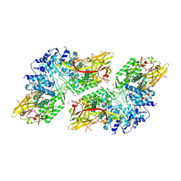
 | | Crystal structures of mannonate dehydratase from Escherichia coli strain K12 complexed with D-mannonate | | 分子名称: | CHLORIDE ION, D-MANNONIC ACID, MANGANESE (II) ION, ... | | 著者 | Qiu, X, Zhu, Y, Yuan, Y, Zhang, Y, Liu, H, Gao, Y, Teng, M, Niu, L. | | 登録日 | 2012-03-23 | | 公開日 | 2013-03-27 | | 最終更新日 | 2024-03-20 | | 実験手法 | X-RAY DIFFRACTION (2.35 Å) | | 主引用文献 | Structural insights into decreased enzymatic activity induced by an insert sequence in mannonate dehydratase from Gram negative bacterium.
J.Struct.Biol., 180, 2012
|
|
4E85
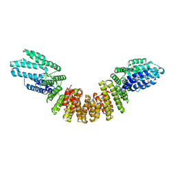 | |
4EAI
 | | Co-crystal structure of an AMPK core with AMP | | 分子名称: | 5'-AMP-activated protein kinase catalytic subunit alpha-1, 5'-AMP-activated protein kinase subunit beta-2, 5'-AMP-activated protein kinase subunit gamma-1, ... | | 著者 | Chen, L, Wang, J, Zhang, Y.-Y, Yan, S.F, Neumann, D, Schlattner, U, Wang, Z.-X, Wu, J.-W. | | 登録日 | 2012-03-22 | | 公開日 | 2012-06-06 | | 最終更新日 | 2023-11-08 | | 実験手法 | X-RAY DIFFRACTION (2.285 Å) | | 主引用文献 | AMP-activated protein kinase undergoes nucleotide-dependent conformational changes
Nat.Struct.Mol.Biol., 19, 2012
|
|
4E48
 | |
4ELY
 | | CCDBVFI:GYRA14EC | | 分子名称: | CHLORIDE ION, CcdB, DNA gyrase subunit A, ... | | 著者 | De Jonge, N, Simic, R, Buts, L, Haesaerts, S, Roelants, K, Garcia-Pino, A, Sterckx, Y, De Greve, H, Lah, J, Loris, R. | | 登録日 | 2012-04-11 | | 公開日 | 2012-05-30 | | 最終更新日 | 2023-09-13 | | 実験手法 | X-RAY DIFFRACTION (1.932 Å) | | 主引用文献 | Alternative interactions define gyrase specificity in the CcdB family.
Mol.Microbiol., 84, 2012
|
|
4E9A
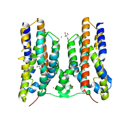
 | | Structure of Peptide Deformylase form Helicobacter Pylori in complex with inhibitor | | 分子名称: | 2-phenylethyl (2E)-3-(3,4-dihydroxyphenyl)prop-2-enoate, 4-(2-HYDROXYETHYL)-1-PIPERAZINE ETHANESULFONIC ACID, COBALT (II) ION, ... | | 著者 | Cui, K, Zhu, L, Lu, W, Huang, J. | | 登録日 | 2012-03-20 | | 公開日 | 2013-04-24 | | 最終更新日 | 2023-11-08 | | 実験手法 | X-RAY DIFFRACTION (1.662 Å) | | 主引用文献 | Identification of Novel Peptide Deformylase Inhibitors from Natural Products
To be Published
|
|
4EAK
 | | Co-crystal structure of an AMPK core with ATP | | 分子名称: | 5'-AMP-activated protein kinase catalytic subunit alpha-1, 5'-AMP-activated protein kinase subunit beta-1, 5'-AMP-activated protein kinase subunit gamma-1, ... | | 著者 | Chen, L, Wang, J, Zhang, Y.-Y, Yan, S.F, Neumann, D, Schlattner, U, Wang, Z.-X, Wu, J.-W. | | 登録日 | 2012-03-22 | | 公開日 | 2012-06-06 | | 最終更新日 | 2023-11-08 | | 実験手法 | X-RAY DIFFRACTION (2.5 Å) | | 主引用文献 | AMP-activated protein kinase undergoes nucleotide-dependent conformational changes
Nat.Struct.Mol.Biol., 19, 2012
|
|
4EGR
 | | 2.50 angstrom resolution structure of 3-phosphoshikimate 1-carboxyvinyltransferase (AroA) from Coxiella burnetii in complex with phosphoenolpyruvate | | 分子名称: | 3-phosphoshikimate 1-carboxyvinyltransferase, PHOSPHOENOLPYRUVATE, SULFATE ION | | 著者 | Krishna, S.N, Light, S.H, Minasov, G, Shuvalova, L, Kwon, K, Anderson, W.F, Center for Structural Genomics of Infectious Diseases (CSGID) | | 登録日 | 2012-03-31 | | 公開日 | 2012-04-11 | | 最終更新日 | 2024-10-09 | | 実験手法 | X-RAY DIFFRACTION (2.5 Å) | | 主引用文献 | 2.50 angstrom resolution structure of 3-phosphoshikimate 1-carboxyvinyltransferase (AroA) from Coxiella burnetii in complex with phosphoenolpyruvate
TO BE PUBLISHED
|
|
4EX6
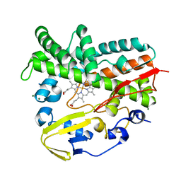
 | | Crystal structure of the alnumycin P phosphatase AlnB | | 分子名称: | AlnB, BORIC ACID, MAGNESIUM ION | | 著者 | Oja, T, Niiranen, L, Sandalova, T, Klika, K.D, Niemi, J, Mantsala, P, Schneider, G, Metsa-Ketela, M. | | 登録日 | 2012-04-30 | | 公開日 | 2013-01-16 | | 最終更新日 | 2023-09-13 | | 実験手法 | X-RAY DIFFRACTION (1.25 Å) | | 主引用文献 | Structural basis for C-ribosylation in the alnumycin A biosynthetic pathway.
Proc.Natl.Acad.Sci.USA, 110, 2013
|
|
4E6B
 | |
4USZ
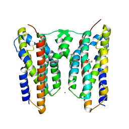
 | | Crystal structure of the first bacterial vanadium dependant iodoperoxidase | | 分子名称: | SODIUM ION, VANADATE ION, VANADIUM-DEPENDENT HALOPEROXIDASE | | 著者 | Rebuffet, E, Delage, L, Fournier, J.B, Rzonca, J, Potin, P, Michel, G, Czjzek, M, Leblanc, C. | | 登録日 | 2014-07-17 | | 公開日 | 2014-10-08 | | 最終更新日 | 2024-01-10 | | 実験手法 | X-RAY DIFFRACTION (2 Å) | | 主引用文献 | The Bacterial Vanadium Iodoperoxidase from the Marine Flavobacteriaceae Zobellia Galactanivorans Reveals Novel Molecular and Evolutionary Features of Halide Specificity in This Enzyme Family.
Appl.Environ.Microbiol., 80, 2014
|
|
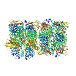
4V9A
 | | Crystal Structure of the 70S ribosome with tetracycline. | | 分子名称: | 16S ribosomal RNA, 23S ribosomal RNA, 30S RIBOSOMAL PROTEIN S10, ... | | 著者 | Jenner, L, Yusupov, M, Yusupova, G. | | 登録日 | 2012-07-18 | | 公開日 | 2014-07-09 | | 最終更新日 | 2023-09-20 | | 実験手法 | X-RAY DIFFRACTION (3.2999 Å) | | 主引用文献 | Structural basis for potent inhibitory activity of the antibiotic tigecycline during protein synthesis.
Proc.Natl.Acad.Sci.USA, 110, 2013
|
|


