7DK4
 
 | |
7DDN
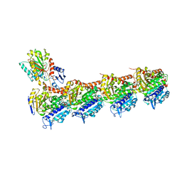
 | | SARS-Cov2 S protein at open state | | 分子名称: | Spike glycoprotein | | 著者 | Cong, Y, Liu, C.X. | | 登録日 | 2020-10-29 | | 公開日 | 2020-11-25 | | 最終更新日 | 2021-01-27 | | 実験手法 | ELECTRON MICROSCOPY (6.3 Å) | | 主引用文献 | Development and structural basis of a two-MAb cocktail for treating SARS-CoV-2 infections.
Nat Commun, 12, 2021
|
|
7DDD
 | | SARS-Cov2 S protein at close state | | 分子名称: | Spike glycoprotein | | 著者 | Cong, Y, Liu, C.X. | | 登録日 | 2020-10-28 | | 公開日 | 2020-11-25 | | 最終更新日 | 2021-01-27 | | 実験手法 | ELECTRON MICROSCOPY (3 Å) | | 主引用文献 | Development and structural basis of a two-MAb cocktail for treating SARS-CoV-2 infections.
Nat Commun, 12, 2021
|
|
7DK7
 | |
7DCX
 | |
7ECY
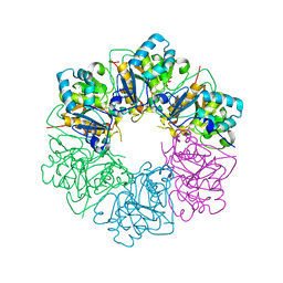
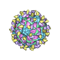 | | EV-D68 in complex with 2H12 Fab (State 3) | | 分子名称: | Capsid protein VP1, Capsid protein VP2, Capsid protein VP3, ... | | 著者 | Xu, C, Cong, Y. | | 登録日 | 2021-03-13 | | 公開日 | 2021-03-31 | | 最終更新日 | 2021-06-02 | | 実験手法 | ELECTRON MICROSCOPY (3.6 Å) | | 主引用文献 | Functional and structural characterization of a two-MAb cocktail for delayed treatment of enterovirus D68 infections.
Nat Commun, 12, 2021
|
|
7EBZ
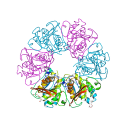
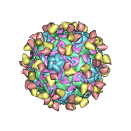 | | EV-D68 in complex with 2H12 Fab (state S1) | | 分子名称: | Capsid protein VP1, Capsid protein VP2, Capsid protein VP3, ... | | 著者 | Xu, C, Cong, Y. | | 登録日 | 2021-03-11 | | 公開日 | 2021-03-31 | | 最終更新日 | 2021-06-02 | | 実験手法 | ELECTRON MICROSCOPY (3.09 Å) | | 主引用文献 | Functional and structural characterization of a two-MAb cocktail for delayed treatment of enterovirus D68 infections.
Nat Commun, 12, 2021
|
|
7EBR
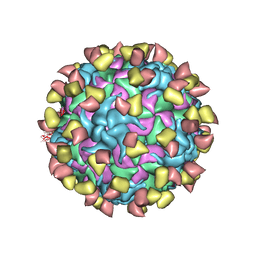 | | EV-D68 in complex with 2H12 Fab (state S2) | | 分子名称: | 2H12 Fab heavy chain, 2H12 Fab light chain, Capsid protein VP1, ... | | 著者 | Xu, C, Cong, Y. | | 登録日 | 2021-03-10 | | 公開日 | 2021-03-31 | | 最終更新日 | 2021-06-02 | | 実験手法 | ELECTRON MICROSCOPY (3.6 Å) | | 主引用文献 | Functional and structural characterization of a two-MAb cocktail for delayed treatment of enterovirus D68 infections.
Nat Commun, 12, 2021
|
|
7EC5
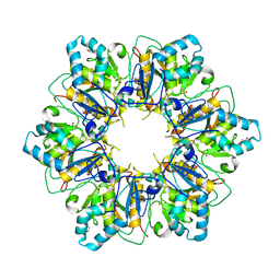
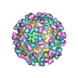 | | EV-D68 in complex with 8F12 Fab | | 分子名称: | 8F12 Fab heavy chain, 8F12 Fab light chain, Capsid protein VP1, ... | | 著者 | Xu, C, Cong, Y. | | 登録日 | 2021-03-11 | | 公開日 | 2021-03-31 | | 最終更新日 | 2021-06-02 | | 実験手法 | ELECTRON MICROSCOPY (2.89 Å) | | 主引用文献 | Functional and structural characterization of a two-MAb cocktail for delayed treatment of enterovirus D68 infections.
Nat Commun, 12, 2021
|
|
7DCD
 | |
7ESV
 | |
7ESQ
 | |
7ESP
 | |
7ESO
 | |
7V4Q
 | |
7V4R
 | |
8JJB
 | | Crystal structure of T2R-TTL-Y61 complex | | 分子名称: | 2-(N-MORPHOLINO)-ETHANESULFONIC ACID, CALCIUM ION, CHLORIDE ION, ... | | 著者 | Yang, J. | | 登録日 | 2023-05-30 | | 公開日 | 2024-03-27 | | 実験手法 | X-RAY DIFFRACTION (2.68 Å) | | 主引用文献 | Structure-based design and synthesis of BML284 derivatives: A novel class of colchicine-site noncovalent tubulin degradation agents.
Eur.J.Med.Chem., 268, 2024
|
|
8JJC
 | | Tubulin-Y62 | | 分子名称: | 2-(N-MORPHOLINO)-ETHANESULFONIC ACID, 4-(6,7-dimethoxy-3,4-dihydro-1~{H}-isoquinolin-2-yl)-6-(3-methoxyphenyl)pyrimidin-2-amine, CALCIUM ION, ... | | 著者 | Yang, J. | | 登録日 | 2023-05-30 | | 公開日 | 2024-03-27 | | 実験手法 | X-RAY DIFFRACTION (2.76 Å) | | 主引用文献 | Structure-based design and synthesis of BML284 derivatives: A novel class of colchicine-site noncovalent tubulin degradation agents.
Eur.J.Med.Chem., 268, 2024
|
|
6QSW
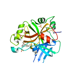 | | Complement factor B protease domain in complex with the reversible inhibitor N-(2-bromo-4-methylnaphthalen-1-yl)-4,5-dihydro-1H-imidazol-2-amine. | | 分子名称: | Complement factor B, SULFATE ION, ~{N}-(2-bromanyl-4-methyl-naphthalen-1-yl)-4,5-dihydro-1~{H}-imidazol-2-amine | | 著者 | Adams, C.M, Sellner, H, Ehara, T, Mac Sweeney, A, Crowley, M, Anderson, K, Karki, R, Mainolfi, N, Valeur, E, Sirockin, F, Gerhartz, B, Erbel, P, Hughes, N, Smith, T.M, Cumin, F, Argikar, U, Mogi, M, Sedrani, R, Wiesmann, C, Jaffee, B, Maibaum, J, Flohr, S, Harrison, R, Eder, J. | | 登録日 | 2019-02-22 | | 公開日 | 2019-03-27 | | 最終更新日 | 2024-01-24 | | 実験手法 | X-RAY DIFFRACTION (1.64 Å) | | 主引用文献 | Small-molecule factor B inhibitor for the treatment of complement-mediated diseases.
Proc.Natl.Acad.Sci.USA, 116, 2019
|
|
8Y95
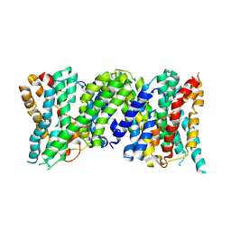
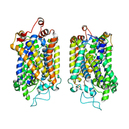 | | Structure of NET-NE in Occluded state | | 分子名称: | CHLORIDE ION, Noradrenaline, SODIUM ION, ... | | 著者 | Zhang, H, Xu, H.E, Jiang, Y. | | 登録日 | 2024-02-06 | | 公開日 | 2024-05-29 | | 最終更新日 | 2024-07-03 | | 実験手法 | ELECTRON MICROSCOPY (3.24 Å) | | 主引用文献 | Dimerization and antidepressant recognition at noradrenaline transporter.
Nature, 630, 2024
|
|
5DDW
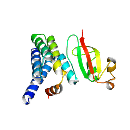
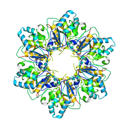
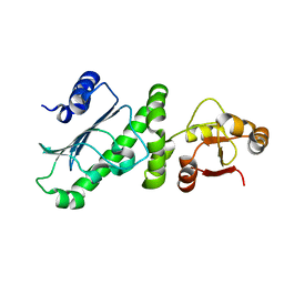
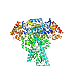 | | Crystal structure of aminotransferase CrmG from Actinoalloteichus sp. WH1-2216-6 in complex with the PMP external aldimine adduct with Caerulomycin M | | 分子名称: | CrmG, GLYCEROL, [5-hydroxy-4-({(E)-[(4-hydroxy-2,2'-bipyridin-6-yl)methylidene]amino}methyl)-6-methylpyridin-3-yl]methyl dihydrogen phosphate | | 著者 | Xu, J, Feng, Z, Liu, J. | | 登録日 | 2015-08-25 | | 公開日 | 2016-08-10 | | 最終更新日 | 2024-03-20 | | 実験手法 | X-RAY DIFFRACTION (2.3 Å) | | 主引用文献 | Biochemical and Structural Insights into the Aminotransferase CrmG in Caerulomycin Biosynthesis
Acs Chem.Biol., 11, 2016
|
|
6KSQ
 | |
6Z3Z
 | |
6KOL
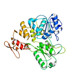
 | | Crystal structure of auracyanin from photosynthetic bacterium Roseiflexus castenholzii | | 分子名称: | Blue (Type 1) copper domain protein, CHLORIDE ION, COPPER (II) ION | | 著者 | Wang, C, Zhang, C.Y, Min, Z.Z, Xin, Y.Y, Xu, X.L. | | 登録日 | 2019-08-12 | | 公開日 | 2020-01-29 | | 最終更新日 | 2023-11-22 | | 実験手法 | X-RAY DIFFRACTION (2.211 Å) | | 主引用文献 | Structural basis underlying the electron transfer features of a blue copper protein auracyanin from the photosynthetic bacterium Roseiflexus castenholzii.
Photosyn. Res., 143, 2020
|
|
6L9S
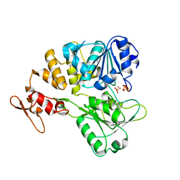
 | | Crystal structure of Na-dithionite reduced auracyanin from photosynthetic bacterium Roseiflexus castenholzii | | 分子名称: | Blue (Type 1) copper domain protein, COPPER (I) ION | | 著者 | Wang, C, Zhang, C.Y, Min, Z.Z, Xu, X.L. | | 登録日 | 2019-11-10 | | 公開日 | 2020-01-29 | | 最終更新日 | 2023-11-22 | | 実験手法 | X-RAY DIFFRACTION (2 Å) | | 主引用文献 | Structural basis underlying the electron transfer features of a blue copper protein auracyanin from the photosynthetic bacterium Roseiflexus castenholzii.
Photosyn. Res., 143, 2020
|
|


