7Y49
 
 | |
7C43
 | |
7C4C
 | |
7C4B
 | |
7C45
 | |
7C47
 | |
7C42
 | |
6K9F
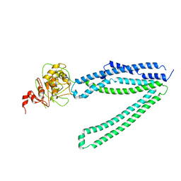
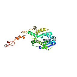
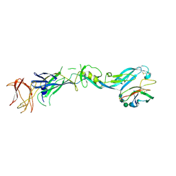
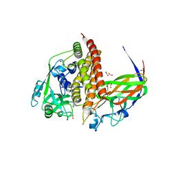
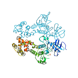
 | | Structure of unknow protein 4 | | 分子名称: | Caspase recruitment domain-containing protein 8 | | 著者 | Gong, Q, Xu, C, Zhang, J, Boo, Z.Z, Wu, B. | | 登録日 | 2019-06-15 | | 公開日 | 2020-09-16 | | 最終更新日 | 2024-03-27 | | 実験手法 | ELECTRON MICROSCOPY (3.7 Å) | | 主引用文献 | Structural basis for distinct inflammasome complex assembly by human NLRP1 and CARD8.
Nat Commun, 12, 2021
|
|
3PA4
 | |
7Y48
 | |
3C6E
 | |
3LJ1
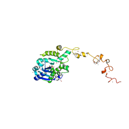
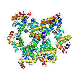
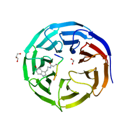
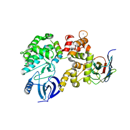
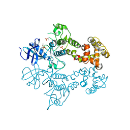 | | IRE1 complexed with Cdk1/2 Inhibitor III | | 分子名称: | 5-AMINO-3-{[4-(AMINOSULFONYL)PHENYL]AMINO}-N-(2,6-DIFLUOROPHENYL)-1H-1,2,4-TRIAZOLE-1-CARBOTHIOAMIDE, Serine/threonine-protein kinase/endoribonuclease IRE1 | | 著者 | Lee, K.P.K, Sicheri, F. | | 登録日 | 2010-01-25 | | 公開日 | 2010-05-12 | | 最終更新日 | 2023-09-06 | | 実験手法 | X-RAY DIFFRACTION (3.33 Å) | | 主引用文献 | Flavonol activation defines an unanticipated ligand-binding site in the kinase-RNase domain of IRE1.
Mol.Cell, 38, 2010
|
|
3SBX
 | |
3SF6
 | |
8GS1
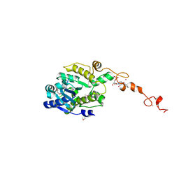
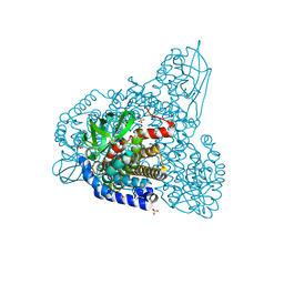
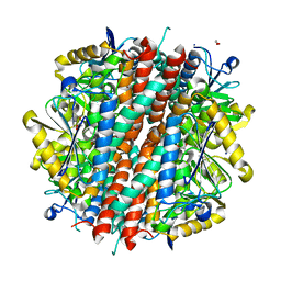
 | | Crystal structure of AziU2-U3 complex from Streptomyces sahachiroi NRRL2485 | | 分子名称: | Azi28, Azi29, FORMIC ACID, ... | | 著者 | Cheng, Y, Li, P, Liu, W, Fang, P. | | 登録日 | 2022-09-04 | | 公開日 | 2023-09-06 | | 最終更新日 | 2023-09-20 | | 実験手法 | X-RAY DIFFRACTION (2.7 Å) | | 主引用文献 | Oxidase Heterotetramer Completes 1-Azabicyclo[3.1.0]hexane Formation with the Association of a Nonribosomal Peptide Synthetase.
J.Am.Chem.Soc., 145, 2023
|
|
6K99
 | | Structure of ASC CARD filament | | 分子名称: | Apoptosis-associated speck-like protein containing a CARD | | 著者 | Gong, Q, Xu, C, Zhang, J, Boo, Z.Z, Wu, B. | | 登録日 | 2019-06-14 | | 公開日 | 2020-09-16 | | 最終更新日 | 2024-03-27 | | 実験手法 | ELECTRON MICROSCOPY (4.1 Å) | | 主引用文献 | Structural basis for distinct inflammasome complex assembly by human NLRP1 and CARD8.
Nat Commun, 12, 2021
|
|
4GA7
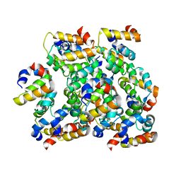
 | | Crystal structure of human serpinB1 mutant | | 分子名称: | Leukocyte elastase inhibitor | | 著者 | Wang, L, Li, Q, Wu, L, Zhang, K, Tong, L, Sun, F, Fan, Z. | | 登録日 | 2012-07-25 | | 公開日 | 2013-01-16 | | 最終更新日 | 2023-11-08 | | 実験手法 | X-RAY DIFFRACTION (2.9 Å) | | 主引用文献 | Identification of SERPINB1 as a physiological inhibitor of human granzyme H
J.Immunol., 190, 2013
|
|
4GAW
 | | Crystal structure of active human granzyme H | | 分子名称: | CHLORIDE ION, Granzyme H, SULFATE ION | | 著者 | Wang, L, Li, Q, Wu, L, Zhang, K, Tong, L, Sun, F, Fan, Z. | | 登録日 | 2012-07-25 | | 公開日 | 2013-01-16 | | 最終更新日 | 2023-11-08 | | 実験手法 | X-RAY DIFFRACTION (3 Å) | | 主引用文献 | Identification of SERPINB1 as a physiological inhibitor of human granzyme H
J.Immunol., 190, 2013
|
|
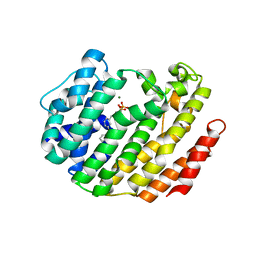
7CFQ
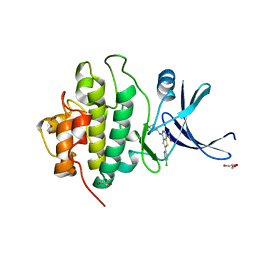
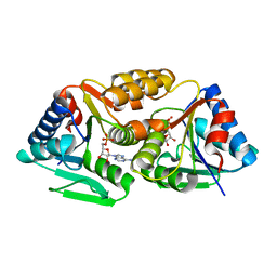
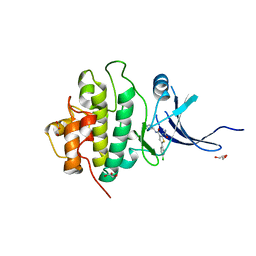
 | | Crystal structure of WDR5 in complex with H3K4me3Q5ser peptide | | 分子名称: | 1,2-ETHANEDIOL, GLYCEROL, H3K4me3Q5ser peptide, ... | | 著者 | Zhao, J, Zhang, X, Zang, J. | | 登録日 | 2020-06-27 | | 公開日 | 2021-07-07 | | 最終更新日 | 2023-11-29 | | 実験手法 | X-RAY DIFFRACTION (1.6 Å) | | 主引用文献 | Structural insights into the recognition of histone H3Q5 serotonylation by WDR5.
Sci Adv, 7, 2021
|
|
7CFP
 | |
3LJ2
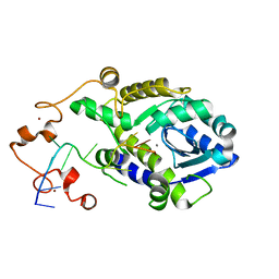
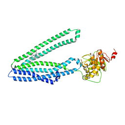
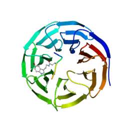
 | | IRE1 complexed with JAK Inhibitor I | | 分子名称: | 2-TERT-BUTYL-9-FLUORO-3,6-DIHYDRO-7H-BENZ[H]-IMIDAZ[4,5-F]ISOQUINOLINE-7-ONE, Serine/threonine-protein kinase/endoribonuclease IRE1 | | 著者 | Lee, K.P.K, Sicheri, F. | | 登録日 | 2010-01-25 | | 公開日 | 2010-05-12 | | 最終更新日 | 2023-09-06 | | 実験手法 | X-RAY DIFFRACTION (3.33 Å) | | 主引用文献 | Flavonol activation defines an unanticipated ligand-binding site in the kinase-RNase domain of IRE1.
Mol.Cell, 38, 2010
|
|
3PA5
 | |
4YGA
 | |
3T3W
 | |
3LEE
 | |


