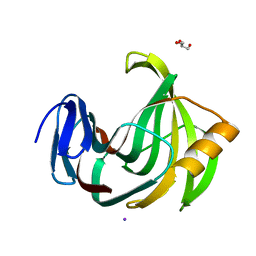
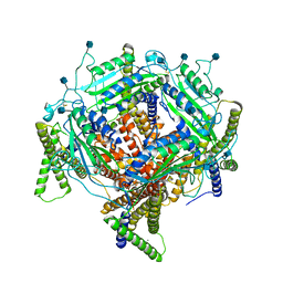
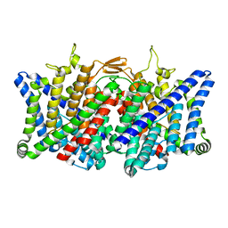
4N25
 
 | | Crystal structure of Protein Arginine Deiminase 2 (250 uM Ca2+) | | 分子名称: | (4R)-2-METHYLPENTANE-2,4-DIOL, (4S)-2-METHYL-2,4-PENTANEDIOL, ACETATE ION, ... | | 著者 | Slade, D.J, Zhang, X, Fang, P, Dreyton, C.J, Zhang, Y, Gross, M.L, Guo, M, Coonrod, S.A, Thompson, P.R. | | 登録日 | 2013-10-04 | | 公開日 | 2015-02-04 | | 最終更新日 | 2023-09-20 | | 実験手法 | X-RAY DIFFRACTION (1.931 Å) | | 主引用文献 | Protein arginine deiminase 2 binds calcium in an ordered fashion: implications for inhibitor design.
Acs Chem.Biol., 10, 2015
|
|
4N2N
 | | Crystal structure of Protein Arginine Deiminase 2 (E354A, 10 mM Ca2+) | | 分子名称: | (4S)-2-METHYL-2,4-PENTANEDIOL, ACETATE ION, CALCIUM ION, ... | | 著者 | Slade, D.J, Zhang, X, Fang, P, Dreyton, C.J, Zhang, Y, Gross, M.L, Guo, M, Coonrod, S.A, Thompson, P.R. | | 登録日 | 2013-10-05 | | 公開日 | 2015-02-04 | | 最終更新日 | 2023-09-20 | | 実験手法 | X-RAY DIFFRACTION (1.8 Å) | | 主引用文献 | Protein arginine deiminase 2 binds calcium in an ordered fashion: implications for inhibitor design.
Acs Chem.Biol., 10, 2015
|
|
6KWD
 | | Crystal Structure Analysis of Endo-beta-1,4-Xylanase II Complexed with Xylotriose | | 分子名称: | Endo-1,4-beta-xylanase 2, GLYCEROL, IODIDE ION, ... | | 著者 | Li, C, Wan, Q. | | 登録日 | 2019-09-06 | | 公開日 | 2020-12-30 | | 最終更新日 | 2023-11-22 | | 実験手法 | X-RAY DIFFRACTION (1.298 Å) | | 主引用文献 | Studying the Role of a Single Mutation of a Family 11 Glycoside Hydrolase Using High-Resolution X-ray Crystallography.
Protein J., 39, 2020
|
|
6L1T
 | | Cryo-EM structure of phosphorylated Tyr39 a-synuclein amyloid fibril | | 分子名称: | Alpha-synuclein | | 著者 | Liu, C, Li, Y.M, Zhao, K, Lim, Y.J, Liu, Z.Y. | | 登録日 | 2019-09-30 | | 公開日 | 2020-08-12 | | 最終更新日 | 2020-09-02 | | 実験手法 | ELECTRON MICROSCOPY (3.22 Å) | | 主引用文献 | Parkinson's disease-related phosphorylation at Tyr39 rearranges alpha-synuclein amyloid fibril structure revealed by cryo-EM.
Proc.Natl.Acad.Sci.USA, 117, 2020
|
|
6KW9
 | |
6KWG
 | |
7D7F
 | | Structure of PKD1L3-CTD/PKD2L1 in calcium-bound state | | 分子名称: | 2-acetamido-2-deoxy-beta-D-glucopyranose, CALCIUM ION, Polycystic kidney disease 2-like 1 protein, ... | | 著者 | Su, Q, Shi, Y.G. | | 登録日 | 2020-10-03 | | 公開日 | 2021-09-01 | | 実験手法 | ELECTRON MICROSCOPY (3 Å) | | 主引用文献 | Structural basis for Ca 2+ activation of the heteromeric PKD1L3/PKD2L1 channel.
Nat Commun, 12, 2021
|
|
6KWF
 | | Crystal Structure Analysis of Endo-beta-1,4-xylanase II Complexed with Xylotriose | | 分子名称: | Endo-1,4-beta-xylanase 2, GLYCEROL, IODIDE ION, ... | | 著者 | Li, C, Wan, Q. | | 登録日 | 2019-09-06 | | 公開日 | 2020-12-30 | | 最終更新日 | 2023-11-22 | | 実験手法 | X-RAY DIFFRACTION (1.22 Å) | | 主引用文献 | Studying the Role of a Single Mutation of a Family 11 Glycoside Hydrolase Using High-Resolution X-ray Crystallography.
Protein J., 39, 2020
|
|
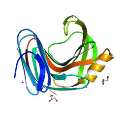
4N28
 | | Crystal structure of Protein Arginine Deiminase 2 (1 mM Ca2+) | | 分子名称: | (4S)-2-METHYL-2,4-PENTANEDIOL, ACETATE ION, CALCIUM ION, ... | | 著者 | Slade, D.J, Zhang, X, Fang, P, Dreyton, C.J, Zhang, Y, Gross, M.L, Guo, M, Coonrod, S.A, Thompson, P.R. | | 登録日 | 2013-10-04 | | 公開日 | 2015-02-04 | | 最終更新日 | 2023-09-20 | | 実験手法 | X-RAY DIFFRACTION (1.879 Å) | | 主引用文献 | Protein arginine deiminase 2 binds calcium in an ordered fashion: implications for inhibitor design.
Acs Chem.Biol., 10, 2015
|
|
4N5G
 | | Crystal Structure of RXRa LBD complexed with a synthetic modulator K8012 | | 分子名称: | 5-(2-{(1Z)-5-fluoro-2-methyl-1-[4-(propan-2-yl)benzylidene]-1H-inden-3-yl}ethyl)-1H-tetrazole, Retinoic acid receptor RXR-alpha | | 著者 | Aleshin, A.E, Su, Y, Zhang, X, Liddington, R.C. | | 登録日 | 2013-10-09 | | 公開日 | 2014-05-14 | | 最終更新日 | 2023-09-20 | | 実験手法 | X-RAY DIFFRACTION (2.11 Å) | | 主引用文献 | Sulindac-Derived RXR alpha Modulators Inhibit Cancer Cell Growth by Binding to a Novel Site.
Chem.Biol., 21, 2014
|
|
7XF5
 | | Full length human CLC-2 channel in apo state | | 分子名称: | Chloride channel protein 2 | | 著者 | Wang, L. | | 登録日 | 2022-04-01 | | 公開日 | 2023-05-03 | | 最終更新日 | 2023-11-15 | | 実験手法 | ELECTRON MICROSCOPY (3.9 Å) | | 主引用文献 | Cryo-EM structures of ClC-2 chloride channel reveal the blocking mechanism of its specific inhibitor AK-42
Nat Commun, 14, 2023
|
|
7XJA
 | | TMD masked refine map of human ClC-2 | | 分子名称: | Chloride channel protein 2 | | 著者 | Wang, L. | | 登録日 | 2022-04-15 | | 公開日 | 2023-05-17 | | 最終更新日 | 2023-11-15 | | 実験手法 | ELECTRON MICROSCOPY (3.5 Å) | | 主引用文献 | Cryo-EM structures of ClC-2 chloride channel reveal the blocking mechanism of its specific inhibitor AK-42
Nat Commun, 14, 2023
|
|
4N2K
 | | Crystal structure of Protein Arginine Deiminase 2 (Q350A, 0 mM Ca2+) | | 分子名称: | (4S)-2-METHYL-2,4-PENTANEDIOL, CALCIUM ION, GLYCEROL, ... | | 著者 | Slade, D.J, Zhang, X, Fang, P, Dreyton, C.J, Zhang, Y, Gross, M.L, Guo, M, Coonrod, S.A, Thompson, P.R. | | 登録日 | 2013-10-05 | | 公開日 | 2015-02-04 | | 最終更新日 | 2023-09-20 | | 実験手法 | X-RAY DIFFRACTION (1.57 Å) | | 主引用文献 | Protein arginine deiminase 2 binds calcium in an ordered fashion: implications for inhibitor design.
Acs Chem.Biol., 10, 2015
|
|
6L1U
 | | Cryo-EM structure of phosphorylated Tyr39 alpha-synuclein amyloid fibril | | 分子名称: | Alpha-synuclein | | 著者 | Liu, C, Li, Y.M, Zhao, K, Lim, Y.J, Liu, Z.Y. | | 登録日 | 2019-09-30 | | 公開日 | 2020-08-12 | | 最終更新日 | 2020-09-02 | | 実験手法 | ELECTRON MICROSCOPY (3.37 Å) | | 主引用文献 | Parkinson's disease-related phosphorylation at Tyr39 rearranges alpha-synuclein amyloid fibril structure revealed by cryo-EM.
Proc.Natl.Acad.Sci.USA, 117, 2020
|
|
6KWC
 | | Crystal Structure Analysis of Endo-beta-1,4-xylanase II | | 分子名称: | Endo-1,4-beta-xylanase 2, GLYCEROL, IODIDE ION | | 著者 | Li, C, Wan, Q. | | 登録日 | 2019-09-06 | | 公開日 | 2021-01-27 | | 最終更新日 | 2023-11-22 | | 実験手法 | X-RAY DIFFRACTION (1.3 Å) | | 主引用文献 | Studying the Role of a Single Mutation of a Family 11 Glycoside Hydrolase Using High-Resolution X-ray Crystallography.
Protein J., 39, 2020
|
|
7YF0
 | | In situ structure of polymerase complex of mammalian reovirus in the core | | 分子名称: | Lambda-2 protein, Mu-2 protein, RNA helicase, ... | | 著者 | Bao, K.Y, Zhang, X.L, Li, D.Y, Zhu, P. | | 登録日 | 2022-07-07 | | 公開日 | 2023-03-29 | | 実験手法 | ELECTRON MICROSCOPY (3.4 Å) | | 主引用文献 | In situ structures of polymerase complex of mammalian reovirus illuminate RdRp activation and transcription regulation.
Proc.Natl.Acad.Sci.USA, 119, 2022
|
|
7YEV
 | | In situ structure of polymerase complex of mammalian reovirus in the pre-elongation state | | 分子名称: | Lambda-2 protein, Mu-2 protein, RNA helicase, ... | | 著者 | Bao, K.Y, Zhang, X.L, Li, D.Y, Zhu, P. | | 登録日 | 2022-07-06 | | 公開日 | 2023-03-29 | | 実験手法 | ELECTRON MICROSCOPY (3.6 Å) | | 主引用文献 | In situ structures of polymerase complex of mammalian reovirus illuminate RdRp activation and transcription regulation.
Proc.Natl.Acad.Sci.USA, 119, 2022
|
|
7YED
 | | In situ structure of polymerase complex of mammalian reovirus in the elongation state | | 分子名称: | GUANOSINE-5'-TRIPHOSPHATE, Lambda-2 protein, MAGNESIUM ION, ... | | 著者 | Bao, K.Y, Zhang, X.L, Li, D.Y, Zhu, P. | | 登録日 | 2022-07-05 | | 公開日 | 2023-04-05 | | 最終更新日 | 2024-07-03 | | 実験手法 | ELECTRON MICROSCOPY (3 Å) | | 主引用文献 | In situ structures of polymerase complex of mammalian reovirus illuminate RdRp activation and transcription regulation.
Proc.Natl.Acad.Sci.USA, 119, 2022
|
|
6M53
 | | Crystal structure of 2, 3-dihydroxybenzoic acid decarboxylase from Fusarium oxysporum | | 分子名称: | 2,3-dihydroxybenzoate decarboxylase, GLYCEROL, ZINC ION | | 著者 | Song, M.K, Feng, J.H, Liu, W.D, Wu, Q.Q, Zhu, D.M. | | 登録日 | 2020-03-09 | | 公開日 | 2020-07-15 | | 最終更新日 | 2023-11-29 | | 実験手法 | X-RAY DIFFRACTION (1.55 Å) | | 主引用文献 | 2,3-Dihydroxybenzoic Acid Decarboxylase from Fusarium oxysporum: Crystal Structures and Substrate Recognition Mechanism.
Chembiochem, 21, 2020
|
|
4JIK
 | |
6JAK
 | | OtsA apo structure | | 分子名称: | Trehalose-6-phosphate synthase | | 著者 | Wang, S, Zhao, Y, Wang, D, Liu, J. | | 登録日 | 2019-01-24 | | 公開日 | 2019-12-04 | | 最終更新日 | 2023-11-22 | | 実験手法 | X-RAY DIFFRACTION (2.41 Å) | | 主引用文献 | Crystal structures of Magnaporthe oryzae trehalose-6-phosphate synthase (MoTps1) suggest a model for catalytic process of Tps1.
Biochem.J., 476, 2019
|
|
6JBI
 | | Structure of Tps1 apo structure | | 分子名称: | Trehalose-6-phosphate synthase | | 著者 | Wang, S, Zhao, Y, Yi, L, Wang, D, Liu, J. | | 登録日 | 2019-01-25 | | 公開日 | 2019-12-04 | | 最終更新日 | 2023-11-22 | | 実験手法 | X-RAY DIFFRACTION (2.5 Å) | | 主引用文献 | Crystal structures of Magnaporthe oryzae trehalose-6-phosphate synthase (MoTps1) suggest a model for catalytic process of Tps1.
Biochem.J., 476, 2019
|
|
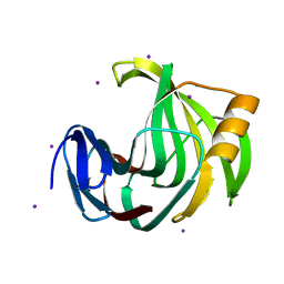
4N20
 | | Crystal structure of Protein Arginine Deiminase 2 (0 mM Ca2+) | | 分子名称: | (4S)-2-METHYL-2,4-PENTANEDIOL, ACETATE ION, CALCIUM ION, ... | | 著者 | Slade, D.J, Zhang, X, Fang, P, Dreyton, C.J, Zhang, Y, Gross, M.L, Guo, M, Coonrod, S.A, Thompson, P.R. | | 登録日 | 2013-10-04 | | 公開日 | 2015-02-04 | | 最終更新日 | 2023-09-20 | | 実験手法 | X-RAY DIFFRACTION (1.657 Å) | | 主引用文献 | Protein arginine deiminase 2 binds calcium in an ordered fashion: implications for inhibitor design.
Acs Chem.Biol., 10, 2015
|
|
4N24
 | | Crystal structure of Protein Arginine Deiminase 2 (100 uM Ca2+) | | 分子名称: | (4R)-2-METHYLPENTANE-2,4-DIOL, (4S)-2-METHYL-2,4-PENTANEDIOL, ACETATE ION, ... | | 著者 | Slade, D.J, Zhang, X, Fang, P, Dreyton, C.J, Zhang, Y, Gross, M.L, Guo, M, Coonrod, S.A, Thompson, P.R. | | 登録日 | 2013-10-04 | | 公開日 | 2015-02-04 | | 最終更新日 | 2023-09-20 | | 実験手法 | X-RAY DIFFRACTION (1.968 Å) | | 主引用文献 | Protein arginine deiminase 2 binds calcium in an ordered fashion: implications for inhibitor design.
Acs Chem.Biol., 10, 2015
|
|
4N2M
 | | Crystal structure of Protein Arginine Deiminase 2 (E354A, 0 mM Ca2+) | | 分子名称: | (4S)-2-METHYL-2,4-PENTANEDIOL, ACETATE ION, CALCIUM ION, ... | | 著者 | Slade, D.J, Zhang, X, Fang, P, Dreyton, C.J, Zhang, Y, Gross, M.L, Guo, M, Coonrod, S.A, Thompson, P.R. | | 登録日 | 2013-10-05 | | 公開日 | 2015-02-04 | | 最終更新日 | 2023-09-20 | | 実験手法 | X-RAY DIFFRACTION (1.599 Å) | | 主引用文献 | Protein arginine deiminase 2 binds calcium in an ordered fashion: implications for inhibitor design.
Acs Chem.Biol., 10, 2015
|
|


