Loading
PDBj
MenuPDBj@FacebookPDBj@TwitterPDBj@YouTubewwPDB FoundationwwPDB
RCSB PDBPDBeBMRBAdv. SearchSearch help
Search by PDB author
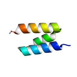
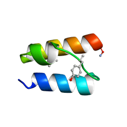
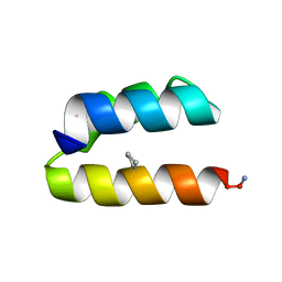
9BB5
DownloadVisualize
BU of 9bb5 by Molmil
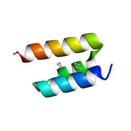
Backbone Modification in the GA Module of Protein PAB: ACPC residues at positions 22 and 26
Descriptor: Peptostreptococcal albumin-binding protein
Authors:Lin, Y, Horne, W.S.
Deposit date:2024-04-05
Release date:2024-06-05
Last modified:2024-08-21
Method:SOLUTION NMR
Cite:Backbone Modification in a Protein Hydrophobic Core.
Chemistry, 30, 2024
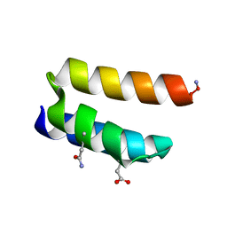
9BB7
DownloadVisualize
BU of 9bb7 by Molmil
Backbone Modification in the GA Module of Protein PAB: ACPC residues at positions 5 and 39, beta3 residue at position 26
Descriptor: Peptostreptococcal albumin-binding protein
Authors:Lin, Y, Horne, W.S.
Deposit date:2024-04-05
Release date:2024-06-05
Last modified:2024-08-21
Method:SOLUTION NMR
Cite:Backbone Modification in a Protein Hydrophobic Core.
Chemistry, 30, 2024
9JKQ
DownloadVisualize
BU of 9jkq by Molmil
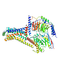
Cryo-EM structure of the METH-bound hTAAR1-Gs complex
Descriptor: (2S)-N-methyl-1-phenylpropan-2-amine, Guanine nucleotide-binding protein G(I)/G(S)/G(O) subunit gamma-2, Guanine nucleotide-binding protein G(I)/G(S)/G(T) subunit beta-1, ...
Authors:Lin, Y, Wang, J, Shi, F.
Deposit date:2024-09-16
Release date:2024-10-16
Method:ELECTRON MICROSCOPY (2.66 Å)
Cite:Molecular Mechanisms of Methamphetamine-Induced Addiction via TAAR1 Activation.
J.Med.Chem., 2024
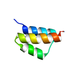
9BB1
DownloadVisualize
BU of 9bb1 by Molmil
Backbone Modification in the GA Module of Protein PAB: Wild-type Sequence
Descriptor: Peptostreptococcal albumin-binding protein
Authors:Lin, Y, Horne, W.S.
Deposit date:2024-04-05
Release date:2024-06-05
Last modified:2024-08-21
Method:SOLUTION NMR
Cite:Backbone Modification in a Protein Hydrophobic Core.
Chemistry, 30, 2024
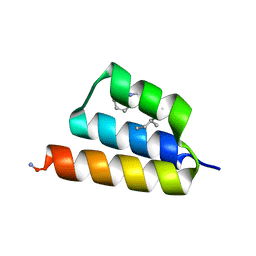
9BB2
DownloadVisualize
BU of 9bb2 by Molmil
Backbone Modification in the GA Module of Protein PAB: beta3-residues at positions 20 and 24
Descriptor: Peptostreptococcal albumin-binding protein
Authors:Lin, Y, Horne, W.S.
Deposit date:2024-04-05
Release date:2024-06-05
Last modified:2024-08-21
Method:SOLUTION NMR
Cite:Backbone Modification in a Protein Hydrophobic Core.
Chemistry, 30, 2024
9BB6
DownloadVisualize
BU of 9bb6 by Molmil
Backbone Modification in the GA Module of Protein PAB: ACPC residues at positions 5 and 13, beta3 residue at position 9
Descriptor: Peptostreptococcal albumin-binding protein
Authors:Lin, Y, Horne, W.S.
Deposit date:2024-04-05
Release date:2024-06-05
Last modified:2024-08-21
Method:SOLUTION NMR
Cite:Backbone Modification in a Protein Hydrophobic Core.
Chemistry, 30, 2024
9BB4
DownloadVisualize
BU of 9bb4 by Molmil
Backbone Modification in the GA Module of Protein PAB: beta3-residues at positions 23 and 26
Descriptor: Peptostreptococcal albumin-binding protein
Authors:Lin, Y, Horne, W.S.
Deposit date:2024-04-05
Release date:2024-06-05
Last modified:2024-08-21
Method:SOLUTION NMR
Cite:Backbone Modification in a Protein Hydrophobic Core.
Chemistry, 30, 2024
9BB3
DownloadVisualize
BU of 9bb3 by Molmil
Backbone Modification in the GA Module of Protein PAB: beta3-residues at positions 22 and 26
Descriptor: Peptostreptococcal albumin-binding protein
Authors:Lin, Y, Horne, W.S.
Deposit date:2024-04-05
Release date:2024-06-05
Last modified:2024-08-21
Method:SOLUTION NMR
Cite:Backbone Modification in a Protein Hydrophobic Core.
Chemistry, 30, 2024
1GOX
DownloadVisualize
BU of 1gox by Molmil
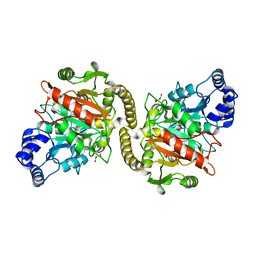
REFINED STRUCTURE OF SPINACH GLYCOLATE OXIDASE AT 2 ANGSTROMS RESOLUTION
Descriptor: (S)-2-HYDROXY-ACID OXIDASE, PEROXISOMAL, FLAVIN MONONUCLEOTIDE
Authors:Lindqvist, Y.
Deposit date:1989-06-14
Release date:1989-10-15
Last modified:2024-06-05
Method:X-RAY DIFFRACTION (2 Å)
Cite:Refined structure of spinach glycolate oxidase at 2 A resolution.
J.Mol.Biol., 209, 1989
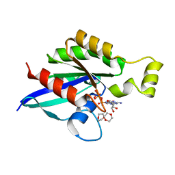
8FPW
DownloadVisualize
BU of 8fpw by Molmil
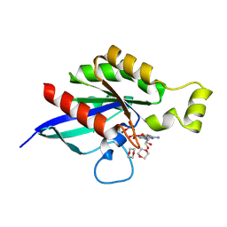
Crystal structure of tumor related RhoA mutant A161V in complex with GDP
Descriptor: 1,4-DIETHYLENE DIOXIDE, GUANOSINE-5'-DIPHOSPHATE, MAGNESIUM ION, ...
Authors:Lin, Y, Zheng, Y.
Deposit date:2023-01-05
Release date:2024-06-05
Last modified:2024-09-04
Method:X-RAY DIFFRACTION (1.4 Å)
Cite:Tumor-derived RHOA mutants interact with effectors in the GDP-bound state.
Nat Commun, 15, 2024
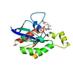
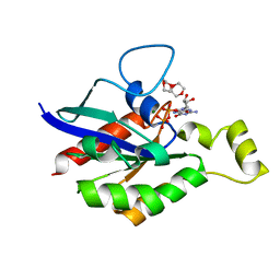
8FPX
DownloadVisualize
BU of 8fpx by Molmil
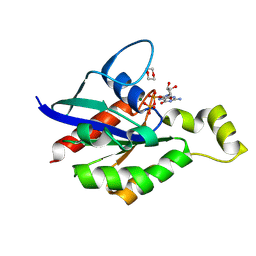
Crystal structure of tumor related RhoA mutant A161P in complex with GDP
Descriptor: 1,4-DIETHYLENE DIOXIDE, GUANOSINE-5'-DIPHOSPHATE, MAGNESIUM ION, ...
Authors:Lin, Y, Zheng, Y.
Deposit date:2023-01-05
Release date:2024-06-05
Last modified:2024-09-04
Method:X-RAY DIFFRACTION (1.47 Å)
Cite:Tumor-derived RHOA mutants interact with effectors in the GDP-bound state.
Nat Commun, 15, 2024
1QHW
DownloadVisualize
BU of 1qhw by Molmil
PURPLE ACID PHOSPHATASE FROM RAT BONE
Descriptor: 2-acetamido-2-deoxy-beta-D-glucopyranose-(1-4)-2-acetamido-2-deoxy-beta-D-glucopyranose, FE (III) ION, PROTEIN (PURPLE ACID PHOSPHATASE), ...
Authors:Lindqvist, Y, Johansson, E, Kaija, H, Vihko, P, Schneider, G.
Deposit date:1999-03-26
Release date:1999-09-15
Last modified:2023-08-16
Method:X-RAY DIFFRACTION (2.2 Å)
Cite:Three-dimensional structure of a mammalian purple acid phosphatase at 2.2 A resolution with a mu-(hydr)oxo bridged di-iron center.
J.Mol.Biol., 291, 1999
6V6U
DownloadVisualize
BU of 6v6u by Molmil
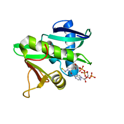
Crystal structure of RhoA-GDP with novel Switch I conformation
Descriptor: 1,4-DIETHYLENE DIOXIDE, GUANOSINE-5'-DIPHOSPHATE, MAGNESIUM ION, ...
Authors:Lin, Y, Zheng, Y.
Deposit date:2019-12-06
Release date:2020-12-23
Last modified:2023-10-11
Method:X-RAY DIFFRACTION (1.16 Å)
Cite:Structure of an inactive conformation of GTP-bound RhoA GTPase.
Structure, 29, 2021
6V6M
DownloadVisualize
BU of 6v6m by Molmil
Crystal structure of an inactive state of GMPPNP-bound RhoA
Descriptor: 1,4-DIETHYLENE DIOXIDE, MAGNESIUM ION, PHOSPHOAMINOPHOSPHONIC ACID-GUANYLATE ESTER, ...
Authors:Lin, Y, Zheng, Y.
Deposit date:2019-12-05
Release date:2020-12-09
Last modified:2023-10-11
Method:X-RAY DIFFRACTION (1.39 Å)
Cite:Structure of an inactive conformation of GTP-bound RhoA GTPase.
Structure, 29, 2021
6V6V
DownloadVisualize
BU of 6v6v by Molmil
Crystal structure of oncogenic RhoA mutant G14V complexed with GDP
Descriptor: 1,4-DIETHYLENE DIOXIDE, GUANOSINE-5'-DIPHOSPHATE, MAGNESIUM ION, ...
Authors:Lin, Y, Zheng, Y.
Deposit date:2019-12-06
Release date:2020-12-23
Last modified:2023-10-11
Method:X-RAY DIFFRACTION (1.4 Å)
Cite:Structure of an inactive conformation of GTP-bound RhoA GTPase.
Structure, 29, 2021
5GCN
DownloadVisualize
BU of 5gcn by Molmil
CATALYTIC DOMAIN OF TETRAHYMENA GCN5 HISTONE ACETYLTRANSFERASE IN COMPLEX WITH COENZYME A
Descriptor: COENZYME A, HISTONE ACETYLTRANSFERASE GCN5
Authors:Lin, Y, Fletcher, C.M, Zhou, J, Allis, C.D, Wagner, G.
Deposit date:1999-03-24
Release date:1999-07-19
Last modified:2024-05-22
Method:SOLUTION NMR
Cite:Solution structure of the catalytic domain of GCN5 histone acetyltransferase bound to coenzyme A
Nature, 400, 1999
3CGP
DownloadVisualize
BU of 3cgp by Molmil
X-ray structure of a pseudouridine-containing yeast spliceosomal U2 snRNA-intron branch site duplex bound to iodide ions
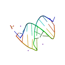
Descriptor: IODIDE ION, RNA (5'-R(*CP*GP*CP*UP*AP*CP*UP*AP*AP*CP*GP*CP*G)-3'), RNA (5'-R(*GP*CP*GP*CP*GP*(PSU)P*AP*GP*UP*AP*GP*C)-3'), ...
Authors:Lin, Y, Kielkopf, C.L.
Deposit date:2008-03-06
Release date:2008-07-01
Last modified:2024-04-03
Method:X-RAY DIFFRACTION (1.57 Å)
Cite:X-ray structures of U2 snRNA-branchpoint duplexes containing conserved pseudouridines.
Biochemistry, 47, 2008
3CGS
DownloadVisualize
BU of 3cgs by Molmil
X-ray structure containing the pseudouridylated U2 snRNA and mammalian intron branch site consensus sequences
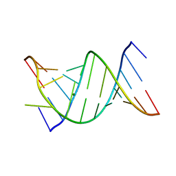
Descriptor: MAGNESIUM ION, RNA (5'-R(*CP*GP*CP*UP*AP*CP*UP*GP*AP*CP*GP*CP*G)-3'), RNA (5'-R(*GP*CP*GP*CP*GP*(PSU)P*AP*GP*UP*AP*GP*C)-3')
Authors:Lin, Y, Kielkopf, C.L.
Deposit date:2008-03-06
Release date:2008-07-01
Last modified:2024-04-03
Method:X-RAY DIFFRACTION (1.65 Å)
Cite:X-ray structures of U2 snRNA-branchpoint duplexes containing conserved pseudouridines.
Biochemistry, 47, 2008
3CGR
DownloadVisualize
BU of 3cgr by Molmil
X-ray structure containing the pseudouridylated U2 snRNA and intron branch site consensus sequences
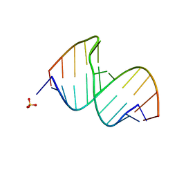
Descriptor: RNA (5'-R(*CP*GP*CP*AP*CP*UP*AP*AP*CP*CP*GP*CP*G)-3'), RNA (5'-R(*GP*CP*GP*CP*GP*GP*(PSU)P*AP*GP*UP*GP*C)-3'), SULFATE ION
Authors:Lin, Y, Kielkopf, C.L.
Deposit date:2008-03-06
Release date:2008-07-01
Last modified:2024-04-03
Method:X-RAY DIFFRACTION (2.1 Å)
Cite:X-ray structures of U2 snRNA-branchpoint duplexes containing conserved pseudouridines.
Biochemistry, 47, 2008
3CGQ
DownloadVisualize
BU of 3cgq by Molmil
X-ray structure of a pseudouridine-containing yeast spliceosomal U2 snRNA-intron branch site duplex
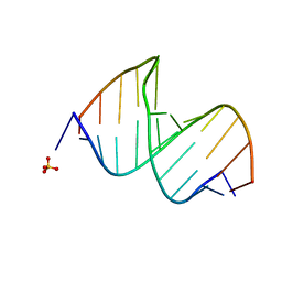
Descriptor: RNA (5'-R(*CP*GP*CP*UP*AP*CP*UP*AP*AP*CP*GP*CP*G)-3'), RNA (5'-R(*GP*CP*GP*CP*GP*(PSU)P*AP*GP*UP*AP*GP*C)-3'), SULFATE ION
Authors:Lin, Y, Kielkopf, C.L.
Deposit date:2008-03-06
Release date:2008-07-01
Last modified:2023-08-30
Method:X-RAY DIFFRACTION (2.55 Å)
Cite:X-ray structures of U2 snRNA-branchpoint duplexes containing conserved pseudouridines.
Biochemistry, 47, 2008
4GYR
DownloadVisualize
BU of 4gyr by Molmil
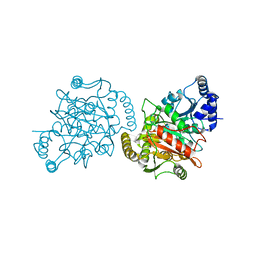
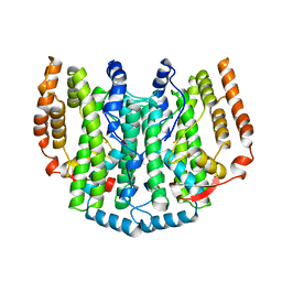
Granulibacter bethesdensis allophanate hydrolase apo
Descriptor: Allophanate hydrolase
Authors:Lin, Y, St Maurice, M.
Deposit date:2012-09-05
Release date:2013-01-30
Last modified:2023-09-13
Method:X-RAY DIFFRACTION (2.8 Å)
Cite:The Structure of Allophanate Hydrolase from Granulibacter bethesdensis Provides Insights into Substrate Specificity in the Amidase Signature Family.
Biochemistry, 52, 2013
4GYS
DownloadVisualize
BU of 4gys by Molmil
Granulibacter bethesdensis allophanate hydrolase co-crystallized with malonate
Descriptor: Allophanate hydrolase, MALONATE ION
Authors:Lin, Y, St Maurice, M.
Deposit date:2012-09-05
Release date:2013-01-30
Last modified:2023-09-13
Method:X-RAY DIFFRACTION (2.201 Å)
Cite:The Structure of Allophanate Hydrolase from Granulibacter bethesdensis Provides Insights into Substrate Specificity in the Amidase Signature Family.
Biochemistry, 52, 2013
1EYF
DownloadVisualize
BU of 1eyf by Molmil
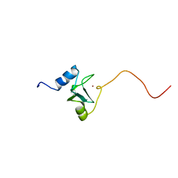
REFINED STRUCTURE OF THE DNA METHYL PHOSPHOTRIESTER REPAIR DOMAIN OF E. COLI ADA
Descriptor: ADA REGULATORY PROTEIN, ZINC ION
Authors:Lin, Y, Dotsch, V, Wintner, T, Peariso, K, Myers, L.C, Penner-Hahn, J.E, Verdine, G.L, Wagner, G.
Deposit date:2000-05-06
Release date:2003-09-09
Last modified:2024-05-22
Method:SOLUTION NMR
Cite:Structural basis for the functional switch of the E. coli Ada protein
Biochemistry, 40, 2001
6QII
DownloadVisualize
BU of 6qii by Molmil
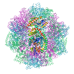
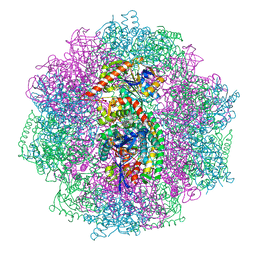
Xenon derivatization of the F420-reducing [NiFe] hydrogenase complex from Methanosarcina barkeri
Descriptor: (R,R)-2,3-BUTANEDIOL, Coenzyme F420 hydrogenase subunit alpha, Coenzyme F420 hydrogenase subunit beta, ...
Authors:Ilina, Y, Lorent, C, Katz, S, Jeoung, J.H, Shima, S, Horch, M, Zebger, I, Dobbek, H.
Deposit date:2019-01-19
Release date:2019-10-23
Last modified:2024-01-24
Method:X-RAY DIFFRACTION (2.28 Å)
Cite:X-ray Crystallography and Vibrational Spectroscopy Reveal the Key Determinants of Biocatalytic Dihydrogen Cycling by [NiFe] Hydrogenases.
Angew.Chem.Int.Ed.Engl., 58, 2019
6QGT
DownloadVisualize
BU of 6qgt by Molmil
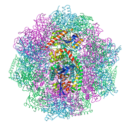
The carbon monoxide inhibition of F420-reducing [NiFe] hydrogenase complex from Methanosarcina barkeri
Descriptor: (4S)-2-METHYL-2,4-PENTANEDIOL, (R,R)-2,3-BUTANEDIOL, Coenzyme F420 hydrogenase subunit beta, ...
Authors:Ilina, Y, Lorent, C, Katz, S, Jeoung, J.H, Shima, S, Horch, M, Zebger, I, Dobbek, H.
Deposit date:2019-01-12
Release date:2019-10-23
Last modified:2024-01-24
Method:X-RAY DIFFRACTION (1.988 Å)
Cite:X-ray Crystallography and Vibrational Spectroscopy Reveal the Key Determinants of Biocatalytic Dihydrogen Cycling by [NiFe] Hydrogenases.
Angew.Chem.Int.Ed.Engl., 58, 2019

226707

PDB entries from 2024-10-30

PDB statisticsPDBj update infoContact PDBjnumon