2OY9
 
 | |
2P0L
 | |
3E9M
 | |
2A9F
 | |
3EUW
 | |
3F5D
 | |
3FD8
 | |
3FII
 | |
3EWN
 | |
3FJY
 | |
3FIJ
 | |
3F6A
 | |
3DLI
 | |
3KOL
 | |
3EHE
 | |
3L3S
 | |
3E3V
 | |
3GV1
 | |
3G7S
 | |
3GAZ
 | |
3GBT
 | |
3EXQ
 | |
3GPV
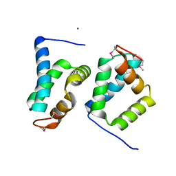
 | | Crystal structure of a transcriptional regulator, MerR family from Bacillus thuringiensis | | Descriptor: | Transcriptional regulator, MerR family | | Authors: | Palani, K, Kumaran, D, Burley, S.K, Swaminathan, S, New York SGX Research Center for Structural Genomics (NYSGXRC) | | Deposit date: | 2009-03-23 | | Release date: | 2009-04-14 | | Last modified: | 2024-11-06 | | Method: | X-RAY DIFFRACTION (1.9 Å) | | Cite: | Crystal structure of a transcriptional regulator, MerR family from Bacillus thuringiensis
To be Published
|
|
3G13
 | |
3GYB
 | |


