Loading
PDBj
MenuPDBj@FacebookPDBj@X(formerly Twitter)PDBj@BlueSkyPDBj@YouTubewwPDB FoundationwwPDBDonate
RCSB PDBPDBeBMRBAdv. SearchSearch help
Search by PDB author
5TS2
DownloadVisualize
BU of 5ts2 by Molmil
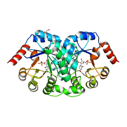
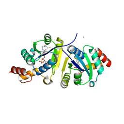
Crystal structure of a phosphopantetheine adenylyltransferase (CoaD, PPAT) from Pseudomonas aeruginosa bound to dephospho coenzyme A
Descriptor: CALCIUM ION, CHLORIDE ION, DEPHOSPHO COENZYME A, ...
Authors:Seattle Structural Genomics Center for Infectious Disease (SSGCID)
Deposit date:2016-10-27
Release date:2016-11-09
Last modified:2023-10-04
Method:X-RAY DIFFRACTION (2.3 Å)
Cite:Crystal structure of a phosphopantetheine adenylyltransferase (CoaD, PPAT) from Pseudomonas aeruginosa bound to dephospho coenzyme A
To Be Published
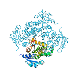
6UDF
DownloadVisualize
BU of 6udf by Molmil
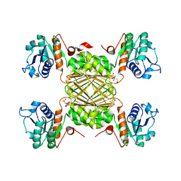
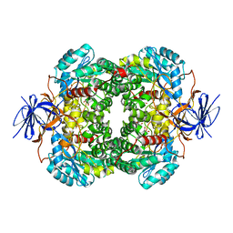
Crystal structure of Enoyl-[acyl-carrier-protein] reductase [NADH] (InhA) from Mycobacterium kansasii
Descriptor: Enoyl-[acyl-carrier-protein] reductase [NADH], FORMIC ACID
Authors:Seattle Structural Genomics Center for Infectious Disease (SSGCID)
Deposit date:2019-09-19
Release date:2019-10-16
Last modified:2023-10-11
Method:X-RAY DIFFRACTION (1.95 Å)
Cite:Crystal stucture of Enoyl-[acyl-carrier-protein] reductase [NADH] (InhA) from Mycobacterium kansasii
to be published
6V91
DownloadVisualize
BU of 6v91 by Molmil
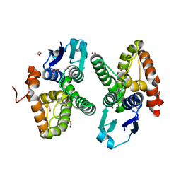
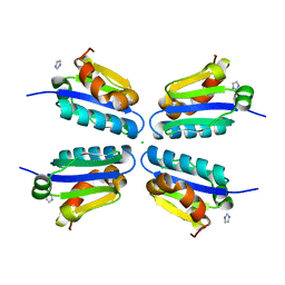
Crystal structure of Stringent starvation protein A (BTH_I2974) from Burkholderia thailandensis
Descriptor: 1,2-ETHANEDIOL, ACETATE ION, FORMIC ACID, ...
Authors:Seattle Structural Genomics Center for Infectious Disease (SSGCID)
Deposit date:2019-12-12
Release date:2020-01-15
Last modified:2024-03-06
Method:X-RAY DIFFRACTION (1.9 Å)
Cite:Crystal structure of Stringent starvation protein A (BTH_I2974) from Burkholderia thailandensis
to be published
8CSO
DownloadVisualize
BU of 8cso by Molmil
Crystal Structure of Orotidine 5'-phosphate decarboxylase from Klebsiella pneumoniae in complex with Uridine-5'-monophosphate
Descriptor: 1,2-ETHANEDIOL, Orotidine 5'-phosphate decarboxylase, URIDINE-5'-MONOPHOSPHATE
Authors:Seattle Structural Genomics Center for Infectious Disease, Seattle Structural Genomics Center for Infectious Disease (SSGCID)
Deposit date:2022-05-13
Release date:2022-06-08
Last modified:2023-10-18
Method:X-RAY DIFFRACTION (2.6 Å)
Cite:Crystal Structure of Orotidine 5'-phosphate decarboxylase from Klebsiella pneumoniae in complex with Guanosine-5'-monophosphate
to be published
8D57
DownloadVisualize
BU of 8d57 by Molmil
Crystal Structure of dihydrodipicolinate reductase from Acinetobacter baumannii
Descriptor: 4-hydroxy-tetrahydrodipicolinate reductase, CITRIC ACID
Authors:Seattle Structural Genomics Center for Infectious Disease, Seattle Structural Genomics Center for Infectious Disease (SSGCID)
Deposit date:2022-06-04
Release date:2022-06-22
Last modified:2023-10-18
Method:X-RAY DIFFRACTION (2.65 Å)
Cite:Crystal Structure of dihydrodipicolinate reductase from Acinetobacter baumannii
to be published
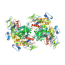
6W2O
DownloadVisualize
BU of 6w2o by Molmil
Crystal structure of uroporphyrinogen III decarboxylate (hemE) from Stenotrophomonas maltophilia
Descriptor: 1,2-ETHANEDIOL, CHLORIDE ION, SULFATE ION, ...
Authors:Seattle Structural Genomics Center for Infectious Disease (SSGCID)
Deposit date:2020-03-06
Release date:2020-04-22
Last modified:2023-10-18
Method:X-RAY DIFFRACTION (1.55 Å)
Cite:Crystal structure of uroporphyrinogen III decarboxylate (hemE) from Stenotrophomonas maltophilia
To Be Published
4NI7
DownloadVisualize
BU of 4ni7 by Molmil
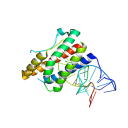
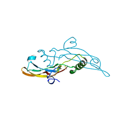
Crystal structure of human interleukin 6 in complex with a modified nucleotide aptamer (SOMAMER SL1025)
Descriptor: Interleukin-6, SODIUM ION, SOMAmer SL1025
Authors:Davies, D, Edwards, T, Gelinas, A, Jarvis, T, Clifton, M.C.
Deposit date:2013-11-05
Release date:2014-01-22
Last modified:2024-11-20
Method:X-RAY DIFFRACTION (2.4 Å)
Cite:Crystal structure of interleukin-6 in complex with a modified nucleic Acid ligand.
J.Biol.Chem., 289, 2014
4NI9
DownloadVisualize
BU of 4ni9 by Molmil
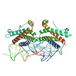
Crystal structure of human interleukin 6 in complex with a modified nucleotide aptamer (SOMAMER SL1025), FORM 2
Descriptor: Interleukin-6, SODIUM ION, SOMAmer SL1025
Authors:Davies, D, Edwards, T, Gelinas, A, Jarvis, T, Clifton, M.C.
Deposit date:2013-11-05
Release date:2014-01-22
Last modified:2024-10-09
Method:X-RAY DIFFRACTION (2.55 Å)
Cite:Crystal structure of interleukin-6 in complex with a modified nucleic Acid ligand.
J.Biol.Chem., 289, 2014
6VJU
DownloadVisualize
BU of 6vju by Molmil
Crystal Structure of Cystathionine beta synthase from Legionella pneumophila with LLP, PLP, and homocysteine
Descriptor: 1,2-ETHANEDIOL, 2-AMINO-4-MERCAPTO-BUTYRIC ACID, ACETATE ION, ...
Authors:Seattle Structural Genomics Center for Infectious Disease (SSGCID)
Deposit date:2020-01-17
Release date:2020-02-05
Last modified:2024-04-03
Method:X-RAY DIFFRACTION (1.3 Å)
Cite:Crystal Structure of Cystathionine beta synthase from Legionella pneumophila with LLP, PLP, and homocysteine
to be published
4PY2
DownloadVisualize
BU of 4py2 by Molmil
Crystal structure of methionyl-tRNA synthetase MetRS from Brucella melitensis in complex with inhibitor 1-{3-[(3,5-DICHLOROBENZYL)AMINO]PROPYL}-3-THIOPHEN-3-YLUREA
Descriptor: 1,2-ETHANEDIOL, 1-{3-[(3,5-dichlorobenzyl)amino]propyl}-3-thiophen-3-ylurea, Methionine--tRNA ligase
Authors:Seattle Structural Genomics Center for Infectious Disease, Seattle Structural Genomics Center for Infectious Disease (SSGCID)
Deposit date:2014-03-25
Release date:2015-04-15
Last modified:2023-09-20
Method:X-RAY DIFFRACTION (2.15 Å)
Cite:Brucella melitensis Methionyl-tRNA-Synthetase (MetRS), a Potential Drug Target for Brucellosis.
Plos One, 11, 2016
4TQT
DownloadVisualize
BU of 4tqt by Molmil
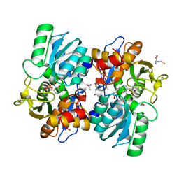
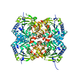
Crystal structure of Dihydropyrimidinase from Brucella suis
Descriptor: 1,2-ETHANEDIOL, D-hydantoinase, ZINC ION
Authors:Seattle Structural Genomics Center for Infectious Disease (SSGCID)
Deposit date:2014-06-12
Release date:2014-08-06
Last modified:2023-11-15
Method:X-RAY DIFFRACTION (2.15 Å)
Cite:Crystal structure of Dihydropyrimidinase from Brucella suis
to be published
4PQ9
DownloadVisualize
BU of 4pq9 by Molmil
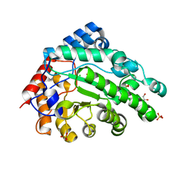
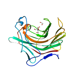
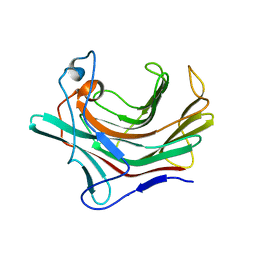
Crystal Structure of a Beta-1,3-glucanase from Mycobacterium marinum
Descriptor: 1,2-ETHANEDIOL, 2-[BIS-(2-HYDROXY-ETHYL)-AMINO]-2-HYDROXYMETHYL-PROPANE-1,3-DIOL, Beta-1,3-glucanase, ...
Authors:Seattle Structural Genomics Center for Infectious Disease, Seattle Structural Genomics Center for Infectious Disease (SSGCID)
Deposit date:2014-02-28
Release date:2014-03-12
Last modified:2023-09-20
Method:X-RAY DIFFRACTION (1.2 Å)
Cite:Crystal Structure of a Beta-1,3-glucanase from Mycobacterium marinum
TO BE PUBLISHED
4Q1T
DownloadVisualize
BU of 4q1t by Molmil
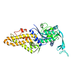
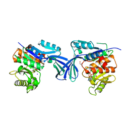
Crystal structure of a glutamate 5-kinase from Burkholderia thailandensis
Descriptor: Glutamate 5-kinase
Authors:Seattle Structural Genomics Center for Infectious Disease (SSGCID)
Deposit date:2014-04-04
Release date:2014-04-23
Last modified:2023-09-20
Method:X-RAY DIFFRACTION (2.15 Å)
Cite:Crystal structure of a glutamate 5-kinase from Burkholderia thailandensis
To be Published
6BKE
DownloadVisualize
BU of 6bke by Molmil
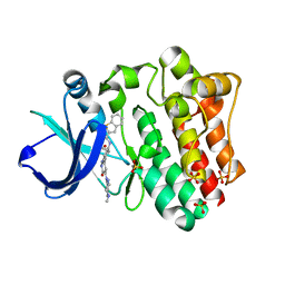
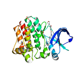
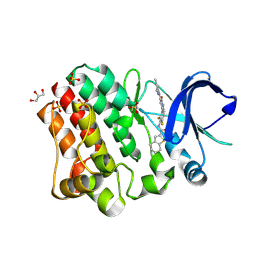
BTK complex with compound 10
Descriptor: N-[2-(2-hydroxyethyl)-3-{5-[(5-methyl-4,5,6,7-tetrahydropyrazolo[1,5-a]pyrazin-2-yl)amino]-6-oxo-1,6-dihydropyridazin-3-yl}phenyl]-1-benzothiophene-2-carboxamide, SULFATE ION, Tyrosine-protein kinase BTK
Authors:Kiefer, J.R, Eigenbrot, C, Yu, C.L, Wang, G.X.
Deposit date:2017-11-08
Release date:2018-11-07
Last modified:2025-04-02
Method:X-RAY DIFFRACTION (1.95 Å)
Cite:Water molecules in protein-ligand interfaces. Evaluation of software tools and SAR comparison.
J. Comput. Aided Mol. Des., 33, 2019
6BIK
DownloadVisualize
BU of 6bik by Molmil
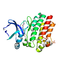
BTK complex with compound 7
Descriptor: 4-tert-butyl-N-[2-(hydroxymethyl)-3-(1-methyl-5-{[5-(morpholine-4-carbonyl)pyridin-2-yl]amino}-6-oxo-1,6-dihydropyridazin-3-yl)phenyl]benzamide, GLYCEROL, SULFATE ION, ...
Authors:Kiefer, J.R, Eigenbrot, C, Yu, C.L.
Deposit date:2017-11-02
Release date:2018-11-07
Last modified:2025-04-02
Method:X-RAY DIFFRACTION (1.901 Å)
Cite:Water molecules in protein-ligand interfaces. Evaluation of software tools and SAR comparison.
J. Comput. Aided Mol. Des., 33, 2019
6BKH
DownloadVisualize
BU of 6bkh by Molmil
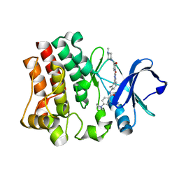
BTK complex with compound 11
Descriptor: N-[2-(hydroxymethyl)-3-{5-[(5-methyl-4,5,6,7-tetrahydropyrazolo[1,5-a]pyrazin-2-yl)amino]-6-oxo-1,6-dihydropyridazin-3-yl}phenyl]-1-benzothiophene-2-carboxamide, SULFATE ION, Tyrosine-protein kinase BTK
Authors:Kiefer, J.R, Eigenbrot, C, Yu, C.L.
Deposit date:2017-11-08
Release date:2018-11-07
Last modified:2025-04-02
Method:X-RAY DIFFRACTION (1.792 Å)
Cite:Water molecules in protein-ligand interfaces. Evaluation of software tools and SAR comparison.
J. Comput. Aided Mol. Des., 33, 2019
6C87
DownloadVisualize
BU of 6c87 by Molmil
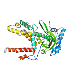
Crystal structure of rab gdp dissociation inhibitor alpha from Naegleria fowleri
Descriptor: Rab GDP dissociation inhibitor alpha, SULFATE ION
Authors:Seattle Structural Genomics Center for Infectious Disease (SSGCID)
Deposit date:2018-01-24
Release date:2018-02-14
Last modified:2023-10-04
Method:X-RAY DIFFRACTION (2.6 Å)
Cite:Crystal structure of rab gdp dissociation inhibitor alpha from Naegleria fowleri
TO BE PUBLISHED
6CFP
DownloadVisualize
BU of 6cfp by Molmil
CRYSTAL STRUCTURE OF POLYMERASE ACID PROTEIN (PA) FROM INFLUENZA A VIRUS, WILSON-SMITH/1933 (H1N1) BOUND TO FRAGMENT HIT BSI-70565 1-{1-[4-FLUOROPHENYL)METHYL]-2-METHYL-1H-IMIDAZOL-4-YL}ETHAN-1-ONE
Descriptor: 1-{1-[(4-fluorophenyl)methyl]-2-methyl-1H-imidazol-4-yl}ethan-1-one, DIMETHYL SULFOXIDE, Ubiquitin-like protein SMT3,Polymerase acidic protein
Authors:Seattle Structural Genomics Center for Infectious Disease (SSGCID)
Deposit date:2018-02-15
Release date:2018-04-11
Last modified:2023-10-04
Method:X-RAY DIFFRACTION (2.45 Å)
Cite:CRYSTAL STRUCTURE OF POLYMERASE ACID PROTEIN (PA) FROM INFLUENZA A VIRUS, WILSON-SMITH/1933 (H1N1) BOUND TO FRAGMENT HIT BSI-70565 1-{1-[4-FLUOROPHENYL)METHYL]-2-METHYL-1H-IMIDAZOL-4-YL}ETHAN-1-ONE
To Be Published
6BLN
DownloadVisualize
BU of 6bln by Molmil
BTK complex with compound 13
Descriptor: GLYCEROL, N-(3-{5-[(1,5-dimethyl-1H-pyrazol-3-yl)amino]-6-oxo-1,6-dihydropyridazin-3-yl}-2,6-difluorophenyl)-4,5,6,7-tetrahydro-1-benzothiophene-2-carboxamide, SULFATE ION, ...
Authors:Kiefer, J.R, Eigenbrot, C, Yu, C.L.
Deposit date:2017-11-10
Release date:2018-11-07
Last modified:2025-04-02
Method:X-RAY DIFFRACTION (1.3 Å)
Cite:Water molecules in protein-ligand interfaces. Evaluation of software tools and SAR comparison.
J. Comput. Aided Mol. Des., 33, 2019
6BQD
DownloadVisualize
BU of 6bqd by Molmil
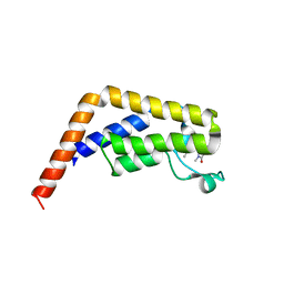
TAF1-BD2 bromodomain in complex with (E)-3-(6-(but-2-en-1-yl)-7-oxo-6,7-dihydro-1H-pyrrolo[2,3-c]pyridin-4-yl)-N,N-dimethylbenzamide
Descriptor: 3-{6-[(2E)-but-2-en-1-yl]-7-oxo-6,7-dihydro-1H-pyrrolo[2,3-c]pyridin-4-yl}-N,N-dimethylbenzamide, Transcription initiation factor TFIID subunit 1
Authors:Murray, J.M, Tang, Y.
Deposit date:2017-11-27
Release date:2019-02-20
Last modified:2023-10-04
Method:X-RAY DIFFRACTION (2.136 Å)
Cite:Water molecules in protein-ligand interfaces. Evaluation of software tools and SAR comparison.
J. Comput. Aided Mol. Des., 33, 2019
6EP9
DownloadVisualize
BU of 6ep9 by Molmil
Crystal structure of BTK kinase domain complexed with N-[2-methyl-3-[4-methyl-6-[4-(4-methylpiperazine-1-carbonyl)anilino]-5-oxo-pyrazin-2-yl]phenyl]-4-(1-piperidyl)benzamide
Descriptor: N-[2-methyl-3-[4-methyl-6-[4-(4-methylpiperazine-1-carbonyl)anilino]-5-oxo-pyrazin-2-yl]phenyl]-4-(1-piperidyl)benzamide, Tyrosine-protein kinase BTK
Authors:Kuglstatter, A, Wong, A.
Deposit date:2017-10-11
Release date:2018-08-22
Last modified:2024-05-08
Method:X-RAY DIFFRACTION (2.01 Å)
Cite:Water molecules in protein-ligand interfaces. Evaluation of software tools and SAR comparison.
J.Comput.Aided Mol.Des., 33, 2019
6XK2
DownloadVisualize
BU of 6xk2 by Molmil
Crystal structure of Ribokinase from Cryptococcus neoformans var. grubii serotype A in complex with ADP
Descriptor: 1,2-ETHANEDIOL, ADENOSINE-5'-DIPHOSPHATE, Ribokinase, ...
Authors:Seattle Structural Genomics Center for Infectious Disease (SSGCID)
Deposit date:2020-06-25
Release date:2020-07-15
Last modified:2023-10-18
Method:X-RAY DIFFRACTION (2.5 Å)
Cite:Crystal structure of Ribokinase from Cryptococcus neoformans var. grubii serotype A in complex with ADP
to be published
6I9J
DownloadVisualize
BU of 6i9j by Molmil
Human transforming growth factor beta2 in a tetragonal crystal form
Descriptor: Transforming growth factor beta-2 proprotein
Authors:Gomis-Ruth, F.X, Marino-Puertas, L, del Amo-Maestro, L, Goulas, T.
Deposit date:2018-11-23
Release date:2019-07-03
Last modified:2024-11-06
Method:X-RAY DIFFRACTION (2 Å)
Cite:Recombinant production, purification, crystallization, and structure analysis of human transforming growth factor beta 2 in a new conformation.
Sci Rep, 9, 2019
5DXD
DownloadVisualize
BU of 5dxd by Molmil
Crystal structure of Putative beta-glucanase (Rv0315 ortholog) from Mycobacterium abscessus
Descriptor: Putative beta-glucanase, SODIUM ION
Authors:Seattle Structural Genomics Center for Infectious Disease (SSGCID)
Deposit date:2015-09-23
Release date:2015-11-04
Last modified:2023-09-27
Method:X-RAY DIFFRACTION (1.7 Å)
Cite:Crystal structure of Putative beta-glucanase (Rv0315 ortholog) from Mycobacterium abcesus
to be published
5DLE
DownloadVisualize
BU of 5dle by Molmil
Crystal structure from a domain (Thr161-F265) from fructose-specific iiabc component (Pts system) from Borrelia burgdorferi
Descriptor: CHLORIDE ION, IMIDAZOLE, Pts system, ...
Authors:Seattle Structural Genomics Center for Infectious Disease (SSGCID)
Deposit date:2015-09-04
Release date:2015-11-04
Last modified:2023-09-27
Method:X-RAY DIFFRACTION (1.55 Å)
Cite:Crystal structure from a domain (Thr161-F265) from fructose-specific iiabc component (Pts system) from Borrelia burgdorferi
to be published

238582

PDB entries from 2025-07-09

PDB statisticsPDBj update infoContact PDBjnumon