Loading
PDBj
MenuPDBj@FacebookPDBj@TwitterPDBj@YouTubewwPDB FoundationwwPDB
RCSB PDBPDBeBMRBAdv. SearchSearch help
Search by PDB author
3DZ1
DownloadVisualize
BU of 3dz1 by Molmil
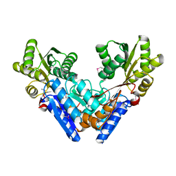
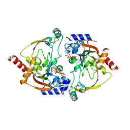
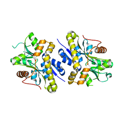
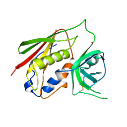
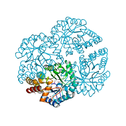
Crystal structure of Dihydrodipicolinate Synthase from Rhodopseudomonas palustris at 1.87A resolution
Descriptor: Dihydrodipicolinate synthase
Authors:Satyanarayana, L, Eswaramoorthy, S, Burley, S.K, Swaminathan, S, New York SGX Research Center for Structural Genomics (NYSGXRC)
Deposit date:2008-07-29
Release date:2008-08-12
Last modified:2021-02-10
Method:X-RAY DIFFRACTION (1.87 Å)
Cite:Crystal structure of Dihydrodipicolinate Synthase from Rhodopseudomonas palustris at 1.87A resolution
To be Published
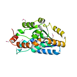
3DZB
DownloadVisualize
BU of 3dzb by Molmil
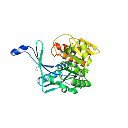
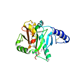
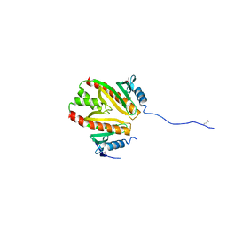
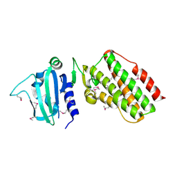
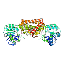
Crystal structure of Prephenate dehydrogenase from Streptococcus thermophilus
Descriptor: Prephenate dehydrogenase
Authors:Zhang, Z, Eswaramoorthy, S, Burley, S.K, Swaminathan, S, New York SGX Research Center for Structural Genomics (NYSGXRC)
Deposit date:2008-07-29
Release date:2008-08-12
Last modified:2024-10-16
Method:X-RAY DIFFRACTION (2.46 Å)
Cite:Crystal structure of Prephenate dehydrogenase from Streptococcus thermophilus
To be Published
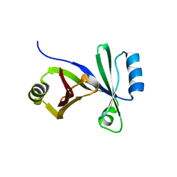
3GPK
DownloadVisualize
BU of 3gpk by Molmil
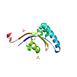
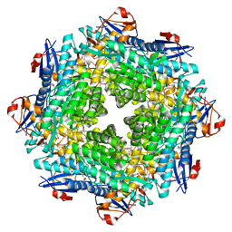
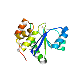
Crystal Structure of PpiC-type peptidyl-prolyl cis-trans isomerase domain at 1.55A resolution.
Descriptor: PpiC-type peptidyl-prolyl cis-trans isomerase, SULFATE ION
Authors:Satyanarayana, L, Eswaramoorthy, S, Burley, S.K, Swaminathan, S, New York SGX Research Center for Structural Genomics (NYSGXRC)
Deposit date:2009-03-23
Release date:2009-03-31
Last modified:2021-02-10
Method:X-RAY DIFFRACTION (1.55 Å)
Cite:Crystal Structure of PpiC-type peptidyl-prolyl cis-trans isomerase domain at 1.55A resolution.
To be Published
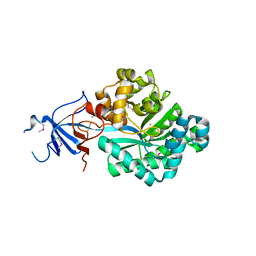
3EAF
DownloadVisualize
BU of 3eaf by Molmil
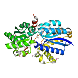
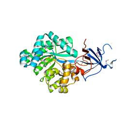
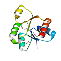
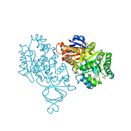
Crystal structure of ABC transporter, substrate binding protein Aeropyrum pernix
Descriptor: ABC transporter, substrate binding protein, GLYCEROL, ...
Authors:Zhang, Z, Eswaramoorthy, S, Burley, S.K, Swaminathan, S, New York SGX Research Center for Structural Genomics (NYSGXRC)
Deposit date:2008-08-25
Release date:2008-09-09
Last modified:2021-02-10
Method:X-RAY DIFFRACTION (2 Å)
Cite:Crystal structure of ABC transporter, substrate binding protein Aeropyrum pernix
To be Published
3G1W
DownloadVisualize
BU of 3g1w by Molmil
Crystal structure of sugar ABC transporter (sugar-binding protein) from Bacillus halodurans
Descriptor: Sugar ABC transporter
Authors:Zhang, Z, Eswaramoorthy, S, Swaminathan, S, Burley, S.K, New York SGX Research Center for Structural Genomics (NYSGXRC)
Deposit date:2009-01-30
Release date:2009-02-17
Last modified:2021-02-10
Method:X-RAY DIFFRACTION (2.02 Å)
Cite:The crystal structure of sugar ABC transporter (sugar-binding protein) from Bacillus halodurans.
To be Published
3H49
DownloadVisualize
BU of 3h49 by Molmil
Crystal structure of a putative Ribokinase (Apo Form) from E.coli at 1.8A resolution
Descriptor: Ribokinase
Authors:Satyanarayana, L, Eswaramoorthy, S, Burley, S.K, Swaminathan, S, New York SGX Research Center for Structural Genomics (NYSGXRC)
Deposit date:2009-04-18
Release date:2009-04-28
Last modified:2021-02-10
Method:X-RAY DIFFRACTION (1.8 Å)
Cite:Crystal structure of a putative Ribokinase (Apo Form) from E.coli at 1.8A resolution.
To be Published
3HDP
DownloadVisualize
BU of 3hdp by Molmil
Crystal structure of the NI(II)-bound Glyoxalase-I from Clostridium acetobutylicum
Descriptor: Glyoxalase-I, NICKEL (II) ION
Authors:Satyanarayana, L, Eswaramoorthy, S, Burley, S.K, Swaminathan, S, New York SGX Research Center for Structural Genomics (NYSGXRC)
Deposit date:2009-05-07
Release date:2009-05-26
Last modified:2023-09-06
Method:X-RAY DIFFRACTION (2.06 Å)
Cite:Crystal structure of the NI(II)-bound Glyoxalase-I from Clostridium acetobutylicum.
To be Published
3HP0
DownloadVisualize
BU of 3hp0 by Molmil
Crystal structure of a Putative polyketide biosynthesis enoyl-CoA hydratase (pksH) from Bacillus subtilis
Descriptor: Putative polyketide biosynthesis enoyl-CoA hydratase homolog pksH
Authors:Satyanarayana, L, Eswaramoorthy, S, Burley, S.K, Swaminathan, S, New York SGX Research Center for Structural Genomics (NYSGXRC)
Deposit date:2009-06-03
Release date:2009-06-23
Last modified:2021-02-10
Method:X-RAY DIFFRACTION (2.32 Å)
Cite:Crystal structure of a Putative polyketide biosynthesis enoyl-CoA hydratase (pksH) from Bacillus subtilis
To be Published
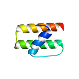
2NN4
DownloadVisualize
BU of 2nn4 by Molmil
Crystal structure of Bacillus subtilis yqgQ, Pfam DUF910
Descriptor: Hypothetical protein yqgQ
Authors:Damodharan, L, Eswaramoorthy, S, Burley, S.K, Swaminathan, S, New York SGX Research Center for Structural Genomics (NYSGXRC)
Deposit date:2006-10-23
Release date:2006-10-31
Last modified:2023-12-27
Method:X-RAY DIFFRACTION (2.1 Å)
Cite:Structure of YqgQ protein from Bacillus subtilis, a conserved hypothetical protein.
Acta Crystallogr.,Sect.F, 66, 2010
2OOF
DownloadVisualize
BU of 2oof by Molmil
The crystal structure of 4-imidazolone-5-propanoate amidohydrolase from environmental sample
Descriptor: 4-imidazolone-5-propanoate amidohydrolase, FE (III) ION
Authors:Tyagi, R, Eswaramoorthy, S, Burley, S.K, Swaminathan, S, New York SGX Research Center for Structural Genomics (NYSGXRC)
Deposit date:2007-01-25
Release date:2007-02-20
Last modified:2023-12-27
Method:X-RAY DIFFRACTION (2.2 Å)
Cite:The crystal structure of 4-imidazolone-5-propanoate amidohydrolase from environmental sample
To be Published
2NYG
DownloadVisualize
BU of 2nyg by Molmil
Crystal structure of YokD protein from Bacillus subtilis
Descriptor: COENZYME A, YokD protein
Authors:Madegowda, M, Eswaramoorthy, S, Burley, S.K, Swaminathan, S, New York SGX Research Center for Structural Genomics (NYSGXRC)
Deposit date:2006-11-20
Release date:2006-12-12
Last modified:2023-12-27
Method:X-RAY DIFFRACTION (2.6 Å)
Cite:Crystal structure of YokD protein from Bacillus subtilis
To be Published
2POF
DownloadVisualize
BU of 2pof by Molmil
Crystal structure of CDP-diacylglycerol pyrophosphatase
Descriptor: CDP-diacylglycerol pyrophosphatase
Authors:Madegowda, M, Eswaramoorthy, S, Burley, S.K, Swaminathan, S, New York SGX Research Center for Structural Genomics (NYSGXRC)
Deposit date:2007-04-26
Release date:2007-05-15
Last modified:2021-02-03
Method:X-RAY DIFFRACTION (1.4 Å)
Cite:Crystal structure of CDP-diacylglycerol pyrophosphatase.
To be Published
2NRJ
DownloadVisualize
BU of 2nrj by Molmil
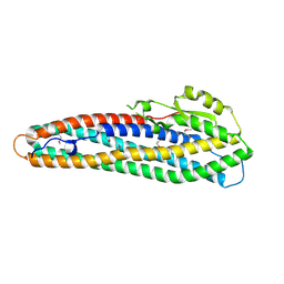
Crystal Structure of Hemolysin binding component from Bacillus cereus
Descriptor: Hbl B protein
Authors:Madegowda, M, Eswaramoorthy, S, Burley, S.K, Swaminathan, S, New York SGX Research Center for Structural Genomics (NYSGXRC)
Deposit date:2006-11-02
Release date:2006-11-14
Last modified:2023-12-27
Method:X-RAY DIFFRACTION (2.03 Å)
Cite:X-ray crystal structure of the B component of Hemolysin BL from Bacillus cereus
Proteins, 71, 2008
2Q09
DownloadVisualize
BU of 2q09 by Molmil
Crystal structure of Imidazolonepropionase from environmental sample with bound inhibitor 3-(2,5-Dioxo-imidazolidin-4-yl)-propionic acid
Descriptor: 3-[(4S)-2,5-DIOXOIMIDAZOLIDIN-4-YL]PROPANOIC ACID, FE (III) ION, Imidazolonepropionase
Authors:Tyagi, R, Eswaramoorthy, S, Burley, S.K, Swaminathan, S, New York SGX Research Center for Structural Genomics (NYSGXRC)
Deposit date:2007-05-21
Release date:2007-06-05
Last modified:2023-11-15
Method:X-RAY DIFFRACTION (1.97 Å)
Cite:A common catalytic mechanism for proteins of the HutI family.
Biochemistry, 47, 2008
2NXO
DownloadVisualize
BU of 2nxo by Molmil
Crystal structure of protein SCO4506 from Streptomyces coelicolor, Pfam DUF178
Descriptor: Hypothetical protein SCO4506
Authors:Tyagi, R, Eswaramoorthy, S, Burley, S.K, Swaminathan, S, New York SGX Research Center for Structural Genomics (NYSGXRC)
Deposit date:2006-11-17
Release date:2006-12-19
Last modified:2023-12-27
Method:X-RAY DIFFRACTION (2.04 Å)
Cite:The crystal structure of a hypothetical protein SCO4506 (gene ID: Q9L0T8) from Streptomyces coelicolor to 2.04 Angstrom resolution
To be Published
2PLG
DownloadVisualize
BU of 2plg by Molmil
Crystal structure of T110839 protein from Synechococcus elongatus
Descriptor: Tll0839 protein
Authors:Madegowda, M, Eswaramoorthy, S, Burley, S.K, Swaminathan, S, New York SGX Research Center for Structural Genomics (NYSGXRC)
Deposit date:2007-04-19
Release date:2007-05-01
Last modified:2021-02-03
Method:X-RAY DIFFRACTION (2.6 Å)
Cite:Crystal structure of T110839 protein from Synechococcus elongatus.
To be Published
2POZ
DownloadVisualize
BU of 2poz by Molmil
Crystal structure of a putative dehydratase from Mesorhizobium loti
Descriptor: Putative dehydratase
Authors:Sugadev, R, Eswaramoorthy, S, Burley, S.K, Swaminathan, S, New York SGX Research Center for Structural Genomics (NYSGXRC)
Deposit date:2007-04-27
Release date:2007-05-15
Last modified:2021-02-03
Method:X-RAY DIFFRACTION (2.04 Å)
Cite:Crystal structure of a putative dehydratase from Mesorhizobium loti.
To be Published
2QVG
DownloadVisualize
BU of 2qvg by Molmil
The crystal structure of a two-component response regulator from Legionella pneumophila
Descriptor: Two component response regulator
Authors:Tyagi, R, Eswaramoorthy, S, Burley, S.K, Swaminathan, S, New York SGX Research Center for Structural Genomics (NYSGXRC)
Deposit date:2007-08-08
Release date:2007-08-28
Last modified:2024-10-09
Method:X-RAY DIFFRACTION (1.5 Å)
Cite:The crystal structure of a two-component response regulator from Legionella pneumophila.
To be Published
2PBE
DownloadVisualize
BU of 2pbe by Molmil
Crystal structure of an aminoglycoside 6-adenyltransferase from Bacillus subtilis
Descriptor: Aminoglycoside 6-adenylyltransferase
Authors:Tyagi, R, Eswaramoorthy, S, Burley, S.K, Swaminathan, S, New York SGX Research Center for Structural Genomics (NYSGXRC)
Deposit date:2007-03-28
Release date:2007-04-10
Last modified:2021-02-03
Method:X-RAY DIFFRACTION (2.65 Å)
Cite:The crystal structure of an aminoglycoside 6-adenyltransferase from Bacillus subtilis
To be Published
1CQV
DownloadVisualize
BU of 1cqv by Molmil
CRYSTAL STRUCTURE OF STAPHYLOCOCCAL ENTEROTOXIN C2 AT 100K CRYSTALLIZED AT PH 5.0
Descriptor: PROTEIN (STAPHYLOCOCCAL ENTEROTOXIN C2), ZINC ION
Authors:Kumaran, D, Swaminathan, S.
Deposit date:1999-08-11
Release date:1999-08-22
Last modified:2023-08-09
Method:X-RAY DIFFRACTION (2.06 Å)
Cite:Structure of staphylococcal enterotoxin C2 at various pH levels.
Acta Crystallogr.,Sect.D, 57, 2001
1RXD
DownloadVisualize
BU of 1rxd by Molmil
Crystal structure of human protein tyrosine phosphatase 4A1
Descriptor: protein tyrosine phosphatase type IVA, member 1; Protein tyrosine phosphatase IVA1
Authors:Sun, J.P, Fedorov, A.A, Almo, S.C, Zhang, Z.Y, Burley, S.K, New York SGX Research Center for Structural Genomics (NYSGXRC)
Deposit date:2003-12-18
Release date:2004-12-28
Last modified:2021-02-03
Method:X-RAY DIFFRACTION (1.9 Å)
Cite:Structural genomics of protein phosphatases.
J.STRUCT.FUNCT.GENOM., 8, 2007
4M51
DownloadVisualize
BU of 4m51 by Molmil
Crystal structure of amidohydrolase nis_0429 (ser145ala mutant) from nitratiruptor sp. sb155-2
Descriptor: 4-(2-HYDROXYETHYL)-1-PIPERAZINE ETHANESULFONIC ACID, Amidohydrolase family protein, BENZOIC ACID, ...
Authors:Patskovsky, Y, Toro, R, Gobble, A, Raushel, F.M, Almo, S.C, Enzyme Function Initiative (EFI)
Deposit date:2013-08-07
Release date:2013-09-04
Last modified:2023-09-20
Method:X-RAY DIFFRACTION (1.08 Å)
Cite:Deamination of 6-aminodeoxyfutalosine in menaquinone biosynthesis by distantly related enzymes.
Biochemistry, 52, 2013
1Z0H
DownloadVisualize
BU of 1z0h by Molmil
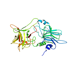
N-terminal helix reorients in recombinant C-fragment of Clostridium botulinum type B
Descriptor: Botulinum neurotoxin type B
Authors:Jayaraman, S, Eswarmoorthy, S, Ashraf, S.A, Smith, L.A, Swaminathan, S.
Deposit date:2005-03-01
Release date:2005-03-15
Last modified:2023-08-23
Method:X-RAY DIFFRACTION (2 Å)
Cite:N-terminal helix reorients in recombinant C-fragment of Clostridium botulinum type B.
Biochem.Biophys.Res.Commun., 330, 2005
2G59
DownloadVisualize
BU of 2g59 by Molmil
Crystal Structure of the Catalytic Domain of Protein Tyrosine Phosphatase from Homo sapiens
Descriptor: PHOSPHATE ION, Receptor-type tyrosine-protein phosphatase O
Authors:Kumaran, D, Swaminathan, S, Burley, S.K, New York SGX Research Center for Structural Genomics (NYSGXRC)
Deposit date:2006-02-22
Release date:2006-03-14
Last modified:2023-08-30
Method:X-RAY DIFFRACTION (2.19 Å)
Cite:Structural genomics of protein phosphatases.
J.Struct.Funct.Genom., 8, 2007
2FH7
DownloadVisualize
BU of 2fh7 by Molmil
Crystal structure of the phosphatase domains of human PTP SIGMA
Descriptor: Receptor-type tyrosine-protein phosphatase S
Authors:Alvarado, J, Udupi, R, Smith, D, Koss, J, Wasserman, S.R, Ozyurt, S, Atwell, S, Powell, A, Kearins, M.C, Rooney, I, Maletic, M, Bain, K.T, Freeman, J.C, Russell, M, Thompson, D.A, Sauder, J.M, Burley, S.K, Almo, S.C, New York SGX Research Center for Structural Genomics (NYSGXRC)
Deposit date:2005-12-23
Release date:2006-01-10
Last modified:2023-08-30
Method:X-RAY DIFFRACTION (2 Å)
Cite:Structural genomics of protein phosphatases.
J.STRUCT.FUNCT.GENOM., 8, 2007

226262

PDB entries from 2024-10-16

PDB statisticsPDBj update infoContact PDBjnumon