Loading
PDBj
MenuPDBj@FacebookPDBj@TwitterPDBj@YouTubewwPDB FoundationwwPDB
RCSB PDBPDBeBMRBAdv. SearchSearch help
Search by PDB author
1VJO
DownloadVisualize
BU of 1vjo by Molmil
Crystal structure of Alanine--glyoxylate aminotransferase (ALR1004) from Nostoc sp. at 1.70 A resolution
Descriptor: PYRIDOXAL-5'-PHOSPHATE, alanine--glyoxylate aminotransferase
Authors:Joint Center for Structural Genomics (JCSG)
Deposit date:2004-03-16
Release date:2004-03-23
Last modified:2023-01-25
Method:X-RAY DIFFRACTION (1.7 Å)
Cite:Crystal structure of an alanine-glyoxylate aminotransferase from Anabaena sp. at 1.70 A resolution reveals a noncovalently linked PLP cofactor
Proteins, 58, 2005
1VR3
DownloadVisualize
BU of 1vr3 by Molmil
Crystal structure of Acireductone dioxygenase (13543033) from Mus musculus at 2.06 A resolution
Descriptor: Acireductone dioxygenase, ISOPROPYL ALCOHOL, NICKEL (II) ION, ...
Authors:Joint Center for Structural Genomics (JCSG)
Deposit date:2005-01-28
Release date:2005-02-15
Last modified:2023-01-25
Method:X-RAY DIFFRACTION (2.06 Å)
Cite:Crystal structure of acireductone dioxygenase (ARD) from Mus musculus at 2.06 angstrom resolution.
Proteins, 64, 2006
1VRM
DownloadVisualize
BU of 1vrm by Molmil
Crystal structure of the apbe protein (tm1553) from thermotoga maritima msb8 at 1.58 A resolution
Descriptor: (4R)-2-METHYLPENTANE-2,4-DIOL, (4S)-2-METHYL-2,4-PENTANEDIOL, UNKNOWN LIGAND, ...
Authors:Joint Center for Structural Genomics (JCSG)
Deposit date:2005-03-10
Release date:2005-03-22
Last modified:2023-01-25
Method:X-RAY DIFFRACTION (1.58 Å)
Cite:Crystal structure of the ApbE protein (TM1553) from Thermotoga maritima at 1.58 A resolution.
Proteins, 64, 2006
1VQ0
DownloadVisualize
BU of 1vq0 by Molmil
Crystal structure of 33 kDa chaperonin (Heat shock protein 33 homolog) (HSP33) (TM1394) from Thermotoga maritima at 2.20 A resolution
Descriptor: 1,2-ETHANEDIOL, 33 kDa chaperonin, CHLORIDE ION, ...
Authors:Joint Center for Structural Genomics (JCSG)
Deposit date:2004-11-30
Release date:2004-12-14
Last modified:2023-09-20
Method:X-RAY DIFFRACTION (2.2 Å)
Cite:Crystal structure of Hsp33 chaperone (TM1394) from Thermotoga maritima at 2.20 A resolution.
Proteins, 61, 2005
1VJ1
DownloadVisualize
BU of 1vj1 by Molmil
Crystal structure of putative NADPH-dependent oxidoreductase from Mus musculus at 2.10 A resolution
Descriptor: putative NADPH-dependent oxidoreductase
Authors:Joint Center for Structural Genomics (JCSG)
Deposit date:2003-12-03
Release date:2003-12-09
Last modified:2023-01-25
Method:X-RAY DIFFRACTION (2.1 Å)
Cite:Crystal structure of a putative NADPH-dependent oxidoreductase (GI: 18204011) from mouse at 2.10 A resolution
Proteins, 56, 2004
1VPZ
DownloadVisualize
BU of 1vpz by Molmil
Crystal structure of a putative carbon storage regulator protein (csra, pa0905) from pseudomonas aeruginosa at 2.05 A resolution
Descriptor: Carbon storage regulator homolog
Authors:Joint Center for Structural Genomics (JCSG)
Deposit date:2004-11-24
Release date:2004-12-14
Last modified:2023-01-25
Method:X-RAY DIFFRACTION (2.05 Å)
Cite:Crystal structure of the global regulatory protein CsrA from Pseudomonas putida at 2.05 A resolution reveals a new fold.
Proteins, 61, 2005
1VQR
DownloadVisualize
BU of 1vqr by Molmil
Crystal structure of a virulence factor (cj0248) from campylobacter jejuni subsp. jejuni at 2.25 A resolution
Descriptor: hypothetical protein Cj0248
Authors:Joint Center for Structural Genomics (JCSG)
Deposit date:2004-12-17
Release date:2004-12-28
Last modified:2023-01-25
Method:X-RAY DIFFRACTION (2.25 Å)
Cite:Crystal structure of virulence factor CJ0248 from Campylobacter jejuni at 2.25 A resolution reveals a new fold.
Proteins, 62, 2006
1VR8
DownloadVisualize
BU of 1vr8 by Molmil
Crystal structure of GTP binding regulator (TM1622) from Thermotoga Maritima at 1.75 A resolution
Descriptor: AZIDE ION, GLYCEROL, GTP binding regulator
Authors:Joint Center for Structural Genomics (JCSG)
Deposit date:2005-02-16
Release date:2005-03-15
Last modified:2023-01-25
Method:X-RAY DIFFRACTION (1.75 Å)
Cite:Crystal structure of an ORFan protein (TM1622) from Thermotoga maritima at 1.75 A resolution reveals a fold similar to the Ran-binding protein Mog1p.
Proteins, 65, 2006
1VK3
DownloadVisualize
BU of 1vk3 by Molmil
Crystal structure of Phosphoribosylformylglycinamidine synthase II (TM1246) from Thermotoga maritima at 2.15 A resolution
Descriptor: CHLORIDE ION, Phosphoribosylformylglycinamidine synthase II
Authors:Joint Center for Structural Genomics (JCSG)
Deposit date:2004-04-23
Release date:2004-05-11
Last modified:2023-01-25
Method:X-RAY DIFFRACTION (2.15 Å)
Cite:Crystal structure of phosphoribosylformylglycinamidine synthase II (smPurL) from Thermotoga maritima at 2.15 A resolution.
Proteins, 63, 2006
1VL4
DownloadVisualize
BU of 1vl4 by Molmil
CRYSTAL STRUCTURE OF A PUTATIVE MODULATOR OF A DNA GYRASE (TM0727) FROM THERMOTOGA MARITIMA MSB8 AT 1.95 A RESOLUTION
Descriptor: pmbA-related protein
Authors:Joint Center for Structural Genomics (JCSG)
Deposit date:2004-07-09
Release date:2004-09-21
Last modified:2024-05-22
Method:X-RAY DIFFRACTION (1.95 Å)
Cite:Crystal structure of a putative modulator of DNA gyrase (pmbA) from Thermotoga maritima at 1.95 A resolution reveals a new fold.
Proteins, 61, 2005
1VKH
DownloadVisualize
BU of 1vkh by Molmil
CRYSTAL STRUCTURE OF A PUTATIVE SERINE HYDROLASE (YDR428C) FROM SACCHAROMYCES CEREVISIAE AT 1.85 A RESOLUTION
Descriptor: CHLORIDE ION, GLYCEROL, putative serine hydrolase
Authors:Joint Center for Structural Genomics (JCSG)
Deposit date:2004-05-20
Release date:2004-06-08
Last modified:2023-01-25
Method:X-RAY DIFFRACTION (1.85 Å)
Cite:Crystal structure of an alpha/beta serine hydrolase (YDR428C) from Saccharomyces cerevisiae at 1.85 A resolution
Proteins, 58, 2005
1ZCZ
DownloadVisualize
BU of 1zcz by Molmil
Crystal structure of Phosphoribosylaminoimidazolecarboxamide formyltransferase / IMP cyclohydrolase (TM1249) from THERMOTOGA MARITIMA at 1.88 A resolution
Descriptor: Bifunctional purine biosynthesis protein purH, POTASSIUM ION, TETRAETHYLENE GLYCOL
Authors:Joint Center for Structural Genomics (JCSG)
Deposit date:2005-04-13
Release date:2005-04-26
Last modified:2023-09-20
Method:X-RAY DIFFRACTION (1.88 Å)
Cite:Crystal structure of AICAR transformylase IMP cyclohydrolase (TM1249) from Thermotoga maritima at 1.88 A resolution.
Proteins, 71, 2008
1ZKG
DownloadVisualize
BU of 1zkg by Molmil
Crystal structure of Transcriptional regulator, TETR family (tm1030) from Thermotoga maritima at 2.30 A resolution
Descriptor: UNKNOWN LIGAND, transcriptional regulator, TetR family
Authors:Joint Center for Structural Genomics (JCSG)
Deposit date:2005-05-02
Release date:2005-07-26
Last modified:2023-01-25
Method:X-RAY DIFFRACTION (2.3 Å)
Cite:Crystal structure of TM1030 from Thermotoga maritima at 2.3 A resolution reveals molecular details of its transcription repressor function.
Proteins, 68, 2007
1Z9F
DownloadVisualize
BU of 1z9f by Molmil
Crystal structure of single stranded DNA-binding protein (TM0604) from Thermotoga maritima at 2.60 A resolution
Descriptor: Single-strand binding protein
Authors:Joint Center for Structural Genomics (JCSG)
Deposit date:2005-04-01
Release date:2005-04-12
Last modified:2023-09-20
Method:X-RAY DIFFRACTION (2.3 Å)
Cite:Crystal structure of a single-stranded DNA-binding protein (TM0604) from Thermotoga maritima at 2.60 A resolution.
Proteins, 63, 2006
1ZX8
DownloadVisualize
BU of 1zx8 by Molmil
CRYSTAL STRUCTURE OF an atypical cyclophilin (peptidylprolyl cis-trans isomerase) (TM1367) FROM THERMOTOGA MARITIMA AT 1.90 A RESOLUTION
Descriptor: NICKEL (II) ION, PENTAETHYLENE GLYCOL, hypothetical protein TM1367
Authors:Joint Center for Structural Genomics (JCSG)
Deposit date:2005-06-07
Release date:2005-07-26
Last modified:2023-01-25
Method:X-RAY DIFFRACTION (1.9 Å)
Cite:Crystal structure of TM1367 from Thermotoga maritima at 1.90 A resolution reveals an atypical member of the cyclophilin (peptidylprolyl isomerase) fold.
Proteins, 63, 2006
1HTM
DownloadVisualize
BU of 1htm by Molmil
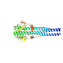
STRUCTURE OF INFLUENZA HAEMAGGLUTININ AT THE PH OF MEMBRANE FUSION
Descriptor: HEMAGGLUTININ HA1 CHAIN, HEMAGGLUTININ HA2 CHAIN
Authors:Bullough, P.A, Hughson, F.M, Skehel, J.J, Wiley, D.C.
Deposit date:1994-11-02
Release date:1995-02-14
Last modified:2024-06-05
Method:X-RAY DIFFRACTION (2.5 Å)
Cite:Structure of influenza haemagglutinin at the pH of membrane fusion.
Nature, 371, 1994
2GVI
DownloadVisualize
BU of 2gvi by Molmil
Crystal structure of a putative formylmethanofuran dehydrogenase subunit e (ta1109) from thermoplasma acidophilum at 1.87 A resolution
Descriptor: 1,2-ETHANEDIOL, ACETIC ACID, UNKNOWN LIGAND, ...
Authors:Joint Center for Structural Genomics (JCSG)
Deposit date:2006-05-02
Release date:2006-06-13
Last modified:2023-01-25
Method:X-RAY DIFFRACTION (1.87 Å)
Cite:Structures of three members of Pfam PF02663 (FmdE) implicated in microbial methanogenesis reveal a conserved alpha+beta core domain and an auxiliary C-terminal treble-clef zinc finger.
Acta Crystallogr.,Sect.F, 66, 2010
2IAY
DownloadVisualize
BU of 2iay by Molmil
Crystal structure of a duf1831 family protein (lp2179) from lactobacillus plantarum at 1.20 A resolution
Descriptor: CHLORIDE ION, GLYCEROL, Hypothetical protein
Authors:Joint Center for Structural Genomics (JCSG)
Deposit date:2006-09-08
Release date:2006-10-10
Last modified:2023-01-25
Method:X-RAY DIFFRACTION (1.2 Å)
Cite:Structure of LP2179, the first representative of Pfam family PF08866, suggests a new fold with a role in amino-acid metabolism.
Acta Crystallogr.,Sect.F, 66, 2010
2ICH
DownloadVisualize
BU of 2ich by Molmil
CRYSTAL STRUCTURE OF A PUTATIVE ATTH (NE1406) FROM NITROSOMONAS EUROPAEA AT 2.00 A RESOLUTION
Descriptor: 2-[N-CYCLOHEXYLAMINO]ETHANE SULFONIC ACID, GLYCEROL, Putative AttH, ...
Authors:Joint Center for Structural Genomics (JCSG)
Deposit date:2006-09-12
Release date:2006-10-03
Last modified:2023-01-25
Method:X-RAY DIFFRACTION (2 Å)
Cite:Structure of the first representative of Pfam family PF09410 (DUF2006) reveals a structural signature of the calycin superfamily that suggests a role in lipid metabolism.
Acta Crystallogr.,Sect.F, 66, 2010
2IIZ
DownloadVisualize
BU of 2iiz by Molmil
Crystal structure of putative melanin biosynthesis protein TyrA with bound heme (NP_716371.1) from Shewanella Oneidensis at 2.30 A resolution
Descriptor: 1,2-ETHANEDIOL, ISOPROPYL ALCOHOL, Melanin biosynthesis protein TyrA, ...
Authors:Joint Center for Structural Genomics (JCSG)
Deposit date:2006-09-28
Release date:2006-11-14
Last modified:2023-11-15
Method:X-RAY DIFFRACTION (2.3 Å)
Cite:Identification and structural characterization of heme binding in a novel dye-decolorizing peroxidase, TyrA.
Proteins, 69, 2007
2GAZ
DownloadVisualize
BU of 2gaz by Molmil
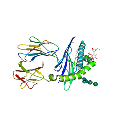
Mycobacterial lipoglycan presentation by CD1d
Descriptor: (2R)-3-[(HYDROXY{[(2R,3R,5S,6R)-3,4,5-TRIHYDROXY-2,6-BIS(ALPHA-D-MANNOPYRANOSYLOXY)CYCLOHEXYL]OXY}PHOSPHORYL)OXY]PROPAN E-1,2-DIYL DIHEXADECANOATE, 2-acetamido-2-deoxy-beta-D-glucopyranose, T-cell surface glycoprotein CD1d1, ...
Authors:Zajonc, D.M.
Deposit date:2006-03-09
Release date:2006-09-26
Last modified:2024-03-13
Method:X-RAY DIFFRACTION (2.61 Å)
Cite:Structural characterization of mycobacterial phosphatidylinositol mannoside binding to mouse CD1d.
J.Immunol., 177, 2006
2FNO
DownloadVisualize
BU of 2fno by Molmil
Crystal structure of a glutathione s-transferase (atu5508) from agrobacterium tumefaciens str. c58 at 2.00 A resolution
Descriptor: AGR_pAT_752p, THIOCYANATE ION
Authors:Joint Center for Structural Genomics (JCSG)
Deposit date:2006-01-11
Release date:2006-02-14
Last modified:2023-01-25
Method:X-RAY DIFFRACTION (2 Å)
Cite:Comparative structural analysis of a novel glutathioneS-transferase (ATU5508) from Agrobacterium tumefaciens at 2.0 A resolution.
Proteins, 65, 2006
2HAG
DownloadVisualize
BU of 2hag by Molmil
Crystal structure of a putative dyp-type peroxidase protein (so_0740) from shewanella oneidensis at 2.75 A resolution
Descriptor: Melanin biosynthesis protein TyrA, putative
Authors:Joint Center for Structural Genomics (JCSG)
Deposit date:2006-06-12
Release date:2006-08-08
Last modified:2023-01-25
Method:X-RAY DIFFRACTION (2.75 Å)
Cite:Crystal structures of two novel dye-decolorizing peroxidases reveal a beta-barrel fold with a conserved heme-binding motif.
Proteins, 69, 2007
2FO4
DownloadVisualize
BU of 2fo4 by Molmil
Enhanced MHC class I binding and immune responses through anchor modification of the non-canonical tumor associated MUC1-8 peptide
Descriptor: (4S)-2-METHYL-2,4-PENTANEDIOL, 2-acetamido-2-deoxy-beta-D-glucopyranose, 8-mer from Mucin-1, ...
Authors:Lazoura, E, Ramsland, P.A.
Deposit date:2006-01-12
Release date:2006-11-14
Last modified:2023-10-25
Method:X-RAY DIFFRACTION (2.7 Å)
Cite:Enhanced major histocompatibility complex class I binding and immune responses through anchor modification of the non-canonical tumour-associated mucin 1-8 peptide
Immunology, 119, 2006
2H1T
DownloadVisualize
BU of 2h1t by Molmil
Crystal structure of a duf1089 family protein (pa1994) from pseudomonas aeruginosa at 1.80 A resolution
Descriptor: (4S)-2-METHYL-2,4-PENTANEDIOL, 1,2-ETHANEDIOL, Hypothetical protein
Authors:Joint Center for Structural Genomics (JCSG)
Deposit date:2006-05-16
Release date:2006-05-30
Last modified:2023-01-25
Method:X-RAY DIFFRACTION (1.8 Å)
Cite:The structure of the first representative of Pfam family PF06475 reveals a new fold with possible involvement in glycolipid metabolism.
Acta Crystallogr.,Sect.F, 66, 2010

225158

PDB entries from 2024-09-18

PDB statisticsPDBj update infoContact PDBjnumon