Loading
PDBj
MenuPDBj@FacebookPDBj@TwitterPDBj@YouTubewwPDB FoundationwwPDB
RCSB PDBPDBeBMRBAdv. SearchSearch help
Search by PDB author
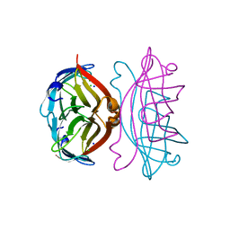
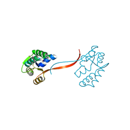
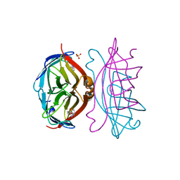
2IZF
DownloadVisualize
BU of 2izf by Molmil
STREPTAVIDIN-BIOTIN PH 4.0 I222 COMPLEX
Descriptor: BIOTIN, STREPTAVIDIN, SULFATE ION
Authors:Katz, B.A.
Deposit date:1997-08-13
Release date:1998-09-16
Last modified:2024-02-21
Method:X-RAY DIFFRACTION (1.58 Å)
Cite:Binding of biotin to streptavidin stabilizes intersubunit salt bridges between Asp61 and His87 at low pH.
J.Mol.Biol., 274, 1997
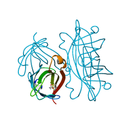
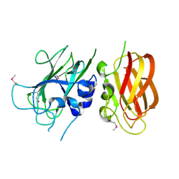
2IZC
DownloadVisualize
BU of 2izc by Molmil
APOSTREPTAVIDIN PH 2.0 I222 COMPLEX
Descriptor: CHLORIDE ION, SODIUM ION, STREPTAVIDIN
Authors:Katz, B.A.
Deposit date:1997-08-13
Release date:1998-09-16
Last modified:2024-02-21
Method:X-RAY DIFFRACTION (1.4 Å)
Cite:Binding of biotin to streptavidin stabilizes intersubunit salt bridges between Asp61 and His87 at low pH.
J.Mol.Biol., 274, 1997
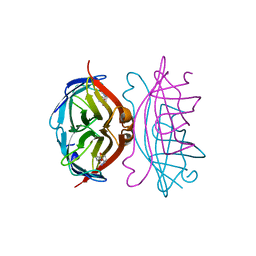
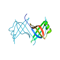
2IZI
DownloadVisualize
BU of 2izi by Molmil
STREPTAVIDIN-BIOTIN PH 2.53 I4122 STRUCTURE
Descriptor: BIOTIN, STREPTAVIDIN
Authors:Katz, B.A.
Deposit date:1997-08-13
Release date:1998-09-16
Last modified:2024-02-21
Method:X-RAY DIFFRACTION (1.5 Å)
Cite:Binding of biotin to streptavidin stabilizes intersubunit salt bridges between Asp61 and His87 at low pH.
J.Mol.Biol., 274, 1997
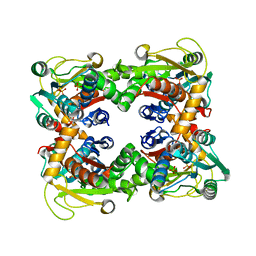
2IZH
DownloadVisualize
BU of 2izh by Molmil
STREPTAVIDIN-BIOTIN PH 10.44 I222 COMPLEX
Descriptor: BIOTIN, STREPTAVIDIN
Authors:Katz, B.A.
Deposit date:1997-08-13
Release date:1998-09-16
Last modified:2024-02-21
Method:X-RAY DIFFRACTION (1.36 Å)
Cite:Binding of biotin to streptavidin stabilizes intersubunit salt bridges between Asp61 and His87 at low pH.
J.Mol.Biol., 274, 1997
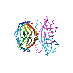
2IZG
DownloadVisualize
BU of 2izg by Molmil
STREPTAVIDIN-BIOTIN PH 2.0 I222 COMPLEX
Descriptor: BIOTIN, STREPTAVIDIN, SULFATE ION
Authors:Katz, B.A.
Deposit date:1997-08-13
Release date:1998-09-16
Last modified:2024-02-21
Method:X-RAY DIFFRACTION (1.36 Å)
Cite:Binding of biotin to streptavidin stabilizes intersubunit salt bridges between Asp61 and His87 at low pH.
J.Mol.Biol., 274, 1997
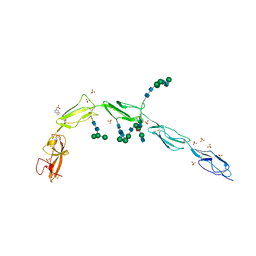
6V06
DownloadVisualize
BU of 6v06 by Molmil
Crystal structure of Beta-2 glycoprotein I purified from plasma (pB2GPI)
Descriptor: 2-acetamido-2-deoxy-beta-D-glucopyranose-(1-2)-beta-D-mannopyranose-(1-6)-beta-D-mannopyranose-(1-4)-2-acetamido-2-deoxy-beta-D-glucopyranose-(1-4)-2-acetamido-2-deoxy-beta-D-glucopyranose, 4-(2-HYDROXYETHYL)-1-PIPERAZINE ETHANESULFONIC ACID, Beta-2-glycoprotein 1, ...
Authors:Chen, Z, Ruben, E.A, Planer, W, Chinnaraj, M, Zuo, X, Pengo, V, Macor, P, Tedesco, F, Pozzi, N.
Deposit date:2019-11-18
Release date:2020-06-17
Last modified:2024-11-06
Method:X-RAY DIFFRACTION (2.4 Å)
Cite:The J-elongated conformation of beta2-glycoprotein I predominates in solution: implications for our understanding of antiphospholipid syndrome.
J.Biol.Chem., 295, 2020
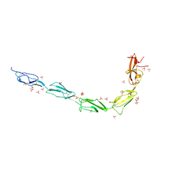
6V09
DownloadVisualize
BU of 6v09 by Molmil
Crystal structure of human recombinant Beta-2 glycoprotein I short tag (ST-B2GPI)
Descriptor: 2-acetamido-2-deoxy-beta-D-glucopyranose, Beta-2-glycoprotein 1, SULFATE ION, ...
Authors:Chen, Z, Ruben, E.A, Planer, W, Chinnaraj, M, Zuo, X, Pengo, V, Macor, P, Tedesco, F, Pozzi, N.
Deposit date:2019-11-18
Release date:2020-06-17
Last modified:2023-10-11
Method:X-RAY DIFFRACTION (2.99 Å)
Cite:The J-elongated conformation of beta2-glycoprotein I predominates in solution: implications for our understanding of antiphospholipid syndrome.
J.Biol.Chem., 295, 2020
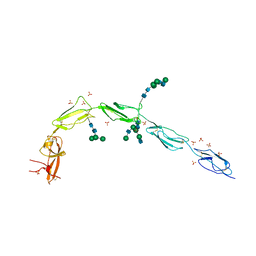
6V08
DownloadVisualize
BU of 6v08 by Molmil
Crystal structure of human recombinant Beta-2 glycoprotein I (hrB2GPI)
Descriptor: 2-acetamido-2-deoxy-beta-D-glucopyranose-(1-4)-2-acetamido-2-deoxy-beta-D-glucopyranose, Beta-2-glycoprotein 1, SULFATE ION, ...
Authors:Chen, Z, Ruben, E.A, Planer, W, Chinnaraj, M, Zuo, X, Pengo, V, Macor, P, Tedesco, F, Pozzi, N.
Deposit date:2019-11-18
Release date:2020-06-17
Last modified:2023-10-11
Method:X-RAY DIFFRACTION (2.58 Å)
Cite:The J-elongated conformation of beta2-glycoprotein I predominates in solution: implications for our understanding of antiphospholipid syndrome.
J.Biol.Chem., 295, 2020
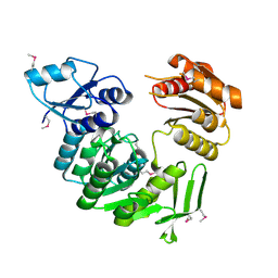
1J6U
DownloadVisualize
BU of 1j6u by Molmil
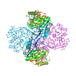
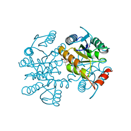
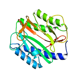
Crystal structure of UDP-N-acetylmuramate-alanine ligase MurC (TM0231) from Thermotoga maritima at 2.3 A resolution
Descriptor: UDP-N-acetylmuramate-alanine ligase MurC
Authors:Joint Center for Structural Genomics (JCSG)
Deposit date:2002-08-29
Release date:2002-11-06
Last modified:2023-01-25
Method:X-RAY DIFFRACTION (2.3 Å)
Cite:Crystal structure of an Udp-n-acetylmuramate-alanine ligase MurC (TM0231) from Thermotoga maritima at 2.3 A resolution.
Proteins, 55, 2004
1ZKG
DownloadVisualize
BU of 1zkg by Molmil
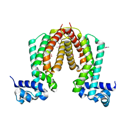
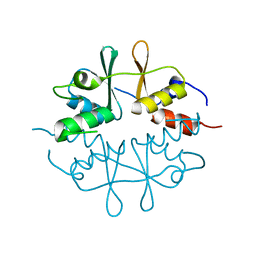
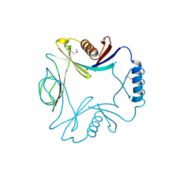
Crystal structure of Transcriptional regulator, TETR family (tm1030) from Thermotoga maritima at 2.30 A resolution
Descriptor: UNKNOWN LIGAND, transcriptional regulator, TetR family
Authors:Joint Center for Structural Genomics (JCSG)
Deposit date:2005-05-02
Release date:2005-07-26
Last modified:2024-10-30
Method:X-RAY DIFFRACTION (2.3 Å)
Cite:Crystal structure of TM1030 from Thermotoga maritima at 2.3 A resolution reveals molecular details of its transcription repressor function.
Proteins, 68, 2007
2AFB
DownloadVisualize
BU of 2afb by Molmil
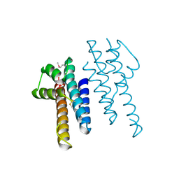
Crystal structure of 2-dehydro-3- deoxygluconokinase (EC 2.7.1.45) (tm0067) from THERMOTOGA MARITIMA at 2.05 A resolution
Descriptor: 2-keto-3-deoxygluconate kinase, CALCIUM ION, NICKEL (II) ION
Authors:Joint Center for Structural Genomics (JCSG)
Deposit date:2005-07-25
Release date:2006-02-14
Last modified:2024-10-16
Method:X-RAY DIFFRACTION (2.05 Å)
Cite:Crystal structure of 2-keto-3-deoxygluconate kinase (TM0067) from Thermotoga maritima at 2.05 A resolution.
Proteins, 70, 2007
1ZX8
DownloadVisualize
BU of 1zx8 by Molmil
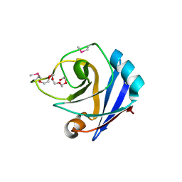
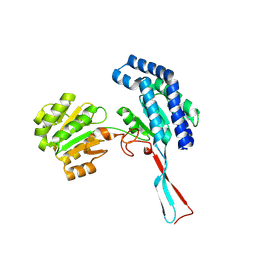
CRYSTAL STRUCTURE OF an atypical cyclophilin (peptidylprolyl cis-trans isomerase) (TM1367) FROM THERMOTOGA MARITIMA AT 1.90 A RESOLUTION
Descriptor: NICKEL (II) ION, PENTAETHYLENE GLYCOL, hypothetical protein TM1367
Authors:Joint Center for Structural Genomics (JCSG)
Deposit date:2005-06-07
Release date:2005-07-26
Last modified:2024-10-30
Method:X-RAY DIFFRACTION (1.9 Å)
Cite:Crystal structure of TM1367 from Thermotoga maritima at 1.90 A resolution reveals an atypical member of the cyclophilin (peptidylprolyl isomerase) fold.
Proteins, 63, 2006
1O5U
DownloadVisualize
BU of 1o5u by Molmil
Crystal structure of a duf861 family protein (tm1112) from thermotoga maritima at 1.83 A resolution
Descriptor: UNKNOWN LIGAND, novel Thermotoga maritima enzyme TM1112
Authors:Joint Center for Structural Genomics (JCSG)
Deposit date:2003-10-06
Release date:2003-11-11
Last modified:2023-09-20
Method:X-RAY DIFFRACTION (1.83 Å)
Cite:Crystal structure of a novel Thermotoga maritima enzyme (TM1112) from the cupin family at 1.83 A resolution
Proteins, 56, 2004
1O4W
DownloadVisualize
BU of 1o4w by Molmil
CRYSTAL STRUCTURE OF a PIN (PILT N-TERMINUS) DOMAIN CONTAINING PROTEIN (AF0591) FROM ARCHAEOGLOBUS FULGIDUS AT 1.90 A RESOLUTION
Descriptor: PIN (PilT N-terminus) domain
Authors:Joint Center for Structural Genomics (JCSG)
Deposit date:2003-07-16
Release date:2003-07-29
Last modified:2024-11-06
Method:X-RAY DIFFRACTION (1.9 Å)
Cite:Crystal structure of a PIN (PilT N-terminus) domain (AF0591) from Archaeoglobus fulgidus at 1.90 A resolution
Proteins, 56, 2004
1O59
DownloadVisualize
BU of 1o59 by Molmil
Crystal structure of Allantoicase (yir029w) from Saccharomyces cerevisiae at 2.40 A resolution
Descriptor: Allantoicase
Authors:Joint Center for Structural Genomics (JCSG)
Deposit date:2003-08-22
Release date:2003-09-02
Last modified:2024-10-30
Method:X-RAY DIFFRACTION (2.4 Å)
Cite:Crystal structure of an allantoicase (YIR029W) from Saccharomyces cerevisiae at 2.4 A resolution
Proteins, 56, 2004
1Z9F
DownloadVisualize
BU of 1z9f by Molmil
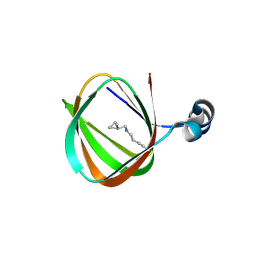
Crystal structure of single stranded DNA-binding protein (TM0604) from Thermotoga maritima at 2.60 A resolution
Descriptor: Single-strand binding protein
Authors:Joint Center for Structural Genomics (JCSG)
Deposit date:2005-04-01
Release date:2005-04-12
Last modified:2023-09-20
Method:X-RAY DIFFRACTION (2.3 Å)
Cite:Crystal structure of a single-stranded DNA-binding protein (TM0604) from Thermotoga maritima at 2.60 A resolution.
Proteins, 63, 2006
1O58
DownloadVisualize
BU of 1o58 by Molmil
Crystal structure of O-acetylserine sulfhydrylase (TM0665) from Thermotoga maritima at 1.80 A resolution
Descriptor: O-acetylserine sulfhydrylase, PHOSPHATE ION
Authors:Joint Center for Structural Genomics (JCSG)
Deposit date:2003-08-20
Release date:2003-09-02
Last modified:2024-05-22
Method:X-RAY DIFFRACTION (1.8 Å)
Cite:Crystal structure of O-acetylserine sulfhydrylase (TM0665) from Thermotoga maritima at 1.8 A resolution
Proteins, 56, 2004
1O1X
DownloadVisualize
BU of 1o1x by Molmil
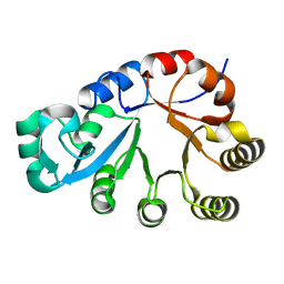
Crystal structure of a ribose 5-phosphate isomerase rpib (tm1080) from thermotoga maritima at 1.90 A resolution
Descriptor: (4S)-2-METHYL-2,4-PENTANEDIOL, ribose-5-phosphate isomerase RpiB
Authors:Joint Center for Structural Genomics (JCSG)
Deposit date:2003-02-12
Release date:2003-04-01
Last modified:2023-01-25
Method:X-RAY DIFFRACTION (1.9 Å)
Cite:Crystal structure of a ribose-5-phosphate isomerase RpiB (TM1080) from Thermotoga maritima at 1.90 A resolution.
Proteins, 56, 2004
1O50
DownloadVisualize
BU of 1o50 by Molmil
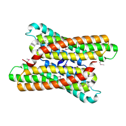
Crystal structure of a cbs domain-containing protein (tm0935) from thermotoga maritima at 1.87 A resolution
Descriptor: CBS domain-containing predicted protein TM0935
Authors:Joint Center for Structural Genomics (JCSG)
Deposit date:2003-07-31
Release date:2003-08-12
Last modified:2024-05-22
Method:X-RAY DIFFRACTION (1.87 Å)
Cite:Crystal structure of a tandem cystathionine-beta-synthase (CBS) domain protein (TM0935) from Thermotoga maritima at 1.87 A resolution
Proteins, 57, 2004
1O5H
DownloadVisualize
BU of 1o5h by Molmil
Crystal structure of formiminotetrahydrofolate cyclodeaminase (TM1560) from Thermotoga maritima at 2.80 A resolution
Descriptor: Formiminotetrahydrofolate cyclodeaminase
Authors:Joint Center for Structural Genomics (JCSG)
Deposit date:2003-09-17
Release date:2003-09-30
Last modified:2024-10-30
Method:X-RAY DIFFRACTION (2.8 Å)
Cite:Crystal structure of a formiminotetrahydrofolate cyclodeaminase (TM1560) from Thermotoga maritima at 2.80 A resolution reveals a new fold
Proteins, 58, 2005
1O1Z
DownloadVisualize
BU of 1o1z by Molmil
Crystal structure of glycerophosphodiester phosphodiesterase (GDPD) (TM1621) from Thermotoga maritima at 1.60 A resolution
Descriptor: SODIUM ION, glycerophosphodiester phosphodiesterase
Authors:Joint Center for Structural Genomics (JCSG)
Deposit date:2003-02-12
Release date:2003-04-01
Last modified:2024-05-22
Method:X-RAY DIFFRACTION (1.6 Å)
Cite:Crystal structure of a glycerophosphodiester phosphodiesterase (GDPD) from Thermotoga maritima (TM1621) at 1.60 A resolution.
Proteins, 56, 2004
1O0X
DownloadVisualize
BU of 1o0x by Molmil
Crystal structure of Methionine aminopeptidase (TM1478) from Thermotoga maritima at 1.90 A resolution
Descriptor: Methionine aminopeptidase
Authors:Joint Center for Structural Genomics (JCSG)
Deposit date:2002-09-13
Release date:2002-11-06
Last modified:2023-09-20
Method:X-RAY DIFFRACTION (1.9 Å)
Cite:Crystal structure of a methionine aminopeptidase (TM1478) from Thermotoga maritima at 1.9 A resolution.
Proteins, 56, 2004
1O22
DownloadVisualize
BU of 1o22 by Molmil
Crystal structure of an orphan protein (TM0875) from Thermotoga maritima at 2.00 A resolution
Descriptor: orphan protein TM0875
Authors:Joint Center for Structural Genomics (JCSG)
Deposit date:2003-02-15
Release date:2003-04-01
Last modified:2024-10-30
Method:X-RAY DIFFRACTION (2 Å)
Cite:Crystal structure of an orphan protein (TM0875) from Thermotoga maritima at 2.00-A resolution reveals a new fold.
Proteins, 56, 2004
1O3U
DownloadVisualize
BU of 1o3u by Molmil
Crystal structure of an hepn domain protein (tm0613) from thermotoga maritima at 1.75 A resolution
Descriptor: conserved hypothetical protein TM0613
Authors:Joint Center for Structural Genomics (JCSG)
Deposit date:2003-03-28
Release date:2003-04-22
Last modified:2024-10-30
Method:X-RAY DIFFRACTION (1.75 Å)
Cite:Crystal structure of an HEPN domain protein (TM0613) from Thermotoga maritima at 1.75 A resolution.
PROTEINS, 54, 2004
1O20
DownloadVisualize
BU of 1o20 by Molmil
Crystal structure of Gamma-glutamyl phosphate reductase (TM0293) from Thermotoga maritima at 2.00 A resolution
Descriptor: Gamma-glutamyl phosphate reductase
Authors:Joint Center for Structural Genomics (JCSG)
Deposit date:2003-02-12
Release date:2003-04-01
Last modified:2023-01-25
Method:X-RAY DIFFRACTION (2 Å)
Cite:Crystal structure of gamma-glutamyl phosphate reductase (TM0293) from Thermotoga maritima at 2.0 A resolution.
Proteins, 54, 2004

227111

PDB entries from 2024-11-06

PDB statisticsPDBj update infoContact PDBjnumon