7K46
 
 | |
7KDN
 | |
5B8I
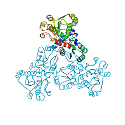
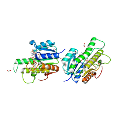
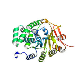
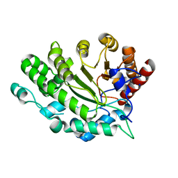
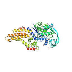
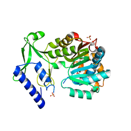
 | | Crystal structure of Calcineurin A and Calcineurin B in complex with FKBP12 and FK506 from Coccidioides immitis RS | | Descriptor: | 1,2-ETHANEDIOL, 2-(N-MORPHOLINO)-ETHANESULFONIC ACID, 8-DEETHYL-8-[BUT-3-ENYL]-ASCOMYCIN, ... | | Authors: | Seattle Structural Genomics Center for Infectious Disease (SSGCID), Fox III, D, Dranow, D.M, Lorimer, D.D, Edwards, T.E. | | Deposit date: | 2015-05-03 | | Release date: | 2015-05-20 | | Last modified: | 2023-09-27 | | Method: | X-RAY DIFFRACTION (1.85 Å) | | Cite: | Harnessing calcineurin-FK506-FKBP12 crystal structures from invasive fungal pathogens to develop antifungal agents.
Nat Commun, 10, 2019
|
|
6TZ8
 | |
3KC6
 | |
4Z0T
 | |
4Z04
 | |
3KHW
 | |
5UZX
 | |
6UK3
 | |
6UJ5
 | |
6UJK
 | |
6ULO
 | |
5UNL
 | |
4ZR8
 | |
4ZN6
 | |
4EXQ
 | |
6ULD
 | |
6TYJ
 | |
5UMH
 | |
5URB
 | |
6UCZ
 | |
6UDE
 | |
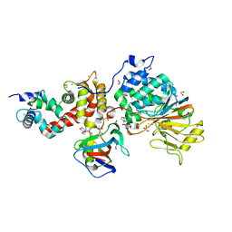
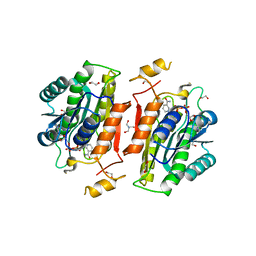
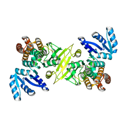
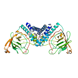
5V0W
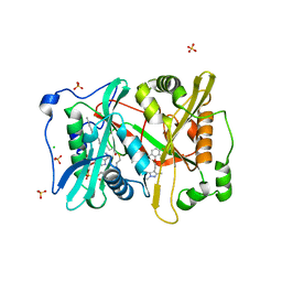 | | Crystal structure of glycylpeptide N-tetradecanoyltransferase from Plasmodium vivax in complex with inhibitor IMP-0001088 | | Descriptor: | 1-[5-[3,4-bis(fluoranyl)-2-[2-(1,3,5-trimethylpyrazol-4-yl)ethoxy]phenyl]-1-methyl-indazol-3-yl]-~{N},~{N}-dimethyl-methanamine, CHLORIDE ION, Glycylpeptide N-tetradecanoyltransferase, ... | | Authors: | Seattle Structural Genomics Center for Infectious Disease (SSGCID) | | Deposit date: | 2017-02-28 | | Release date: | 2018-03-07 | | Last modified: | 2025-01-15 | | Method: | X-RAY DIFFRACTION (1.8 Å) | | Cite: | Structure of Plasmodium vivax N-myristoyltransferase with inhibitor IMP-1088: exploring an NMT inhibitor for antimalarial therapy
Acta Crystallogr.,Sect.F, 81, 2025
|
|
4F3N
 | |


