Loading
PDBj
MenuPDBj@FacebookPDBj@TwitterPDBj@YouTubewwPDB FoundationwwPDB
RCSB PDBPDBeBMRBAdv. SearchSearch help
Search by PDB author
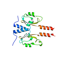
5WZ4
DownloadVisualize
BU of 5wz4 by Molmil
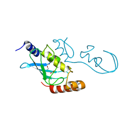
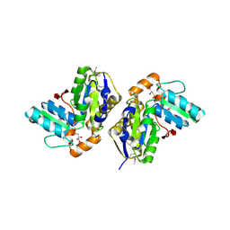
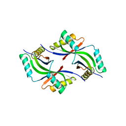
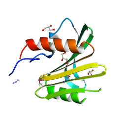
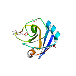
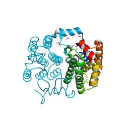
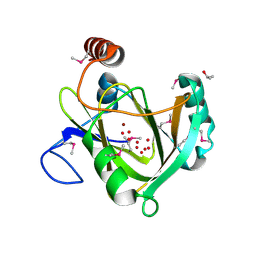
Crystal structure of Mycobacterium tuberculosis VapC20 (Rv2549c), Sarcin-Ricin loop cleaving toxin
Descriptor: 23S rRNA-specific endonuclease VapC20
Authors:Thakur, K.G, Deep, A.
Deposit date:2017-01-17
Release date:2017-10-25
Last modified:2024-10-16
Method:X-RAY DIFFRACTION (1.775 Å)
Cite:Crystal structure of Mycobacterium tuberculosis VapC20 toxin and its interactions with cognate antitoxin, VapB20, suggest a model for toxin-antitoxin assembly.
FEBS J., 284, 2017
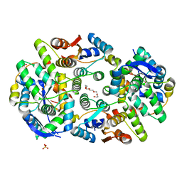
6F2P
DownloadVisualize
BU of 6f2p by Molmil
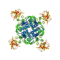
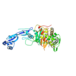
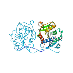
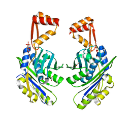
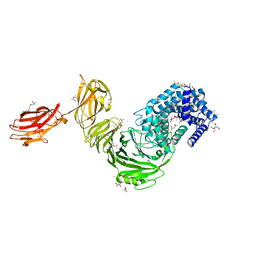
Structure of Paenibacillus xanthan lyase to 2.6 A resolution
Descriptor: (4R)-2-METHYLPENTANE-2,4-DIOL, (4S)-2-METHYL-2,4-PENTANEDIOL, 2-[BIS-(2-HYDROXY-ETHYL)-AMINO]-2-HYDROXYMETHYL-PROPANE-1,3-DIOL, ...
Authors:Lo Leggio, L, Kadziola, A.
Deposit date:2017-11-27
Release date:2018-12-05
Last modified:2024-01-17
Method:X-RAY DIFFRACTION (2.6 Å)
Cite:Structure and Dynamics of a Promiscuous Xanthan Lyase from Paenibacillus nanensis and the Design of Variants with Increased Stability and Activity.
Cell Chem Biol, 26, 2019
7YRT
DownloadVisualize
BU of 7yrt by Molmil
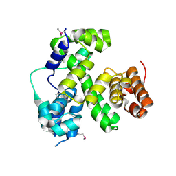
Crystal structure of PAS like domain of FlrB, the histidine kinase involved in flagellar synthesis of Vibrio cholerae
Descriptor: histidine kinase
Authors:Dasgupta, J, Mukherjee, P.
Deposit date:2022-08-10
Release date:2022-10-05
Last modified:2024-05-01
Method:X-RAY DIFFRACTION (2.27 Å)
Cite:PAS domain of flagellar histidine kinase FlrB has a unique architecture and binds heme as a sensory ligand in an unconventional fashion.
Structure, 32, 2024
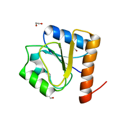
8H5V
DownloadVisualize
BU of 8h5v by Molmil
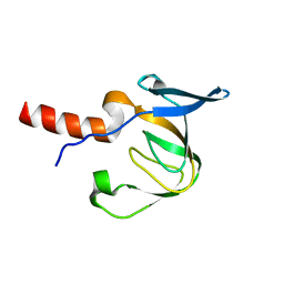
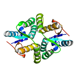
Crystal structure of the FleQ domain of Vibrio cholerae FlrA
Descriptor: 1,2-ETHANEDIOL, SULFATE ION, flagellar regulatory protein A
Authors:Dasgupta, J, Chakraborty, S.
Deposit date:2022-10-14
Release date:2022-11-02
Last modified:2024-05-15
Method:X-RAY DIFFRACTION (2 Å)
Cite:The N-terminal FleQ domain of the Vibrio cholerae flagellar master regulator FlrA plays pivotal structural roles in stabilizing its active state.
Febs Lett., 597, 2023
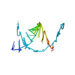
8SKQ
DownloadVisualize
BU of 8skq by Molmil
RNA oligonucleotide containing an alpha-(L)-threofuranosyl nucleic acid (TNA)
Descriptor: TNA-containing RNA oligonucleotide
Authors:Harp, J.M, Egli, M.
Deposit date:2023-04-20
Release date:2024-02-14
Method:X-RAY DIFFRACTION (1.35 Å)
Cite:Shorter Is Better: The alpha-(l)-Threofuranosyl Nucleic Acid Modification Improves Stability, Potency, Safety, and Ago2 Binding and Mitigates Off-Target Effects of Small Interfering RNAs.
J.Am.Chem.Soc., 145, 2023
6A7V
DownloadVisualize
BU of 6a7v by Molmil
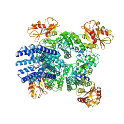
Crystal structure of Mycobacterium tuberculosis VapBC11 toxin-antitoxin complex
Descriptor: Antitoxin VapB11, PENTAETHYLENE GLYCOL, Ribonuclease VapC11, ...
Authors:Deep, A, Thakur, K.G.
Deposit date:2018-07-04
Release date:2018-10-10
Last modified:2023-11-22
Method:X-RAY DIFFRACTION (1.67 Å)
Cite:Structural, functional and biological insights into the role of Mycobacterium tuberculosis VapBC11 toxin-antitoxin system: targeting a tRNase to tackle mycobacterial adaptation.
Nucleic Acids Res., 46, 2018
5YWR
DownloadVisualize
BU of 5ywr by Molmil
Crystal Structure of RING E3 ligase ZNRF1 in complex with Ube2N (Ubc13)
Descriptor: E3 ubiquitin-protein ligase ZNRF1, FORMIC ACID, TRIETHYLENE GLYCOL, ...
Authors:Behera, A.P, Naskar, P, Datta, A.B.
Deposit date:2017-11-30
Release date:2018-06-06
Last modified:2024-03-27
Method:X-RAY DIFFRACTION (1.47 Å)
Cite:Structural insights into the nanomolar affinity of RING E3 ligase ZNRF1 for Ube2N and its functional implications.
Biochem. J., 475, 2018
5BKI
DownloadVisualize
BU of 5bki by Molmil
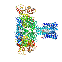
Blocker-free closed MthK channel in nanodisc
Descriptor: (1R)-2-{[(S)-{[(2S)-2,3-dihydroxypropyl]oxy}(hydroxy)phosphoryl]oxy}-1-[(hexadecanoyloxy)methyl]ethyl (9Z)-octadec-9-enoate, CADMIUM ION, Calcium-gated potassium channel MthK, ...
Authors:Fan, C, Nimigean, C.M.
Deposit date:2021-03-19
Release date:2022-09-21
Last modified:2023-10-04
Method:ELECTRON MICROSCOPY (3.1 Å)
Cite:Calcium-gated potassium channel blockade via membrane-facing fenestrations.
Nat.Chem.Biol., 2023
5BKK
DownloadVisualize
BU of 5bkk by Molmil
bbTBA-bound closed MthK channel in nanodisc
Descriptor: (1R)-2-{[(S)-{[(2S)-2,3-dihydroxypropyl]oxy}(hydroxy)phosphoryl]oxy}-1-[(hexadecanoyloxy)methyl]ethyl (9Z)-octadec-9-enoate, (~{R})-phenyl-[4-[(tributyl-$l^{4}-azanyl)methyl]phenyl]methanol, Calcium-gated potassium channel MthK, ...
Authors:Fan, C, Nimigean, C.M.
Deposit date:2021-03-19
Release date:2022-09-21
Last modified:2023-10-04
Method:ELECTRON MICROSCOPY (3.5 Å)
Cite:Calcium-gated potassium channel blockade via membrane-facing fenestrations.
Nat.Chem.Biol., 2023
5BKJ
DownloadVisualize
BU of 5bkj by Molmil
TPeA-bound closed MthK channel in nanodisc
Descriptor: (1R)-2-{[(S)-{[(2S)-2,3-dihydroxypropyl]oxy}(hydroxy)phosphoryl]oxy}-1-[(hexadecanoyloxy)methyl]ethyl (9Z)-octadec-9-enoate, 1-(tripentyl-$l^{4}-azanyl)pentane, Calcium-gated potassium channel MthK, ...
Authors:Fan, C, Nimigean, C.M.
Deposit date:2021-03-19
Release date:2022-09-21
Last modified:2023-10-04
Method:ELECTRON MICROSCOPY (3.5 Å)
Cite:Calcium-gated potassium channel blockade via membrane-facing fenestrations.
Nat.Chem.Biol., 2023
7LY1
DownloadVisualize
BU of 7ly1 by Molmil
Crystal structure of Pseudomonas aeruginosa PBP3 in complex with vaborbactam
Descriptor: Peptidoglycan D,D-transpeptidase FtsI, Vaborbactam
Authors:van den Akker, F, Kumar, V.
Deposit date:2021-03-05
Release date:2021-09-01
Last modified:2023-10-18
Method:X-RAY DIFFRACTION (2.2 Å)
Cite:Structural analysis of the boronic acid beta-lactamase inhibitor vaborbactam binding to Pseudomonas aeruginosa penicillin-binding protein 3.
Plos One, 16, 2021
2HC8
DownloadVisualize
BU of 2hc8 by Molmil
Structure of the A. fulgidus CopA A-domain
Descriptor: Cation-transporting ATPase, P-type
Authors:Sazinsky, M.H, Agawal, S, Arguello, J.M, Rosenzweig, A.C.
Deposit date:2006-06-15
Release date:2006-09-05
Last modified:2024-02-14
Method:X-RAY DIFFRACTION (1.65 Å)
Cite:Structure of the Actuator Domain from the Archaeoglobus fulgidus Cu(+)-ATPase(,).
Biochemistry, 45, 2006
2FEA
DownloadVisualize
BU of 2fea by Molmil
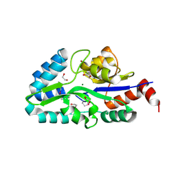
Crystal structure of MtnX phosphatase from Bacillus Subtilis at 2.00 A resolution
Descriptor: 1,2-ETHANEDIOL, 2-hydroxy-3-keto-5-methylthiopentenyl-1-phosphate phosphatase, MAGNESIUM ION, ...
Authors:Joint Center for Structural Genomics (JCSG)
Deposit date:2005-12-15
Release date:2005-12-27
Last modified:2023-01-25
Method:X-RAY DIFFRACTION (2 Å)
Cite:Crystal structure of MtnX phosphatase from Bacillus subtilis at 2.0 A resolution provides a structural basis for bipartite phosphomonoester hydrolysis of 2-hydroxy-3-keto-5-methylthiopentenyl-1-phosphate.
Proteins, 69, 2007
1VRM
DownloadVisualize
BU of 1vrm by Molmil
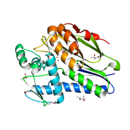
Crystal structure of the apbe protein (tm1553) from thermotoga maritima msb8 at 1.58 A resolution
Descriptor: (4R)-2-METHYLPENTANE-2,4-DIOL, (4S)-2-METHYL-2,4-PENTANEDIOL, UNKNOWN LIGAND, ...
Authors:Joint Center for Structural Genomics (JCSG)
Deposit date:2005-03-10
Release date:2005-03-22
Last modified:2023-01-25
Method:X-RAY DIFFRACTION (1.58 Å)
Cite:Crystal structure of the ApbE protein (TM1553) from Thermotoga maritima at 1.58 A resolution.
Proteins, 64, 2006
1VR0
DownloadVisualize
BU of 1vr0 by Molmil
Crystal structure of putative 2-phosphosulfolactate phosphatase (15026306) from Clostridium acetobutylicum at 2.6 A resolution
Descriptor: (2R)-3-SULFOLACTIC ACID, MAGNESIUM ION, Probable 2-phosphosulfolactate phosphatase
Authors:Joint Center for Structural Genomics (JCSG)
Deposit date:2005-01-26
Release date:2005-02-15
Last modified:2024-10-09
Method:X-RAY DIFFRACTION (2.49 Å)
Cite:Crystal structure of 2-phosphosulfolactate phosphatase (ComB) from Clostridium acetobutylicum at 2.6 A resolution reveals a new fold with a novel active site.
Proteins, 65, 2006
1VQ3
DownloadVisualize
BU of 1vq3 by Molmil
Crystal structure of Phosphoribosylformylglycinamidine synthase, purS subunit (EC 6.3.5.3) (TM1244) from Thermotoga maritima at 1.90 A resolution
Descriptor: Phosphoribosylformylglycinamidine synthase, purS subunit
Authors:Joint Center for Structural Genomics (JCSG)
Deposit date:2004-12-15
Release date:2004-12-28
Last modified:2023-09-20
Method:X-RAY DIFFRACTION (1.9 Å)
Cite:Crystal structure of phosphoribosylformyl-glycinamidine synthase II, PurS subunit (TM1244) from Thermotoga maritima at 1.90 A resolution.
Proteins, 65, 2006
1VR8
DownloadVisualize
BU of 1vr8 by Molmil
Crystal structure of GTP binding regulator (TM1622) from Thermotoga Maritima at 1.75 A resolution
Descriptor: AZIDE ION, GLYCEROL, GTP binding regulator
Authors:Joint Center for Structural Genomics (JCSG)
Deposit date:2005-02-16
Release date:2005-03-15
Last modified:2023-01-25
Method:X-RAY DIFFRACTION (1.75 Å)
Cite:Crystal structure of an ORFan protein (TM1622) from Thermotoga maritima at 1.75 A resolution reveals a fold similar to the Ran-binding protein Mog1p.
Proteins, 65, 2006
1ZX8
DownloadVisualize
BU of 1zx8 by Molmil
CRYSTAL STRUCTURE OF an atypical cyclophilin (peptidylprolyl cis-trans isomerase) (TM1367) FROM THERMOTOGA MARITIMA AT 1.90 A RESOLUTION
Descriptor: NICKEL (II) ION, PENTAETHYLENE GLYCOL, hypothetical protein TM1367
Authors:Joint Center for Structural Genomics (JCSG)
Deposit date:2005-06-07
Release date:2005-07-26
Last modified:2024-10-30
Method:X-RAY DIFFRACTION (1.9 Å)
Cite:Crystal structure of TM1367 from Thermotoga maritima at 1.90 A resolution reveals an atypical member of the cyclophilin (peptidylprolyl isomerase) fold.
Proteins, 63, 2006
2FNO
DownloadVisualize
BU of 2fno by Molmil
Crystal structure of a glutathione s-transferase (atu5508) from agrobacterium tumefaciens str. c58 at 2.00 A resolution
Descriptor: AGR_pAT_752p, THIOCYANATE ION
Authors:Joint Center for Structural Genomics (JCSG)
Deposit date:2006-01-11
Release date:2006-02-14
Last modified:2023-01-25
Method:X-RAY DIFFRACTION (2 Å)
Cite:Comparative structural analysis of a novel glutathioneS-transferase (ATU5508) from Agrobacterium tumefaciens at 2.0 A resolution.
Proteins, 65, 2006
2GHR
DownloadVisualize
BU of 2ghr by Molmil
Crystal structure of homoserine o-succinyltransferase (NP_981826.1) from Bacillus cereus ATCC 10987 at 2.40 A resolution
Descriptor: Homoserine O-succinyltransferase, SULFATE ION
Authors:Joint Center for Structural Genomics (JCSG)
Deposit date:2006-03-27
Release date:2006-04-11
Last modified:2023-01-25
Method:X-RAY DIFFRACTION (2.4 Å)
Cite:Crystal structure of homoserine O-succinyltransferase from Bacillus cereus at 2.4 A resolution
Proteins, 68, 2007
3N0C
DownloadVisualize
BU of 3n0c by Molmil
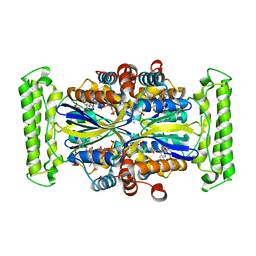
TM0449 mutant crystal grown by hanging drop method
Descriptor: 2'-DEOXYURIDINE 5'-MONOPHOSPHATE, FLAVIN-ADENINE DINUCLEOTIDE, Thymidylate synthase thyX
Authors:Mathews, I.I.
Deposit date:2010-05-13
Release date:2011-05-25
Last modified:2023-09-06
Method:X-RAY DIFFRACTION (2.3 Å)
Cite:Diffraction study of protein crystals grown in cryoloops and micromounts.
J.Appl.Crystallogr., 43, 2010
3N0B
DownloadVisualize
BU of 3n0b by Molmil
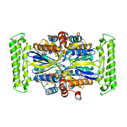
TM0449 mutant crystals grown in loops/micromounts
Descriptor: 2'-DEOXYURIDINE 5'-MONOPHOSPHATE, FLAVIN-ADENINE DINUCLEOTIDE, Thymidylate synthase thyX
Authors:Mathews, I.I.
Deposit date:2010-05-13
Release date:2011-05-25
Last modified:2023-09-06
Method:X-RAY DIFFRACTION (2.3 Å)
Cite:Diffraction study of protein crystals grown in cryoloops and micromounts.
J.Appl.Crystallogr., 43, 2010
1VR3
DownloadVisualize
BU of 1vr3 by Molmil
Crystal structure of Acireductone dioxygenase (13543033) from Mus musculus at 2.06 A resolution
Descriptor: Acireductone dioxygenase, ISOPROPYL ALCOHOL, NICKEL (II) ION, ...
Authors:Joint Center for Structural Genomics (JCSG)
Deposit date:2005-01-28
Release date:2005-02-15
Last modified:2023-01-25
Method:X-RAY DIFFRACTION (2.06 Å)
Cite:Crystal structure of acireductone dioxygenase (ARD) from Mus musculus at 2.06 angstrom resolution.
Proteins, 64, 2006
1VQ0
DownloadVisualize
BU of 1vq0 by Molmil
Crystal structure of 33 kDa chaperonin (Heat shock protein 33 homolog) (HSP33) (TM1394) from Thermotoga maritima at 2.20 A resolution
Descriptor: 1,2-ETHANEDIOL, 33 kDa chaperonin, CHLORIDE ION, ...
Authors:Joint Center for Structural Genomics (JCSG)
Deposit date:2004-11-30
Release date:2004-12-14
Last modified:2023-09-20
Method:X-RAY DIFFRACTION (2.2 Å)
Cite:Crystal structure of Hsp33 chaperone (TM1394) from Thermotoga maritima at 2.20 A resolution.
Proteins, 61, 2005
1VQR
DownloadVisualize
BU of 1vqr by Molmil
Crystal structure of a virulence factor (cj0248) from campylobacter jejuni subsp. jejuni at 2.25 A resolution
Descriptor: hypothetical protein Cj0248
Authors:Joint Center for Structural Genomics (JCSG)
Deposit date:2004-12-17
Release date:2004-12-28
Last modified:2024-10-09
Method:X-RAY DIFFRACTION (2.25 Å)
Cite:Crystal structure of virulence factor CJ0248 from Campylobacter jejuni at 2.25 A resolution reveals a new fold.
Proteins, 62, 2006

226707

PDB entries from 2024-10-30

PDB statisticsPDBj update infoContact PDBjnumon