Loading
PDBj
MenuPDBj@FacebookPDBj@TwitterPDBj@YouTubewwPDB FoundationwwPDB
RCSB PDBPDBeBMRBAdv. SearchSearch help
Search by PDB author
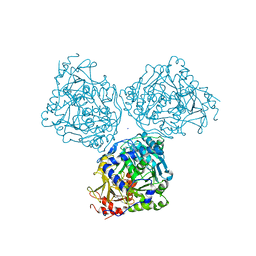
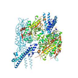
7KF3
DownloadVisualize
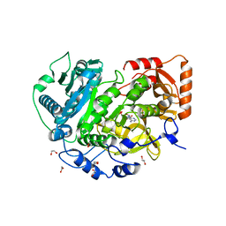
BU of 7kf3 by Molmil
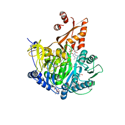
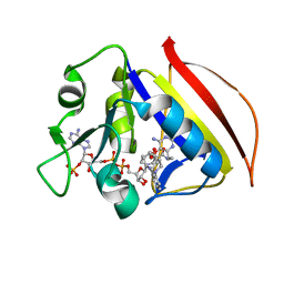
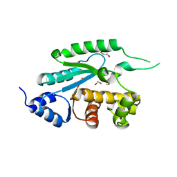
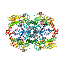
Crystal structure of GDP-mannose 4,6-dehydratase from Brucella abortus (strain 2308) in complex with Guanosine-diphosphate-rhamnose
Descriptor: GDP-mannose 4,6-dehydratase, GUANOSINE-5'-DIPHOSPHATE-RHAMNOSE, NADP NICOTINAMIDE-ADENINE-DINUCLEOTIDE PHOSPHATE
Authors:Seattle Structural Genomics Center for Infectious Disease (SSGCID)
Deposit date:2020-10-13
Release date:2020-11-04
Last modified:2023-10-18
Method:X-RAY DIFFRACTION (2.35 Å)
Cite:Crystal structure of GDP-mannose 4,6-dehydratase from Brucella abortus (strain 2308) in complex with Guanosine-diphosphate-rhamnose
to be published
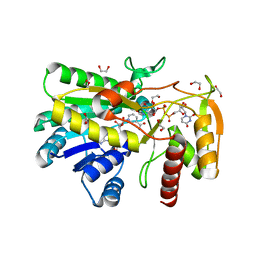
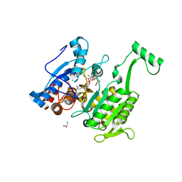
7L4G
DownloadVisualize
BU of 7l4g by Molmil
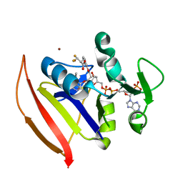
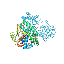
Crystal Structure of Acetyl-CoA synthetase in complex with acetyl adenylate from Cryptococcus neoformans H99
Descriptor: 1,2-ETHANEDIOL, Acetyl-coenzyme A synthetase, PHOSPHATE ION, ...
Authors:Seattle Structural Genomics Center for Infectious Disease (SSGCID)
Deposit date:2020-12-19
Release date:2020-12-30
Last modified:2023-10-18
Method:X-RAY DIFFRACTION (2.2 Å)
Cite:Crystal Structure of Acetyl-CoA synthetase in complex with acetyl adenylate from Cryptococcus neoformans H99
to be published
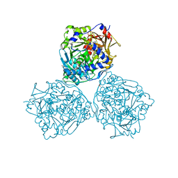
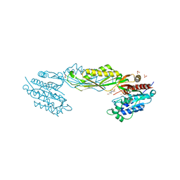
7KDS
DownloadVisualize
BU of 7kds by Molmil
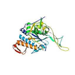
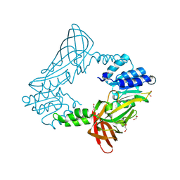
Crystal structure of Acetyl-CoA synthetase 2 in complex with Adenosine-5'-propylphosphate from Candida albicans
Descriptor: ADENOSINE-5'-MONOPHOSPHATE-PROPYL ESTER, Acetyl-coenzyme A synthetase 2, SODIUM ION
Authors:Seattle Structural Genomics Center for Infectious Disease (SSGCID)
Deposit date:2020-10-09
Release date:2020-11-11
Last modified:2023-10-18
Method:X-RAY DIFFRACTION (2.9 Å)
Cite:Crystal structure of Acetyl-CoA synthetase 2 in complex with Adenosine-5'-propylphosphate from Candida albicans
To Be Published
7KN1
DownloadVisualize
BU of 7kn1 by Molmil
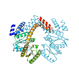
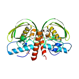
Crystal structure of UDP-glucose-4-epimerase (galE) from Stenotrophomonas maltophila with bound NAD and formylated UDP-arabinopyranose
Descriptor: 1,2-ETHANEDIOL, NICOTINAMIDE-ADENINE-DINUCLEOTIDE, UDP-4-deoxy-4-formamido-beta-L-arabinopyranose, ...
Authors:Seattle Structural Genomics Center for Infectious Disease (SSGCID)
Deposit date:2020-11-03
Release date:2020-11-11
Last modified:2023-10-18
Method:X-RAY DIFFRACTION (1.45 Å)
Cite:Crystal structure of UDP-glucose-4-epimerase (galE) from Stenotrophomonas maltophila with bound NAD and formylated UDP-arabinopyranose
To Be Published
7KQZ
DownloadVisualize
BU of 7kqz by Molmil
Crystal Structure of Acetyl-CoA synthetase in complex with adenosine-5'-ethylphosphate from Coccidioides immitis RS
Descriptor: 1,2-ETHANEDIOL, 5'-O-[(S)-ethoxy(hydroxy)phosphoryl]adenosine, Acetyl-coenzyme A synthetase
Authors:Seattle Structural Genomics Center for Infectious Disease (SSGCID)
Deposit date:2020-11-18
Release date:2020-12-16
Last modified:2023-10-18
Method:X-RAY DIFFRACTION (2.15 Å)
Cite:Crystal Structure of Acetyl-CoA synthetase in complex with adenosine-5'-ethylphosphate from Coccidioides immitis RS
to be published
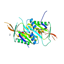
7L2A
DownloadVisualize
BU of 7l2a by Molmil
Molybdopterin cofactor biosynthesis protein E from Burkholderia multivorans ATCC 17616
Descriptor: 1,2-ETHANEDIOL, DIMETHYL SULFOXIDE, IMIDAZOLE, ...
Authors:Seattle Structural Genomics Center for Infectious Disease, Seattle Structural Genomics Center for Infectious Disease (SSGCID)
Deposit date:2020-12-16
Release date:2020-12-30
Last modified:2024-10-16
Method:X-RAY DIFFRACTION (1.85 Å)
Cite:Molybdopterin cofactor biosynthesis protein E from Burkholderia multivorans ATCC 17616
to be published
7L3Q
DownloadVisualize
BU of 7l3q by Molmil
Crystal Structure of Acetyl-CoA synthetase in complex with adenosine-5'-methylphosphate and Co-enzyme A from Coccidioides immitis RS
Descriptor: 1,2-ETHANEDIOL, 5'-O-[(R)-hydroxy(methoxy)phosphoryl]adenosine, Acetyl-coenzyme A synthetase, ...
Authors:Seattle Structural Genomics Center for Infectious Disease (SSGCID)
Deposit date:2020-12-18
Release date:2020-12-30
Last modified:2023-10-18
Method:X-RAY DIFFRACTION (2.15 Å)
Cite:Crystal Structure of Acetyl-CoA synthetase in complex with adenosine-5'-methylphosphate and Co-enzyme A from Coccidioides immitis RS
to be published
7KI9
DownloadVisualize
BU of 7ki9 by Molmil
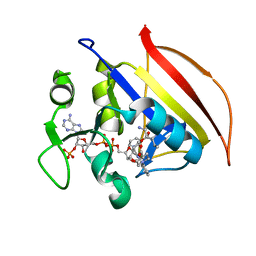
Crystal Structure of Dihydrofolate reductase (DHFR) from Mycobacterium ulcerans Agy99 in complex with NADP and inhibitor SDDC-0001912
Descriptor: 1-[2-(3-{[(2,4-diamino-6-ethylpyrimidin-5-yl)oxy]methyl}azetidin-1-yl)phenyl]piperidine-4-carboxylic acid, Dihydrofolate reductase, NADP NICOTINAMIDE-ADENINE-DINUCLEOTIDE PHOSPHATE
Authors:Seattle Structural Genomics Center for Infectious Disease (SSGCID)
Deposit date:2020-10-23
Release date:2021-07-07
Last modified:2023-10-18
Method:X-RAY DIFFRACTION (1.25 Å)
Cite:Crystal Structure of Dihydrofolate reductase (DHFR) from Mycobacterium ulcerans Agy99 in complex with NADP and inhibitor SDDC-0001912
to be published
7KM9
DownloadVisualize
BU of 7km9 by Molmil
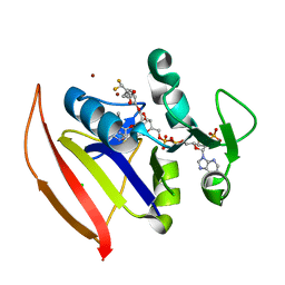
Crystal Structure of Dihydrofolate reductase (DHFR) from Mycobacterium ulcerans Agy99 in complex with NADP and inhibitor SDDC-0001913, tetragonal crystal from
Descriptor: (2R)-2-[(3-{3-[(2,4-diamino-6-ethylpyrimidin-5-yl)oxy]propoxy}phenyl)methyl]-3,3-difluoropropanoic acid, BROMIDE ION, CHLORIDE ION, ...
Authors:Seattle Structural Genomics Center for Infectious Disease (SSGCID)
Deposit date:2020-11-02
Release date:2021-07-07
Last modified:2023-10-18
Method:X-RAY DIFFRACTION (1.95 Å)
Cite:Crystal Structure of Dihydrofolate reductase (DHFR) from Mycobacterium ulcerans Agy99 in complex with NADP and inhibitor SDDC-0001913, tetragonal crystal form
to be published
7KM8
DownloadVisualize
BU of 7km8 by Molmil
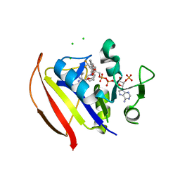
Crystal Structure of Dihydrofolate reductase (DHFR) from Mycobacterium ulcerans Agy99 in complex with NADP and inhibitor SDDC-0001914, orthorhombic crystal from
Descriptor: (2S)-2-[(3-{3-[(2,4-diamino-6-ethylpyrimidin-5-yl)oxy]propoxy}phenyl)methyl]-3,3-difluoropropanoic acid, CHLORIDE ION, Dihydrofolate reductase, ...
Authors:Seattle Structural Genomics Center for Infectious Disease (SSGCID)
Deposit date:2020-11-02
Release date:2021-07-07
Last modified:2023-10-18
Method:X-RAY DIFFRACTION (1.55 Å)
Cite:Crystal Structure of Dihydrofolate reductase (DHFR) from Mycobacterium ulcerans Agy99 in complex with NADP and inhibitor SDDC-0001914, orthorhombic crystal form
to be published
7KI8
DownloadVisualize
BU of 7ki8 by Molmil
Crystal Structure of Dihydrofolate reductase (DHFR) from Mycobacterium ulcerans Agy99 in complex with NADP and inhibitor SDDC-0001580
Descriptor: 1-[2-({3-[(2,4-diamino-6-ethylpyrimidin-5-yl)oxy]propyl}amino)phenyl]piperidine-4-carboxylic acid, Dihydrofolate reductase, NADP NICOTINAMIDE-ADENINE-DINUCLEOTIDE PHOSPHATE
Authors:Seattle Structural Genomics Center for Infectious Disease (SSGCID)
Deposit date:2020-10-23
Release date:2021-07-07
Last modified:2023-10-18
Method:X-RAY DIFFRACTION (1.25 Å)
Cite:Crystal Structure of Dihydrofolate reductase (DHFR) from Mycobacterium ulcerans Agy99 in complex with NADP and inhibitor SDDC-0001580
to be published
7KM7
DownloadVisualize
BU of 7km7 by Molmil
Crystal Structure of Dihydrofolate reductase (DHFR) from Mycobacterium ulcerans Agy99 in complex with NADP and inhibitor SDDC-0001914, tetragonal crystal from
Descriptor: (2S)-2-[(3-{3-[(2,4-diamino-6-ethylpyrimidin-5-yl)oxy]propoxy}phenyl)methyl]-3,3-difluoropropanoic acid, BROMIDE ION, Dihydrofolate reductase, ...
Authors:Seattle Structural Genomics Center for Infectious Disease (SSGCID)
Deposit date:2020-11-02
Release date:2021-07-07
Last modified:2023-10-18
Method:X-RAY DIFFRACTION (1.8 Å)
Cite:Crystal Structure of Dihydrofolate reductase (DHFR) from Mycobacterium ulcerans Agy99 in complex with NADP and inhibitor SDDC-0001914, tetragonal crystal form
to be published
3OC9
DownloadVisualize
BU of 3oc9 by Molmil
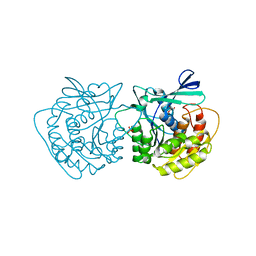
Crystal structure of putative UDP-N-acetylglucosamine pyrophosphorylase from Entamoeba histolytica
Descriptor: 1,2-ETHANEDIOL, SULFATE ION, UDP-N-acetylglucosamine pyrophosphorylase
Authors:Seattle Structural Genomics Center for Infectious Disease (SSGCID)
Deposit date:2010-08-09
Release date:2010-08-18
Last modified:2023-09-06
Method:X-RAY DIFFRACTION (1.8 Å)
Cite:Structure of uridine diphosphate N-acetylglucosamine pyrophosphorylase from Entamoeba histolytica.
Acta Crystallogr F Struct Biol Commun, 71, 2015
3P32
DownloadVisualize
BU of 3p32 by Molmil
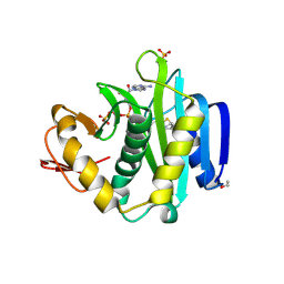
Hydrolysis of GTP to GDP by an MCM-associated and MeaB- and MMAA-like G-protein from Mycobacterium tuberculosis
Descriptor: GUANOSINE-5'-DIPHOSPHATE, Probable GTPase Rv1496/MT1543, TRIETHYLENE GLYCOL
Authors:Seattle Structural Genomics Center for Infectious Disease (SSGCID)
Deposit date:2010-10-04
Release date:2010-11-10
Last modified:2023-09-06
Method:X-RAY DIFFRACTION (1.9 Å)
Cite:Crystal structures of Mycobacterial MeaB and MMAA-like GTPases.
J.Struct.Funct.Genom., 16, 2015
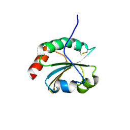
6BLJ
DownloadVisualize
BU of 6blj by Molmil
Crystal structure of cytoplasmic Serine-tRNA ligase from Naegleria fowleri in complex with AMP
Descriptor: 1,2-ETHANEDIOL, ADENOSINE MONOPHOSPHATE, Serine-tRNA ligase
Authors:Seattle Structural Genomics Center for Infectious Disease (SSGCID)
Deposit date:2017-11-10
Release date:2017-12-20
Last modified:2023-10-04
Method:X-RAY DIFFRACTION (2.1 Å)
Cite:Crystal structure of cytoplasmic Serine-tRNA ligase from Neisseria fowlerii in complex with AMP
TO BE PUBLISHED
6BKV
DownloadVisualize
BU of 6bkv by Molmil
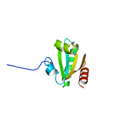
Crystal structure of Thioredoxin from Helicobacter pylori (strain G27)
Descriptor: Thioredoxin
Authors:Seattle Structural Genomics Center for Infectious Disease (SSGCID)
Deposit date:2017-11-09
Release date:2017-12-06
Last modified:2024-10-16
Method:X-RAY DIFFRACTION (2.35 Å)
Cite:Crystal structure of Thioredoxin from Helicobacter pylori (strain G27)
To be Published
6C0E
DownloadVisualize
BU of 6c0e by Molmil
Crystal Structure of Isocitrate Dehydrogenase from Legionella pneumophila with bound NADPH with an alpha-ketoglutarate adduct
Descriptor: (3~{S})-3-[(4~{S})-3-aminocarbonyl-1-[(2~{R},3~{R},4~{S},5~{R})-5-[[[[(2~{R},3~{R},4~{R},5~{R})-5-(6-aminopurin-9-yl)-3-oxidanyl-4-phosphonooxy-oxolan-2-yl]methoxy-oxidanyl-phosphoryl]oxy-oxidanyl-phosphoryl]oxymethyl]-3,4-bis(oxidanyl)oxolan-2-yl]-4~{H}-pyridin-4-yl]-2-oxidanylidene-pentanedioic acid, CHLORIDE ION, GLYCINE, ...
Authors:Seattle Structural Genomics Center for Infectious Disease, Seattle Structural Genomics Center for Infectious Disease (SSGCID)
Deposit date:2017-12-29
Release date:2018-02-07
Last modified:2023-10-04
Method:X-RAY DIFFRACTION (1.7 Å)
Cite:Crystal Structure of Isocitrate Dehydrogenase from Legionella pneumophila with bound NADPH with an ??-ketoglutarate adduct
to be published
6C0D
DownloadVisualize
BU of 6c0d by Molmil
Crystal structure of an Amidase (hydantoinase/carbamoylase family) from Burkholderia phymatum
Descriptor: 1,2-ETHANEDIOL, 4-(2-HYDROXYETHYL)-1-PIPERAZINE ETHANESULFONIC ACID, Amidase, ...
Authors:Seattle Structural Genomics Center for Infectious Disease (SSGCID)
Deposit date:2017-12-29
Release date:2018-03-21
Last modified:2024-04-03
Method:X-RAY DIFFRACTION (1.8 Å)
Cite:Crystal structure of an Amidase (hydantoinase/carbamoylase family) from Burkholderia phymatum
to be published
6BQY
DownloadVisualize
BU of 6bqy by Molmil
Crystal Structure of Tyrosine-tRNA Synthetase from Acinetobacter baumannii
Descriptor: 1,2-ETHANEDIOL, Tyrosine--tRNA ligase
Authors:Seattle Structural Genomics Center for Infectious Disease (SSGCID)
Deposit date:2017-11-29
Release date:2017-12-13
Last modified:2023-10-04
Method:X-RAY DIFFRACTION (2.1 Å)
Cite:Crystal Structure of Glutamate-tRNA Synthetase from Helicobacter pylori
To be Published
6BS7
DownloadVisualize
BU of 6bs7 by Molmil
Crystal structure of Adenylosuccinate synthetase from Legionella pneumophila Philadelphia 1
Descriptor: Adenylosuccinate synthetase
Authors:Seattle Structural Genomics Center for Infectious Disease (SSGCID)
Deposit date:2017-12-01
Release date:2018-01-17
Last modified:2023-10-04
Method:X-RAY DIFFRACTION (1.95 Å)
Cite:Crystal structure of Adenylosuccinate synthetase from Legionella pneumophila
to be published
6CU3
DownloadVisualize
BU of 6cu3 by Molmil
Crystal structure of a protein arginine N-methyltransferase from Naegleria fowleri
Descriptor: 1,2-ETHANEDIOL, protein arginine N-methyltransferase
Authors:Seattle Structural Genomics Center for Infectious Disease (SSGCID)
Deposit date:2018-03-23
Release date:2018-04-04
Last modified:2024-10-23
Method:X-RAY DIFFRACTION (2.5 Å)
Cite:Crystal structure of a protein arginine N-methyltransferase from Naegleria fowleri
To Be Published
6CK7
DownloadVisualize
BU of 6ck7 by Molmil
Crystal structure of a peptide deformylase from Legionella pneumophila bound to actinonin
Descriptor: ACTINONIN, Peptide deformylase, SULFATE ION, ...
Authors:Seattle Structural Genomics Center for Infectious Disease (SSGCID)
Deposit date:2018-02-27
Release date:2018-03-07
Last modified:2023-10-04
Method:X-RAY DIFFRACTION (1.65 Å)
Cite:Crystal structure of a peptide deformylase from Legionella pneumophila bound to actinonin
To be Published
6CKP
DownloadVisualize
BU of 6ckp by Molmil
Crystal structure of a thioredoxin domain 2 from Brucella melitensis at 1.15 Angstrom resolution
Descriptor: Thioredoxin
Authors:Seattle Structural Genomics Center for Infectious Disease (SSGCID)
Deposit date:2018-02-28
Release date:2018-04-04
Last modified:2024-10-30
Method:X-RAY DIFFRACTION (1.15 Å)
Cite:Crystal structure of a thioredoxin domain 2 from Brucella melitensis at 1.15 Angstrom resolution
To be Published
6CK0
DownloadVisualize
BU of 6ck0 by Molmil
Crystal Structure of Biotin Acetyl Coenzyme A Carboxylase Synthetase from Helicobacter pylori with bound Biotinylated ATP
Descriptor: 1,2-ETHANEDIOL, 5'-O-[(S)-({5-[(2R,3aS,4S,6aR)-2-hydroxyhexahydro-1H-thieno[3,4-d]imidazol-4-yl]pentanoyl}oxy){[(S)-hydroxy(phosphonooxy)phosphoryl]oxy}phosphoryl]adenosine, Biotin acetyl coenzyme A carboxylase synthetase, ...
Authors:Seattle Structural Genomics Center for Infectious Disease (SSGCID)
Deposit date:2018-02-27
Release date:2018-04-04
Last modified:2023-10-04
Method:X-RAY DIFFRACTION (2.25 Å)
Cite:Crystal Structure of Biotin Acetyl Coenzyme A Carboxylase Synthetase from Helicobacter pylori with bound Biotinylated ATP
to be published
6CW5
DownloadVisualize
BU of 6cw5 by Molmil
Crystal structure of Ribokinase from Cryptococcus neoformans var. grubii serotype A
Descriptor: GLYCEROL, PHOSPHATE ION, Ribokinase
Authors:Seattle Structural Genomics Center for Infectious Disease (SSGCID)
Deposit date:2018-03-29
Release date:2018-04-18
Last modified:2023-10-04
Method:X-RAY DIFFRACTION (1.8 Å)
Cite:Crystal structure of Ribokinase from Cryptococcus neoformans var. grubii serotype A
to be published

227111

PDB entries from 2024-11-06

PDB statisticsPDBj update infoContact PDBjnumon