Loading
PDBj
MenuPDBj@FacebookPDBj@TwitterPDBj@YouTubewwPDB FoundationwwPDB
RCSB PDBPDBeBMRBAdv. SearchSearch help
Search by PDB author
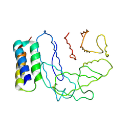
6BMT
DownloadVisualize
BU of 6bmt by Molmil
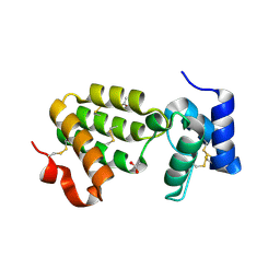
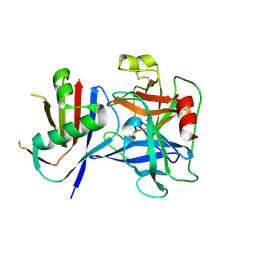
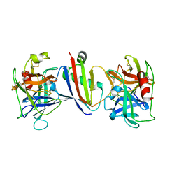
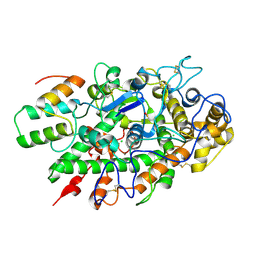
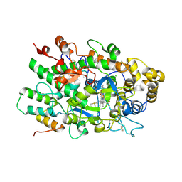
Crystal Structure of a Recombinant form of Human Myeloperoxidase Bound to an Inhibitor from Staphylococcus delphini
Descriptor: 2-acetamido-2-deoxy-beta-D-glucopyranose, CALCIUM ION, CHLORIDE ION, ...
Authors:Ploscariu, N.T, Geisbrecht, B.V.
Deposit date:2017-11-15
Release date:2018-01-03
Last modified:2024-10-30
Method:X-RAY DIFFRACTION (2.403 Å)
Cite:Identification and structural characterization of a novel myeloperoxidase inhibitor from Staphylococcus delphini.
Arch. Biochem. Biophys., 645, 2018
2NOJ
DownloadVisualize
BU of 2noj by Molmil
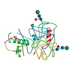
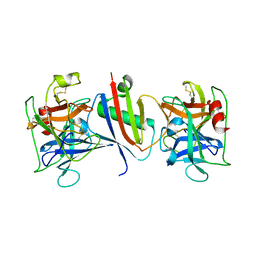
Crystal structure of Ehp / C3d complex
Descriptor: Complement C3, Efb homologous protein
Authors:Hammel, M, Geisbrecht, B.V.
Deposit date:2006-10-25
Release date:2007-08-14
Last modified:2023-08-30
Method:X-RAY DIFFRACTION (2.7 Å)
Cite:Characterization of Ehp, a Secreted Complement Inhibitory Protein from Staphylococcus aureus.
J.Biol.Chem., 282, 2007
7S0R
DownloadVisualize
BU of 7s0r by Molmil
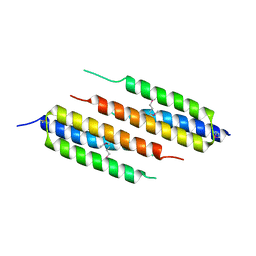
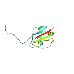
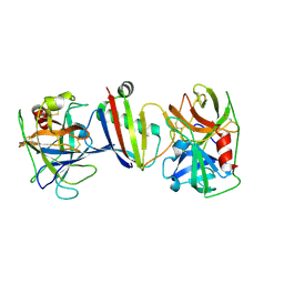
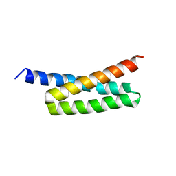
Crystal Structure of a Complement Factor H-binding Fragment within the B75KN Region of the Group B Streptococcus Beta Antigen C Protein
Descriptor: C protein beta antigen
Authors:Xu, X, Geisbrecht, B.V.
Deposit date:2021-08-31
Release date:2021-12-15
Last modified:2022-06-29
Method:X-RAY DIFFRACTION (2.5 Å)
Cite:Group B Streptococcus Surface Protein beta : Structural Characterization of a Complement Factor H-Binding Motif and Its Contribution to Immune Evasion.
J Immunol., 208, 2022
1SZH
DownloadVisualize
BU of 1szh by Molmil
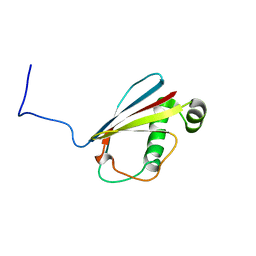
Crystal Structure of C. elegans HER-1
Descriptor: ACETATE ION, Her-1 protein
Authors:Hamaoka, B.Y, Dann III, C.E, Geisbrecht, B.V, Leahy, D.J.
Deposit date:2004-04-05
Release date:2004-08-10
Last modified:2024-10-30
Method:X-RAY DIFFRACTION (1.5 Å)
Cite:Crystal structure of Caenorhabditis elegans HER-1 and characterization of the interaction between HER-1 and TRA-2A.
Proc.Natl.Acad.Sci.USA, 101, 2004
3I86
DownloadVisualize
BU of 3i86 by Molmil
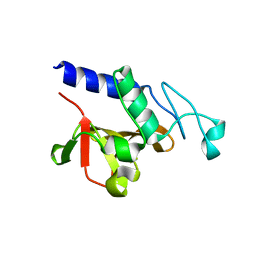
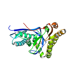
Crystal structure of the P60 Domain from M. avium subspecies paratuberculosis antigen MAP1204
Descriptor: ISOPROPYL ALCOHOL, Putative uncharacterized protein
Authors:Ramyar, K.X, Lingle, C.K, McWhorter, W.J, Bouyain, S, Bannantine, J.P, Geisbrecht, B.V.
Deposit date:2009-07-09
Release date:2010-07-21
Last modified:2023-09-06
Method:X-RAY DIFFRACTION (2.4 Å)
Cite:Atypical structural features of two P60 family members from Mycobacterium avium subspecies paratuberculosis
To be Published
3GT2
DownloadVisualize
BU of 3gt2 by Molmil
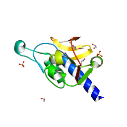
Crystal Structure of the P60 Domain from M. avium paratuberculosis Antigen MAP1272c
Descriptor: 1,2-ETHANEDIOL, Putative uncharacterized protein, SULFATE ION
Authors:Ramyar, K.X, Lingle, C.K, McWhorter, W.J, Bouyain, S, Bannantine, J.P, Geisbrecht, B.V.
Deposit date:2009-03-27
Release date:2010-04-07
Last modified:2024-10-30
Method:X-RAY DIFFRACTION (1.75 Å)
Cite:Crystal Structures of Two P60-Family Antigens from Mycobacterium Avium Paratuberculosis
To be Published
8D4O
DownloadVisualize
BU of 8d4o by Molmil
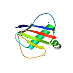
Crystal Structure of the Neutrophil Serine Protease Inhibitor Eap1 from S. aureus
Descriptor: Extracellular Adherence Protein, SODIUM ION
Authors:Gido, C.D, Geisbrecht, B.V.
Deposit date:2022-06-02
Release date:2022-12-21
Last modified:2024-04-03
Method:X-RAY DIFFRACTION (1.45 Å)
Cite:Characterization of two distinct neutrophil serine protease-binding modes within a Staphylococcus aureus innate immune evasion protein family.
J.Biol.Chem., 299, 2023
8D4V
DownloadVisualize
BU of 8d4v by Molmil
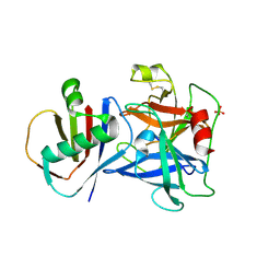
Crystal Structure of Cathepsin G Inhibited by Eap2 from S. aureus
Descriptor: Cathepsin G, C-terminal truncated form, Extracellular Adherence Protein, ...
Authors:Gido, C.D, Herdendorf, T.J, Geisbrecht, B.V.
Deposit date:2022-06-02
Release date:2022-12-21
Last modified:2023-10-25
Method:X-RAY DIFFRACTION (1.85 Å)
Cite:Characterization of two distinct neutrophil serine protease-binding modes within a Staphylococcus aureus innate immune evasion protein family.
J.Biol.Chem., 299, 2023
8D4U
DownloadVisualize
BU of 8d4u by Molmil
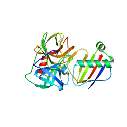
Crystal Structure of Neutrophil Elastase Inhibited by Eap2 from S. aureus
Descriptor: 2-acetamido-2-deoxy-beta-D-glucopyranose, 2-acetamido-2-deoxy-beta-D-glucopyranose-(1-4)-[alpha-L-fucopyranose-(1-6)]2-acetamido-2-deoxy-beta-D-glucopyranose, Extracellular Adherence Protein, ...
Authors:Gido, C.D, Herdendorf, T.J, Geisbrecht, B.V.
Deposit date:2022-06-02
Release date:2022-12-21
Last modified:2023-10-25
Method:X-RAY DIFFRACTION (1.9 Å)
Cite:Characterization of two distinct neutrophil serine protease-binding modes within a Staphylococcus aureus innate immune evasion protein family.
J.Biol.Chem., 299, 2023
8D4Q
DownloadVisualize
BU of 8d4q by Molmil
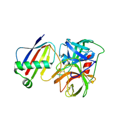
Crystal Structure of Neutrophil Elastase Inhibited by Eap1 from S. aureus
Descriptor: 2-acetamido-2-deoxy-beta-D-glucopyranose-(1-4)-[alpha-L-fucopyranose-(1-6)]2-acetamido-2-deoxy-beta-D-glucopyranose, Extracellular Adherence Protein, Neutrophil elastase
Authors:Gido, C.D, Herdendorf, T.J, Geisbrecht, B.V.
Deposit date:2022-06-02
Release date:2022-12-21
Last modified:2024-10-23
Method:X-RAY DIFFRACTION (2.2 Å)
Cite:Characterization of two distinct neutrophil serine protease-binding modes within a Staphylococcus aureus innate immune evasion protein family.
J.Biol.Chem., 299, 2023
8D4S
DownloadVisualize
BU of 8d4s by Molmil
Crystal Structure of Cathepsin G Inhibited by Eap1 from S. aureus
Descriptor: 2-acetamido-2-deoxy-beta-D-glucopyranose, Cathepsin G, C-terminal truncated form, ...
Authors:Gido, C.D, Herdendorf, T.J, Geisbrecht, B.V.
Deposit date:2022-06-02
Release date:2022-12-21
Last modified:2023-10-25
Method:X-RAY DIFFRACTION (1.95 Å)
Cite:Characterization of two distinct neutrophil serine protease-binding modes within a Staphylococcus aureus innate immune evasion protein family.
J.Biol.Chem., 299, 2023
4NZL
DownloadVisualize
BU of 4nzl by Molmil
Extracellular proteins of Staphylococcus aureus inhibit the neutrophil serine proteases
Descriptor: Neutrophil elastase, Uncharacterized protein, beta-D-mannopyranose-(1-4)-2-acetamido-2-deoxy-beta-D-glucopyranose-(1-4)-[beta-L-fucopyranose-(1-6)]2-acetamido-2-deoxy-beta-D-glucopyranose
Authors:Stapels, D.A.C, von Koeckritz-Blickwede, M, Ramyar, K.X, Bischoff, M, Milder, F, Ruyken, M, Scheepmaker, L, McWhorter, W.J, Herrmann, M, van Kessel, K.P.M, Geisbrecht, B.V, Rooijakkers, S.H.M.
Deposit date:2013-12-12
Release date:2014-08-20
Last modified:2024-10-09
Method:X-RAY DIFFRACTION (1.85 Å)
Cite:Staphylococcus aureus secretes a unique class of neutrophil serine protease inhibitors.
Proc.Natl.Acad.Sci.USA, 111, 2014
8GDG
DownloadVisualize
BU of 8gdg by Molmil
Solution structure of the Neutrophil Serine Protease Inhibitor, EapH2
Descriptor: Cell surface like-protein Map-w
Authors:Mishra, N.B, Geisbrecht, B.V, Prakash, O.
Deposit date:2023-03-05
Release date:2023-04-26
Last modified:2024-05-15
Method:SOLUTION NMR
Cite:Simultaneous inhibition of two neutrophil serine proteases by the S. aureus innate immune evasion protein EapH2.
J.Biol.Chem., 299, 2023
8GDH
DownloadVisualize
BU of 8gdh by Molmil
Solution structure of the Neutrophil Serine Protease Inhibitor, EapH1
Descriptor: Cell surface protein map-w
Authors:Mishra, N.B, Geisbrecht, B.V, Prakash, O.
Deposit date:2023-03-05
Release date:2023-04-26
Last modified:2024-05-15
Method:SOLUTION NMR
Cite:Simultaneous inhibition of two neutrophil serine proteases by the S. aureus innate immune evasion protein EapH2.
J.Biol.Chem., 299, 2023
8G24
DownloadVisualize
BU of 8g24 by Molmil
Crystal Structure of Cathepsin-G and Neutrophil Elastase Inhibited by S. aureus EapH2 at pH 5.5
Descriptor: 2-acetamido-2-deoxy-beta-D-glucopyranose-(1-4)-2-acetamido-2-deoxy-beta-D-glucopyranose, Cathepsin-G, MAP domain-containing protein, ...
Authors:Mishra, N.B, Geisbrecht, B.V.
Deposit date:2023-02-03
Release date:2023-04-26
Last modified:2024-10-23
Method:X-RAY DIFFRACTION (1.82 Å)
Cite:Simultaneous inhibition of two neutrophil serine proteases by the S. aureus innate immune evasion protein EapH2.
J.Biol.Chem., 299, 2023
8G25
DownloadVisualize
BU of 8g25 by Molmil
Crystal Structure of Cathepsin-G and Neutrophil Elastase Inhibited by S. aureus EapH2 at pH 7.5
Descriptor: Cathepsin-G, MAP domain-containing protein, Neutrophil elastase
Authors:Mishra, N.B, Geisbrecht, B.V.
Deposit date:2023-02-03
Release date:2023-04-26
Last modified:2023-10-25
Method:X-RAY DIFFRACTION (1.8 Å)
Cite:Simultaneous inhibition of two neutrophil serine proteases by the S. aureus innate immune evasion protein EapH2.
J.Biol.Chem., 299, 2023
8G26
DownloadVisualize
BU of 8g26 by Molmil
Crystal Structure of Cathepsin-G and Neutrophil Elastase Inhibited by S. aureus EapH2 at pH 8.5
Descriptor: 2-acetamido-2-deoxy-beta-D-glucopyranose, 2-acetamido-2-deoxy-beta-D-glucopyranose-(1-4)-2-acetamido-2-deoxy-beta-D-glucopyranose, Cathepsin-G, ...
Authors:Mishra, N.B, Geisbrecht, B.V.
Deposit date:2023-02-03
Release date:2023-04-26
Last modified:2023-10-25
Method:X-RAY DIFFRACTION (1.85 Å)
Cite:Simultaneous inhibition of two neutrophil serine proteases by the S. aureus innate immune evasion protein EapH2.
J.Biol.Chem., 299, 2023
5VJ0
DownloadVisualize
BU of 5vj0 by Molmil
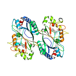
Crystal Structure of heme-containing DyP Type Peroxidase from Enterobacter lignolyticus
Descriptor: Dyp-type peroxidase family, PROTOPORPHYRIN IX CONTAINING FE
Authors:Meekins, D.A, Li, P, Geisbrecht, B.V.
Deposit date:2017-04-17
Release date:2017-08-16
Last modified:2023-10-04
Method:X-RAY DIFFRACTION (1.93 Å)
Cite:Mechanistic Insights into Dye-Decolorizing Peroxidase Revealed by Solvent Isotope and Viscosity Effects.
ACS Catal, 7, 2017
8TE5
DownloadVisualize
BU of 8te5 by Molmil
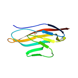
Crystal structure of a multiple lysine-to-arginine substitution mutant of the human CRIg C3b-binding domain
Descriptor: V-set and immunoglobulin domain-containing protein 4
Authors:Abram, T.G, Geisbrecht, B.V.
Deposit date:2023-07-05
Release date:2023-07-12
Last modified:2024-01-24
Method:X-RAY DIFFRACTION (1.502 Å)
Cite:New Insights into the Complement Receptor of the Ig Superfamily Obtained from Structural and Functional Studies on Two Mutants.
Immunohorizons, 7, 2023
8TE6
DownloadVisualize
BU of 8te6 by Molmil
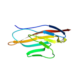
Crystal structure of a multiple lysine-to-arginine substitution mutant of the human CRIg C3b-binding domain
Descriptor: V-set and immunoglobulin domain-containing protein 4
Authors:Abram, T.G, Geisbrecht, B.V.
Deposit date:2023-07-05
Release date:2023-07-12
Last modified:2024-01-24
Method:X-RAY DIFFRACTION (1.251 Å)
Cite:New Insights into the Complement Receptor of the Ig Superfamily Obtained from Structural and Functional Studies on Two Mutants.
Immunohorizons, 7, 2023
5UZU
DownloadVisualize
BU of 5uzu by Molmil
Immune evasion by a Staphylococcal Peroxidase Inhibitor that blocks myeloperoxidase
Descriptor: 2-acetamido-2-deoxy-beta-D-glucopyranose, 2-acetamido-2-deoxy-beta-D-glucopyranose-(1-4)-2-acetamido-2-deoxy-beta-D-glucopyranose, CALCIUM ION, ...
Authors:de Jong, N, Geisbrecht, B.V, van Strijp, J, Haas, P, Nijland, R, Ramyar, K, Fevre, C, Guerra, F, Voyich-Kane, J, van Kessel, C, Garcia, B.
Deposit date:2017-02-27
Release date:2017-07-26
Last modified:2023-11-15
Method:X-RAY DIFFRACTION (2.403 Å)
Cite:Immune evasion by a staphylococcal inhibitor of myeloperoxidase.
Proc. Natl. Acad. Sci. U.S.A., 114, 2017
7JTQ
DownloadVisualize
BU of 7jtq by Molmil
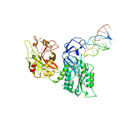
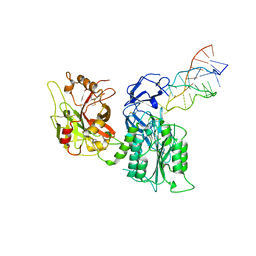
Human Complement Factor B Inhibited by a Slow Off-Rate Modified Aptamer of 31 Bases
Descriptor: Complement factor B, DNA (32-MER)
Authors:Xu, X, Geisbrecht, B.V.
Deposit date:2020-08-18
Release date:2020-12-09
Last modified:2023-10-18
Method:X-RAY DIFFRACTION (3.5 Å)
Cite:Inhibition of the Complement Alternative Pathway by Chemically Modified DNA Aptamers That Bind with Picomolar Affinity to Factor B.
J Immunol., 206, 2021
7JTN
DownloadVisualize
BU of 7jtn by Molmil
Human Complement Factor B Inhibited by a Slow Off-Rate Modified Aptamer of 29 Bases
Descriptor: Complement factor B, DNA (30-MER)
Authors:Xu, X, Geisbrecht, B.V.
Deposit date:2020-08-18
Release date:2020-12-09
Last modified:2023-10-18
Method:X-RAY DIFFRACTION (3.1 Å)
Cite:Inhibition of the Complement Alternative Pathway by Chemically Modified DNA Aptamers That Bind with Picomolar Affinity to Factor B.
J Immunol., 206, 2021
4DU7
DownloadVisualize
BU of 4du7 by Molmil
Crystal structure of Staphylococcus epidermidis mevalonate diphosphate decarboxylase complexed with substrate mevalonate diphosphate
Descriptor: (3R)-3-HYDROXY-5-{[(R)-HYDROXY(PHOSPHONOOXY)PHOSPHORYL]OXY}-3-METHYLPENTANOIC ACID, FORMIC ACID, Mevalonate diphosphate decarboxylase
Authors:Barta, M.L, McWhorter, W.J, Geisbrecht, B.V.
Deposit date:2012-02-21
Release date:2012-07-11
Last modified:2023-09-13
Method:X-RAY DIFFRACTION (2.201 Å)
Cite:Structural basis for nucleotide binding and reaction catalysis in mevalonate diphosphate decarboxylase.
Biochemistry, 51, 2012
4H6H
DownloadVisualize
BU of 4h6h by Molmil
Crystal Structure of Staphylococcal Complement Inhibitor SCIN-B(4-85)
Descriptor: Fibrinogen-binding protein
Authors:Garcia, B.L, Geisbrecht, B.V.
Deposit date:2012-09-19
Release date:2012-12-19
Last modified:2023-09-20
Method:X-RAY DIFFRACTION (2.5024 Å)
Cite:A Structurally Dynamic N-terminal Helix Is a Key Functional Determinant in Staphylococcal Complement Inhibitor (SCIN) Proteins.
J.Biol.Chem., 288, 2013

226707

PDB entries from 2024-10-30

PDB statisticsPDBj update infoContact PDBjnumon