Loading
PDBj
MenuPDBj@FacebookPDBj@TwitterPDBj@YouTubewwPDB FoundationwwPDB
RCSB PDBPDBeBMRBAdv. SearchSearch help
Search by PDB author
8EYY
DownloadVisualize
BU of 8eyy by Molmil
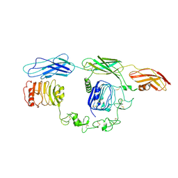
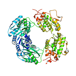
Cryo-EM structure of 4 insulins bound full-length mouse IR mutant with physically decoupled alpha CTs (C684S/C685S/C687S, denoted as IR-3CS) Asymmetric conformation 2
Descriptor: Insulin, Insulin receptor
Authors:Li, J, Wu, J.Y, Hall, C, Bai, X.C, Choi, E.
Deposit date:2022-10-29
Release date:2022-11-09
Last modified:2024-10-30
Method:ELECTRON MICROSCOPY (4.9 Å)
Cite:Molecular basis for the role of disulfide-linked alpha CTs in the activation of insulin-like growth factor 1 receptor and insulin receptor.
Elife, 11, 2022
7TYJ
DownloadVisualize
BU of 7tyj by Molmil
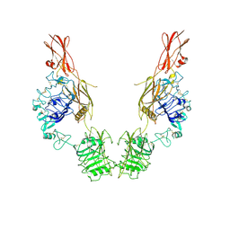
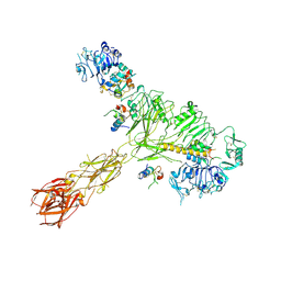
Cryo-EM Structure of insulin receptor-related receptor (IRR) in apo-state captured at pH 7. The 3D refinement was focused on one of two halves with C1 symmetry applied
Descriptor: Insulin receptor-related protein
Authors:Wang, L.W, Hall, C, Li, J, Choi, E, Bai, X.C.
Deposit date:2022-02-13
Release date:2023-02-15
Last modified:2024-10-16
Method:ELECTRON MICROSCOPY (3.3 Å)
Cite:Structural basis of the alkaline pH-dependent activation of insulin receptor-related receptor.
Nat.Struct.Mol.Biol., 30, 2023
7TYK
DownloadVisualize
BU of 7tyk by Molmil
Cryo-EM Structure of insulin receptor-related receptor (IRR) in apo-state captured at pH 7. The 3D refinement was applied with C2 symmetry
Descriptor: Insulin receptor-related protein
Authors:Wang, L.W, Hall, C, Li, J, Choi, E, Bai, X.C.
Deposit date:2022-02-13
Release date:2023-02-15
Last modified:2023-05-31
Method:ELECTRON MICROSCOPY (3.5 Å)
Cite:Structural basis of the alkaline pH-dependent activation of insulin receptor-related receptor.
Nat.Struct.Mol.Biol., 30, 2023
7TYM
DownloadVisualize
BU of 7tym by Molmil
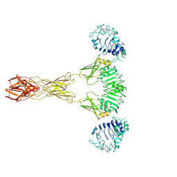
Cryo-EM Structure of insulin receptor-related receptor (IRR) in active-state captured at pH 9. The 3D refinement was applied with C2 symmetry
Descriptor: Insulin receptor-related protein
Authors:Wang, L.W, Hall, C, Li, J, Choi, E, Bai, X.C.
Deposit date:2022-02-13
Release date:2023-02-15
Last modified:2024-10-30
Method:ELECTRON MICROSCOPY (3.4 Å)
Cite:Structural basis of the alkaline pH-dependent activation of insulin receptor-related receptor.
Nat.Struct.Mol.Biol., 30, 2023
7D2M
DownloadVisualize
BU of 7d2m by Molmil
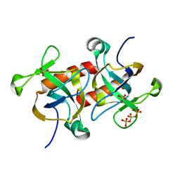
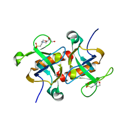
Crystal structure of MazF (Form-II) from Deinococcus radiodurans
Descriptor: Endoribonuclease MazF, SULFATE ION, TRIPHOSPHATE
Authors:Dhanasingh, I, Lee, S.H.
Deposit date:2020-09-17
Release date:2021-02-10
Last modified:2023-11-29
Method:X-RAY DIFFRACTION (1.79 Å)
Cite:Functional and structural characterization of Deinococcus radiodurans R1 MazEF toxin-antitoxin system, Dr0416-Dr0417.
J.Microbiol, 59, 2021
7D28
DownloadVisualize
BU of 7d28 by Molmil
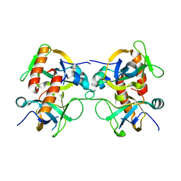
Crystal structure of MazF (Form-I) from Deinococcus radiodurans
Descriptor: 2-(N-MORPHOLINO)-ETHANESULFONIC ACID, Endoribonuclease MazF, GLYCEROL
Authors:Dhanasingh, I, Lee, S.H.
Deposit date:2020-09-16
Release date:2021-02-10
Last modified:2023-11-29
Method:X-RAY DIFFRACTION (1.5 Å)
Cite:Functional and structural characterization of Deinococcus radiodurans R1 MazEF toxin-antitoxin system, Dr0416-Dr0417.
J.Microbiol, 59, 2021
7D2Q
DownloadVisualize
BU of 7d2q by Molmil
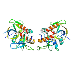
Crystal structure of MazE-MazF (Form-I) from Deinococcus radiodurans
Descriptor: AbrB/MazE/SpoVT family DNA-binding domain-containing protein, Endoribonuclease MazF
Authors:Dhanasingh, I, Lee, S.H.
Deposit date:2020-09-17
Release date:2021-02-10
Last modified:2023-11-29
Method:X-RAY DIFFRACTION (1.99 Å)
Cite:Functional and structural characterization of Deinococcus radiodurans R1 MazEF toxin-antitoxin system, Dr0416-Dr0417.
J.Microbiol, 59, 2021
7D2P
DownloadVisualize
BU of 7d2p by Molmil
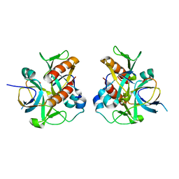
Crystal structure of MazE-MazF (Form-II) from Deinococcus radiodurans
Descriptor: AbrB/MazE/SpoVT family DNA-binding domain-containing protein, Endoribonuclease MazF
Authors:Dhanasingh, I, Lee, S.H.
Deposit date:2020-09-17
Release date:2021-02-10
Last modified:2023-11-29
Method:X-RAY DIFFRACTION (2.07 Å)
Cite:Functional and structural characterization of Deinococcus radiodurans R1 MazEF toxin-antitoxin system, Dr0416-Dr0417.
J.Microbiol, 59, 2021
7D2N
DownloadVisualize
BU of 7d2n by Molmil
Crystal structure of MazE-MazF (Form-III) from Deinococcus radiodurans
Descriptor: AbrB/MazE/SpoVT family DNA-binding domain-containing protein, Endoribonuclease MazF
Authors:Dhanasingh, I, Lee, S.H.
Deposit date:2020-09-17
Release date:2021-02-10
Last modified:2023-11-29
Method:X-RAY DIFFRACTION (1.6 Å)
Cite:Functional and structural characterization of Deinococcus radiodurans R1 MazEF toxin-antitoxin system, Dr0416-Dr0417.
J.Microbiol, 59, 2021
8RON
DownloadVisualize
BU of 8ron by Molmil
Crystal structure of human FAD synthase, isoform 2
Descriptor: Isoform 2 of FAD synthase
Authors:Leo, G, Capaldi, S.
Deposit date:2024-01-11
Release date:2024-04-10
Last modified:2024-07-24
Method:X-RAY DIFFRACTION (2.6 Å)
Cite:Structural insights into the bifunctional enzyme human FAD synthase.
Structure, 32, 2024
8ROM
DownloadVisualize
BU of 8rom by Molmil
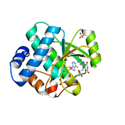
Crystal structure of human FAD synthase PAPS domain in complex with FAD
Descriptor: FAD synthase, FLAVIN-ADENINE DINUCLEOTIDE, PHOSPHATE ION
Authors:Leo, G, Capaldi, S.
Deposit date:2024-01-11
Release date:2024-04-10
Last modified:2024-07-24
Method:X-RAY DIFFRACTION (1.69 Å)
Cite:Structural insights into the bifunctional enzyme human FAD synthase.
Structure, 32, 2024
<12


226707

PDB entries from 2024-10-30

PDB statisticsPDBj update infoContact PDBjnumon