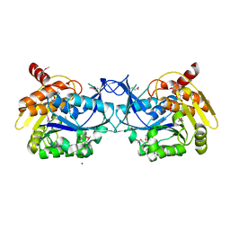
Loading
PDBj
MenuPDBj@FacebookPDBj@TwitterPDBj@YouTubewwPDB FoundationwwPDB
RCSB PDBPDBeBMRBAdv. SearchSearch help
Search by PDB author
3HV1
DownloadVisualize
BU of 3hv1 by Molmil
Crystal structure of a polar amino acid ABC uptake transporter substrate binding protein from Streptococcus thermophilus
Descriptor: Polar amino acid ABC uptake transporter substrate binding protein
Authors:Palani, K, Burley, S.K, Swaminathan, S, New York SGX Research Center for Structural Genomics (NYSGXRC)
Deposit date:2009-06-15
Release date:2009-06-23
Last modified:2021-02-10
Method:X-RAY DIFFRACTION (1.9 Å)
Cite:Crystal structure of a polar amino acid ABC uptake transporter substrate binding protein from Streptococcus thermophilus
To be Published
3HYO
DownloadVisualize
BU of 3hyo by Molmil
Crystal structure of pyridoxal kinase from Lactobacillus plantarum in complex with ADP
Descriptor: ADENOSINE-5'-DIPHOSPHATE, MAGNESIUM ION, Pyridoxal kinase
Authors:Bagaria, A, Kumaran, D, Burley, S.K, Swaminathan, S, New York SGX Research Center for Structural Genomics (NYSGXRC)
Deposit date:2009-06-22
Release date:2009-06-30
Last modified:2023-11-22
Method:X-RAY DIFFRACTION (1.85 Å)
Cite:Crystal structure of pyridoxal kinase from Lactobacillus plantarum in complex with ADP
To be Published
3DXI
DownloadVisualize
BU of 3dxi by Molmil
Crystal structure of the N-terminal domain of a putative aldolase (BVU_2661) from Bacteroides vulgatus
Descriptor: Putative aldolase
Authors:Eswaramoorthy, S, Pabalan, A.A, Burley, S.K, Swaminathan, S, New York SGX Research Center for Structural Genomics (NYSGXRC)
Deposit date:2008-07-24
Release date:2008-08-26
Last modified:2021-02-10
Method:X-RAY DIFFRACTION (2.04 Å)
Cite:Crystal structure of the N-terminal domain of a putative aldolase (BVU_2661) from Bacteroides vulgatus
To be Published
3DZ1
DownloadVisualize
BU of 3dz1 by Molmil
Crystal structure of Dihydrodipicolinate Synthase from Rhodopseudomonas palustris at 1.87A resolution
Descriptor: Dihydrodipicolinate synthase
Authors:Satyanarayana, L, Eswaramoorthy, S, Burley, S.K, Swaminathan, S, New York SGX Research Center for Structural Genomics (NYSGXRC)
Deposit date:2008-07-29
Release date:2008-08-12
Last modified:2021-02-10
Method:X-RAY DIFFRACTION (1.87 Å)
Cite:Crystal structure of Dihydrodipicolinate Synthase from Rhodopseudomonas palustris at 1.87A resolution
To be Published
3HUT
DownloadVisualize
BU of 3hut by Molmil
Crystal structure of a putative branched-chain amino acid ABC transporter from Rhodospirillum rubrum
Descriptor: putative branched-chain amino acid ABC transporter
Authors:Satyanarayana, L, Burley, S.K, Swaminathan, S, New York SGX Research Center for Structural Genomics (NYSGXRC)
Deposit date:2009-06-15
Release date:2009-06-30
Last modified:2021-02-10
Method:X-RAY DIFFRACTION (1.93 Å)
Cite:Crystal structure of a putative branched-chain amino acid ABC transporter from Rhodospirillum rubrum
To be Published
1RTT
DownloadVisualize
BU of 1rtt by Molmil
Crystal structure determination of a putative NADH-dependent reductase using sulfur anomalous signal
Descriptor: SULFATE ION, conserved hypothetical protein
Authors:Agarwal, R, Swaminathan, S, Burley, S.K, New York SGX Research Center for Structural Genomics (NYSGXRC)
Deposit date:2003-12-10
Release date:2004-08-17
Last modified:2024-02-14
Method:X-RAY DIFFRACTION (1.28 Å)
Cite:Structure determination of an FMN reductase from Pseudomonas aeruginosa PA01 using sulfur anomalous signal.
ACTA CRYSTALLOGR.,SECT.D, 62, 2006
3I5I
DownloadVisualize
BU of 3i5i by Molmil
The crystal structure of squid myosin S1 in the presence of SO4 2-
Descriptor: CALCIUM ION, Myosin catalytic light chain LC-1, mantle muscle, ...
Authors:Yang, Y, Gourinath, S, Kovacs, M, Nyitray, L, Reutzel, R, Himmel, D.M, O'Neall-Hennessey, E, Reshetnikova, L, Szent-Gyorgyi, A.G, Brown, J.H, Cohen, C.
Deposit date:2009-07-05
Release date:2009-07-28
Last modified:2023-09-06
Method:X-RAY DIFFRACTION (3.3 Å)
Cite:Rigor-like structures from muscle myosins reveal key mechanical elements in the transduction pathways of this allosteric motor.
Structure, 15, 2007
3I3V
DownloadVisualize
BU of 3i3v by Molmil
Crystal Structure of probable secreted solute-binding lipoprotein from Streptomyces coelicolor
Descriptor: Probable secreted solute-binding lipoprotein
Authors:Damodharan, L, Burley, S.K, Swaminathan, S, New York SGX Research Center for Structural Genomics (NYSGXRC)
Deposit date:2009-07-01
Release date:2009-07-14
Last modified:2021-02-10
Method:X-RAY DIFFRACTION (2.3 Å)
Cite:Crystal Structure of probable secreted solute-binding lipoprotein from Streptomyces coelicolor
To be Published
1RVI
DownloadVisualize
BU of 1rvi by Molmil
SOLUTION STRUCTURE OF THE DNA DODECAMER CGTTTTAAAACG
Descriptor: 5'-D(*CP*GP*TP*TP*TP*TP*AP*AP*AP*AP*CP*G)-3'
Authors:Stefl, R, Wu, H, Ravindranathan, S, Sklenar, V, Feigon, J.
Deposit date:2003-12-13
Release date:2004-02-10
Last modified:2024-05-22
Method:SOLUTION NMR
Cite:DNA A-tract bending in three dimensions: Solving the dA4T4 vs. dT4A4 conundrum.
Proc.Natl.Acad.Sci.USA, 101, 2004
1RW0
DownloadVisualize
BU of 1rw0 by Molmil
Crystal structure of protein yfiH from Salmonella enterica serovar Typhi, Pfam DUF152
Descriptor: conserved hypothetical protein
Authors:Seetharaman, J, Swaminathan, S, Burley, S.K, New York SGX Research Center for Structural Genomics (NYSGXRC)
Deposit date:2003-12-15
Release date:2004-08-17
Last modified:2023-08-23
Method:X-RAY DIFFRACTION (2 Å)
Cite:Crystal Structure of Hypothetical protein yfiH
To be Published
2GOK
DownloadVisualize
BU of 2gok by Molmil
Crystal structure of the imidazolonepropionase from Agrobacterium tumefaciens at 1.87 A resolution
Descriptor: CHLORIDE ION, FE (III) ION, GLYCEROL, ...
Authors:Tyagi, R, Kumaran, D, Swaminathan, S, Burley, S.K, New York SGX Research Center for Structural Genomics (NYSGXRC)
Deposit date:2006-04-13
Release date:2006-04-25
Last modified:2024-02-14
Method:X-RAY DIFFRACTION (1.87 Å)
Cite:X-ray structure of imidazolonepropionase from Agrobacterium tumefaciens at 1.87 A resolution.
Proteins, 69, 2007
4RKI
DownloadVisualize
BU of 4rki by Molmil
Crystal structure of sliding beta clamp from Helicobacter pylori
Descriptor: DNA polymerase III subunit beta
Authors:Satyawali, P, Tarique, K.F, Abdul Rehman, S.A, Gourinath, S.
Deposit date:2014-10-13
Release date:2016-01-13
Last modified:2023-09-20
Method:X-RAY DIFFRACTION (2.05 Å)
Cite:Structural insight into beta-Clamp and its interaction with DNA Ligase in Helicobacter pylori.
Sci Rep, 6, 2016
3HS3
DownloadVisualize
BU of 3hs3 by Molmil
Crystal structure of periplasmic binding ribose operon repressor protein from Lactobacillus acidophilus
Descriptor: Ribose operon repressor
Authors:Agarwal, R, Burley, S.K, Swaminathan, S, New York SGX Research Center for Structural Genomics (NYSGXRC)
Deposit date:2009-06-10
Release date:2009-06-23
Last modified:2024-02-21
Method:X-RAY DIFFRACTION (1.6 Å)
Cite:Crystal structure of periplasmic binding ribose operon repressor protein from Lactobacillus acidophilus
To be Published
3HZ6
DownloadVisualize
BU of 3hz6 by Molmil
Crystal structure of xylulokinase from Chromobacterium violaceum
Descriptor: ADENOSINE-5'-DIPHOSPHATE, D-XYLULOSE, GLYCEROL, ...
Authors:Zhang, Z, Burley, S.K, Swaminathan, S, New York SGX Research Center for Structural Genomics (NYSGXRC)
Deposit date:2009-06-23
Release date:2009-07-21
Last modified:2021-02-10
Method:X-RAY DIFFRACTION (1.65 Å)
Cite:Crystal structure of xylulokinase from Chromobacterium violaceum
To be Published
3IFR
DownloadVisualize
BU of 3ifr by Molmil
The crystal structure of xylulose kinase from Rhodospirillum rubrum
Descriptor: Carbohydrate kinase, FGGY, PHOSPHATE ION
Authors:Zhang, Z, Burley, S.K, Swaminathan, S, New York SGX Research Center for Structural Genomics (NYSGXRC)
Deposit date:2009-07-24
Release date:2009-08-25
Last modified:2021-02-10
Method:X-RAY DIFFRACTION (2.3 Å)
Cite:The crystal structure of xylulose kinase from Rhodospirillum rubrum
To be Published
3IQ0
DownloadVisualize
BU of 3iq0 by Molmil
Crystal structure of a putative Ribokinase II in complex with ATP and Mg+2 from E.coli
Descriptor: ADENOSINE-5'-TRIPHOSPHATE, MAGNESIUM ION, putative Ribokinase II
Authors:Satyanarayana, L, Burley, S.K, Swaminathan, S, New York SGX Research Center for Structural Genomics (NYSGXRC)
Deposit date:2009-08-18
Release date:2009-09-08
Last modified:2021-02-10
Method:X-RAY DIFFRACTION (1.79 Å)
Cite:Crystal structure of a putative Ribokinase (II)in complex with ATP and Mg+2 from E.coli
To be Published
1RVH
DownloadVisualize
BU of 1rvh by Molmil
SOLUTION STRUCTURE OF THE DNA DODECAMER GCAAAATTTTGC
Descriptor: 5'-D(*GP*CP*AP*AP*AP*AP*TP*TP*TP*TP*GP*C)-3'
Authors:Stefl, R, Wu, H, Ravindranathan, S, Sklenar, V, Feigon, J.
Deposit date:2003-12-13
Release date:2004-02-10
Last modified:2024-05-22
Method:SOLUTION NMR
Cite:DNA A-tract bending in three dimensions: Solving the dA4T4 vs. dT4A4 conundrum.
Proc.Natl.Acad.Sci.USA, 101, 2004
1RRM
DownloadVisualize
BU of 1rrm by Molmil
Crystal Structure of Lactaldehyde reductase
Descriptor: ADENOSINE-5-DIPHOSPHORIBOSE, FE (II) ION, Lactaldehyde reductase, ...
Authors:Kumaran, D, Swaminathan, S, Burley, S.K, New York SGX Research Center for Structural Genomics (NYSGXRC)
Deposit date:2003-12-08
Release date:2004-08-17
Last modified:2021-02-03
Method:X-RAY DIFFRACTION (1.6 Å)
Cite:Crystal structure of Lactaldehyde reductase
To be Published
3LJS
DownloadVisualize
BU of 3ljs by Molmil
Crystal structure of Fructokinase from Xylella fastidiosa
Descriptor: Fructokinase, PHOSPHATE ION
Authors:Satyanarayana, L, Burley, S.K, Swaminathan, S, New York SGX Research Center for Structural Genomics (NYSGXRC)
Deposit date:2010-01-26
Release date:2010-03-09
Last modified:2021-02-10
Method:X-RAY DIFFRACTION (1.97 Å)
Cite:Crystal structure of Fructokinase from Xylella fastidiosa
To be Published
3LM4
DownloadVisualize
BU of 3lm4 by Molmil
Crystal Structure of 2,3-Dihydroxy Biphenyl dioxygenase from Rhodococcus sp. (strain RHA1)
Descriptor: (2Z,4E)-2-HYDROXY-6-OXO-6-PHENYLHEXA-2,4-DIENOIC ACID, Catechol 2,3-dioxygenase, FE (III) ION, ...
Authors:Syed Ibrahim, B, Kumaran, D, Burley, S.K, Swaminathan, S, New York SGX Research Center for Structural Genomics (NYSGXRC)
Deposit date:2010-01-29
Release date:2010-02-23
Last modified:2021-02-10
Method:X-RAY DIFFRACTION (1.8 Å)
Cite:Crystal Structure of 2,3-Dihydroxy Biphenyl dioxygenase from Rhodococcus sp. (strain RHA1)
To be Published
3HDC
DownloadVisualize
BU of 3hdc by Molmil
The crystal structure of thioredoxin protein from Geobacter metallireducens
Descriptor: Thioredoxin family protein
Authors:Zhang, Z, Burley, S.K, Swaminathan, S, New York SGX Research Center for Structural Genomics (NYSGXRC)
Deposit date:2009-05-07
Release date:2009-05-26
Last modified:2021-02-10
Method:X-RAY DIFFRACTION (1.771 Å)
Cite:The crystal structure of thioredoxin protein from Geobacter metallireducens
To be Published
3FFZ
DownloadVisualize
BU of 3ffz by Molmil
Domain organization in Clostridium butulinum neurotoxin type E is unique: Its implication in faster translocation
Descriptor: ACETATE ION, Botulinum neurotoxin type E, SODIUM ION, ...
Authors:Kumaran, D, Eswaramoorthy, S, Swaminathan, S.
Deposit date:2008-12-04
Release date:2008-12-16
Last modified:2023-09-06
Method:X-RAY DIFFRACTION (2.65 Å)
Cite:Domain organization in Clostridium botulinum neurotoxin type E is unique: its implication in faster translocation.
J.Mol.Biol., 386, 2009
3HZ4
DownloadVisualize
BU of 3hz4 by Molmil
Crystal Structure of Thioredoxin from Methanosarcina mazei
Descriptor: 1,2-ETHANEDIOL, Thioredoxin
Authors:Syed Ibrahim, B, Burley, S.K, Swaminathan, S, New York SGX Research Center for Structural Genomics (NYSGXRC)
Deposit date:2009-06-23
Release date:2009-06-30
Last modified:2021-02-10
Method:X-RAY DIFFRACTION (2.3 Å)
Cite:Crystal Structure of Thioredoxin from Methanosarcina mazei
To be Published
3LDT
DownloadVisualize
BU of 3ldt by Molmil
Crystal structure of an Outer membrane protein(OmpA)from Legionella pneumophila
Descriptor: GLYCEROL, Outer membrane protein, OmpA family protein
Authors:Satyanarayana, L, Burley, S.K, Swaminathan, S, New York SGX Research Center for Structural Genomics (NYSGXRC)
Deposit date:2010-01-13
Release date:2010-02-02
Last modified:2021-02-10
Method:X-RAY DIFFRACTION (2.3 Å)
Cite:Crystal structure of an Outer membrane protein(OmpA)from Legionella pneumophila
To be Published
3LHL
DownloadVisualize
BU of 3lhl by Molmil
Crystal structure of a putative agmatinase from Clostridium difficile
Descriptor: (4S)-2-METHYL-2,4-PENTANEDIOL, MANGANESE (II) ION, PHOSPHATE ION, ...
Authors:Palani, K, Burley, S.K, Swaminathan, S, New York SGX Research Center for Structural Genomics (NYSGXRC)
Deposit date:2010-01-22
Release date:2010-02-23
Last modified:2021-02-10
Method:X-RAY DIFFRACTION (2.3 Å)
Cite:Crystal structure of a putative agmatinase from Clostridium difficile
To be Published

223790

PDB entries from 2024-08-14

PDB statisticsPDBj update infoContact PDBjnumon