Loading
PDBj
MenuPDBj@FacebookPDBj@TwitterPDBj@YouTubewwPDB FoundationwwPDB
RCSB PDBPDBeBMRBAdv. SearchSearch help
Search by PDB author
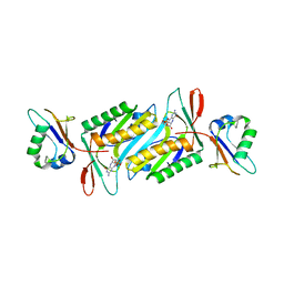
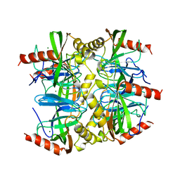
1QEZ
DownloadVisualize
BU of 1qez by Molmil
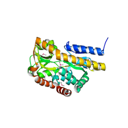
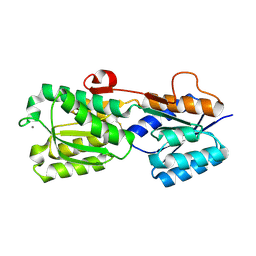
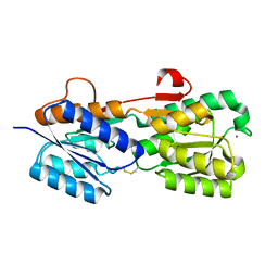
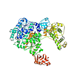
SULFOLOBUS ACIDOCALDARIUS INORGANIC PYROPHOSPHATASE: AN ARCHAEL PYROPHOSPHATASE.
Descriptor: MAGNESIUM ION, PROTEIN (INORGANIC PYROPHOSPHATASE)
Authors:Leppanen, V.-M, Nummelin, H, Hansen, T, Lahti, R, Schafer, G, Goldman, A.
Deposit date:1999-04-06
Release date:1999-04-14
Last modified:2023-08-16
Method:X-RAY DIFFRACTION (2.7 Å)
Cite:Sulfolobus acidocaldarius inorganic pyrophosphatase: structure, thermostability, and effect of metal ion in an archael pyrophosphatase.
Protein Sci., 8, 1999
2I74
DownloadVisualize
BU of 2i74 by Molmil
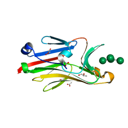
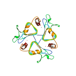
Crystal structure of mouse Peptide N-Glycanase C-terminal domain in complex with mannopentaose
Descriptor: ACETATE ION, GLYCEROL, PNGase, ...
Authors:Zhou, X, Zhao, G, Wang, L, Li, G, Lennarz, W.J, Schindelin, H.
Deposit date:2006-08-30
Release date:2006-10-24
Last modified:2023-08-30
Method:X-RAY DIFFRACTION (1.75 Å)
Cite:Structural and biochemical studies of the C-terminal domain of mouse peptide-N-glycanase identify it as a mannose-binding module.
Proc.Natl.Acad.Sci.Usa, 103, 2006
2HPG
DownloadVisualize
BU of 2hpg by Molmil
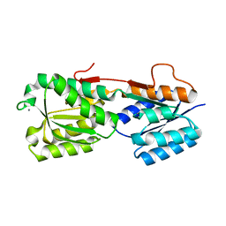
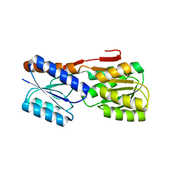
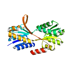
The crystal structure of a thermophilic TRAP periplasmic binding protein
Descriptor: ABC transporter, periplasmic substrate-binding protein
Authors:Cuneo, M.J, Hellinga, H.W.
Deposit date:2006-07-17
Release date:2007-03-20
Last modified:2024-10-30
Method:X-RAY DIFFRACTION (1.9 Å)
Cite:Structural analysis of a periplasmic binding protein in the tripartite ATP-independent transporter family reveals a tetrameric assembly that may have a role in ligand transport.
J.Biol.Chem., 283, 2008
2HPH
DownloadVisualize
BU of 2hph by Molmil
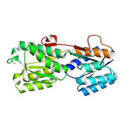
High resolution structure of E. coli glucose/galactose binding protein bound with glucose
Descriptor: CALCIUM ION, D-galactose-binding periplasmic protein, beta-D-glucopyranose
Authors:Cuneo, M.J, Hellinga, H.W.
Deposit date:2006-07-17
Release date:2007-03-20
Last modified:2023-08-30
Method:X-RAY DIFFRACTION (1.33 Å)
Cite:High resolution structure of E. coli glucose/galactose binding protein bound with glucose
To be Published
1NVI
DownloadVisualize
BU of 1nvi by Molmil
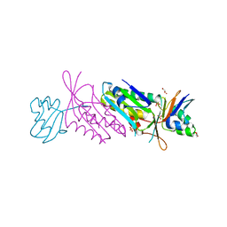
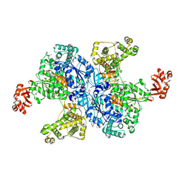
Orthorhombic Crystal Form of Molybdopterin Synthase
Descriptor: GLYCEROL, Molybdopterin converting factor subunit 1, Molybdopterin converting factor subunit 2, ...
Authors:Rudolph, M.J, Wuebbens, M.M, Turque, O, Rajagopalan, K.V, Schindelin, H.
Deposit date:2003-02-03
Release date:2003-05-06
Last modified:2023-08-16
Method:X-RAY DIFFRACTION (1.9 Å)
Cite:Structural Studies of Molybdopterin Synthase Provide Insights into its Catalytic Mechanism
J.Biol.Chem., 278, 2003
2IPM
DownloadVisualize
BU of 2ipm by Molmil
Crystal structure of a disulfide mutant glucose binding protein
Descriptor: CALCIUM ION, D-galactose-binding periplasmic protein, beta-D-glucopyranose
Authors:Cuneo, M.J, Hellinga, H.W.
Deposit date:2006-10-12
Release date:2007-03-20
Last modified:2023-08-30
Method:X-RAY DIFFRACTION (1.12 Å)
Cite:The free energy cost of domain reorganization in E. coli glucose binding protein
To be Published
2IPN
DownloadVisualize
BU of 2ipn by Molmil
Crystal structure of a disulfide mutant glucose binding protein
Descriptor: CALCIUM ION, D-galactose-binding periplasmic protein, beta-D-glucopyranose
Authors:Cuneo, M.J, Hellinga, H.W.
Deposit date:2006-10-12
Release date:2007-03-20
Last modified:2024-10-30
Method:X-RAY DIFFRACTION (1.15 Å)
Cite:The free energy cost of domain reorganization in E. coli glucose binding protein
To be Published
1THJ
DownloadVisualize
BU of 1thj by Molmil
CARBONIC ANHYDRASE FROM METHANOSARCINA
Descriptor: CARBONIC ANHYDRASE, ZINC ION
Authors:Kisker, C, Schindelin, H, Rees, D.C.
Deposit date:1996-04-02
Release date:1996-10-14
Last modified:2024-02-14
Method:X-RAY DIFFRACTION (2.8 Å)
Cite:A left-hand beta-helix revealed by the crystal structure of a carbonic anhydrase from the archaeon Methanosarcina thermophila.
EMBO J., 15, 1996
2IOY
DownloadVisualize
BU of 2ioy by Molmil
Crystal structure of Thermoanaerobacter tengcongensis ribose binding protein
Descriptor: Periplasmic sugar-binding protein, beta-D-ribopyranose
Authors:Cuneo, M.J, Hellinga, H.W.
Deposit date:2006-10-11
Release date:2007-03-20
Last modified:2024-02-21
Method:X-RAY DIFFRACTION (1.9 Å)
Cite:The backbone structure of the thermophilic Thermoanaerobacter tengcongensis ribose binding protein is essentially identical to its mesophilic E. coli homolog.
Bmc Struct.Biol., 8, 2008
2IPL
DownloadVisualize
BU of 2ipl by Molmil
Crystal structure of a disulfide mutant glucose binding protein
Descriptor: CALCIUM ION, D-galactose-binding periplasmic protein, beta-D-glucopyranose
Authors:Cuneo, M.J, Hellinga, H.W.
Deposit date:2006-10-12
Release date:2007-03-20
Last modified:2023-08-30
Method:X-RAY DIFFRACTION (1.2 Å)
Cite:The free energy cost of domain reorganization in E. coli glucose binding protein
To be Published
4NNJ
DownloadVisualize
BU of 4nnj by Molmil
Crystal structure of Uba1 in complex with ubiquitin-AMP and thioesterified ubiquitin
Descriptor: ADENOSINE MONOPHOSPHATE, GLYCEROL, SULFATE ION, ...
Authors:Schaefer, A, Schindelin, H.
Deposit date:2013-11-18
Release date:2014-05-07
Last modified:2024-10-16
Method:X-RAY DIFFRACTION (2.4 Å)
Cite:Structure of the ubiquitin-activating enzyme loaded with two ubiquitin molecules.
Acta Crystallogr.,Sect.D, 70, 2014
3C6Q
DownloadVisualize
BU of 3c6q by Molmil
Apo and ligand-bound form of a thermophilic glucose/xylose binding protein
Descriptor: Sugar ABC transporter, periplasmic sugar-binding protein, beta-D-xylopyranose
Authors:Cuneo, M.J, Hellinga, H.W.
Deposit date:2008-02-05
Release date:2008-02-26
Last modified:2023-08-30
Method:X-RAY DIFFRACTION (2.3 Å)
Cite:Open to closed transition of a thermophilic glucose binding protein
To be Published
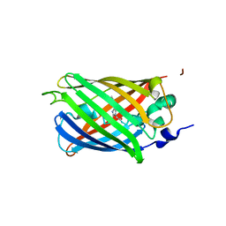
3SRY
DownloadVisualize
BU of 3sry by Molmil
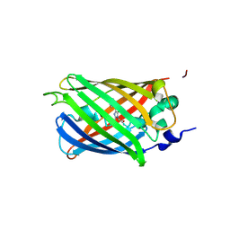
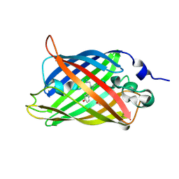
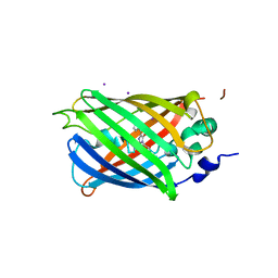
Engineered high-affinity halide-binding protein derived from YFP: halide-free
Descriptor: Green fluorescent protein
Authors:Wang, W, Grimley, J.S, Beese, L.S, Hellinga, H.W.
Deposit date:2011-07-07
Release date:2012-07-11
Last modified:2024-11-06
Method:X-RAY DIFFRACTION (1.159 Å)
Cite:Determination of engineered chloride-binding site structures in fluorescent proteins reveals principles of halide recognition
To be Published
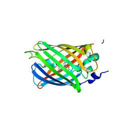
3SVB
DownloadVisualize
BU of 3svb by Molmil
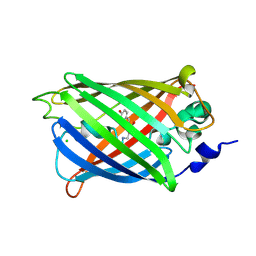
Engineered medium-affinity halide-binding protein derived from YFP: fluoride complex
Descriptor: 1,2-ETHANEDIOL, Green fluorescent protein
Authors:Wang, W, Grimley, J.S, Beese, L.S, Hellinga, H.W.
Deposit date:2011-07-12
Release date:2012-07-18
Last modified:2024-10-30
Method:X-RAY DIFFRACTION (1.3 Å)
Cite:Determination of engineered chloride-binding site structures in fluorescent proteins reveals principles of halide recognition
To be Published
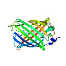
3SST
DownloadVisualize
BU of 3sst by Molmil
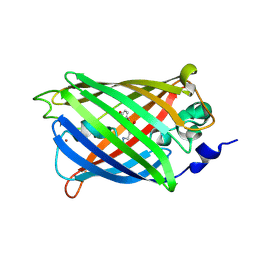
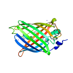
Engineered low-affinity halide-binding protein derived from YFP: chloride complex
Descriptor: CHLORIDE ION, Green fluorescent protein
Authors:Wang, W, Grimley, J.S, Beese, L.S, Hellinga, H.W.
Deposit date:2011-07-08
Release date:2012-07-11
Last modified:2023-11-15
Method:X-RAY DIFFRACTION (1.403 Å)
Cite:Determination of engineered chloride-binding site structures in fluorescent proteins reveals principles of halide recognition
To be Published
3SVE
DownloadVisualize
BU of 3sve by Molmil
Engineered low-affinity halide-binding protein derived from YFP: bromide complex
Descriptor: BROMIDE ION, Green fluorescent protein
Authors:Wang, W, Grimley, J.S, Beese, L.S, Hellinga, H.W.
Deposit date:2011-07-12
Release date:2012-07-18
Last modified:2024-10-09
Method:X-RAY DIFFRACTION (1.492 Å)
Cite:Determination of engineered chloride-binding site structures in fluorescent proteins reveals principles of halide recognition
To be Published
4EYI
DownloadVisualize
BU of 4eyi by Molmil
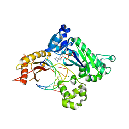
Human DNA polymerase iota incorporating dATP opposite N-(deoxyguanosin-8-yl)-1-aminopyrene lesion
Descriptor: 2'-DEOXYADENOSINE 5'-TRIPHOSPHATE, DNA polymerase iota, DNA primer, ...
Authors:Kirouac, K, Ling, H.
Deposit date:2012-05-01
Release date:2013-01-09
Last modified:2024-02-28
Method:X-RAY DIFFRACTION (2.9 Å)
Cite:Replication of a carcinogenic nitropyrene DNA lesion by human Y-family DNA polymerase.
Nucleic Acids Res., 41, 2013
2QIE
DownloadVisualize
BU of 2qie by Molmil
Staphylococcus aureus molybdopterin synthase in complex with precursor Z
Descriptor: (2R,4AR,5AR,11AR,12AS)-8-AMINO-2-HYDROXY-4A,5A,9,11,11A,12A-HEXAHYDRO[1,3,2]DIOXAPHOSPHININO[4',5':5,6]PYRANO[3,2-G]PTERIDINE-10,12(4H,6H)-DIONE 2-OXIDE, Molybdopterin synthase small subunit, Molybdopterin-converting factor subunit 2
Authors:Daniels, J.N, Schindelin, H.
Deposit date:2007-07-04
Release date:2008-02-19
Last modified:2023-08-30
Method:X-RAY DIFFRACTION (2.5 Å)
Cite:Crystal structure of a molybdopterin synthase-precursor Z complex: insight into its sulfur transfer mechanism and its role in molybdenum cofactor deficiency.
Biochemistry, 47, 2008
3SS0
DownloadVisualize
BU of 3ss0 by Molmil
Engineered high-affinity halide-binding protein derived from YFP: fluoride complex
Descriptor: Green fluorescent protein
Authors:Wang, W, Grimley, J.S, Beese, L.S, Hellinga, H.W.
Deposit date:2011-07-07
Release date:2012-07-11
Last modified:2024-10-16
Method:X-RAY DIFFRACTION (1.492 Å)
Cite:Determination of engineered chloride-binding site structures in fluorescent proteins reveals principles of halide recognition
To be Published
3SSH
DownloadVisualize
BU of 3ssh by Molmil
Engineered high-affinity halide-binding protein derived from YFP: chloride complex
Descriptor: CHLORIDE ION, Green fluorescent protein
Authors:Wang, W, Grimley, J.S, Beese, L.S, Hellinga, H.W.
Deposit date:2011-07-08
Release date:2012-07-11
Last modified:2024-10-16
Method:X-RAY DIFFRACTION (1.277 Å)
Cite:Determination of engineered chloride-binding site structures in fluorescent proteins reveals principles of halide recognition
To be Published
3SVC
DownloadVisualize
BU of 3svc by Molmil
Engineered medium-affinity halide-binding protein derived from YFP: chloride complex
Descriptor: 1,2-ETHANEDIOL, CHLORIDE ION, Green fluorescent protein
Authors:Wang, W, Grimley, J.S, Beese, L.S, Hellinga, H.W.
Deposit date:2011-07-12
Release date:2012-07-18
Last modified:2023-11-15
Method:X-RAY DIFFRACTION (1.31 Å)
Cite:Determination of engineered chloride-binding site structures in fluorescent proteins reveals principles of halide recognition
To be Published
3CMM
DownloadVisualize
BU of 3cmm by Molmil
Crystal Structure of the Uba1-Ubiquitin Complex
Descriptor: PROLINE, Ubiquitin, Ubiquitin-activating enzyme E1 1
Authors:Lee, I, Schindelin, H.
Deposit date:2008-03-23
Release date:2008-08-05
Last modified:2024-02-21
Method:X-RAY DIFFRACTION (2.7 Å)
Cite:Structural insights into E1-catalyzed ubiquitin activation and transfer to conjugating enzymes.
Cell(Cambridge,Mass.), 134, 2008
3SSL
DownloadVisualize
BU of 3ssl by Molmil
Engineered high-affinity halide-binding protein derived from YFP: iodide complex
Descriptor: Green fluorescent protein, IODIDE ION
Authors:Wang, W, Grimley, J.S, Beese, L.S, Hellinga, H.W.
Deposit date:2011-07-08
Release date:2012-07-11
Last modified:2023-11-15
Method:X-RAY DIFFRACTION (1.449 Å)
Cite:Determination of engineered chloride-binding site structures in fluorescent proteins reveals principles of halide recognition
To be published
3SSY
DownloadVisualize
BU of 3ssy by Molmil
Engineered low-affinity halide-binding protein derived from YFP: iodide complex
Descriptor: Green fluorescent protein, IODIDE ION
Authors:Wang, W, Grimley, J.S, Beese, L.S, Hellinga, H.W.
Deposit date:2011-07-08
Release date:2012-07-11
Last modified:2023-11-15
Method:X-RAY DIFFRACTION (1.769 Å)
Cite:Determination of engineered chloride-binding site structures in fluorescent proteins reveals principles of halide recognition
To be Published
1MVZ
DownloadVisualize
BU of 1mvz by Molmil
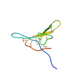
NMR solution structure of a Bowman Birk inhibitor isolated from snail medic seeds (Medicago Scutellata)
Descriptor: Bowman-Birk type protease inhibitor, (MSTI)
Authors:Catalano, M, Ragona, L, Molinari, H, Tava, A, Zetta, L.
Deposit date:2002-09-27
Release date:2003-04-22
Last modified:2022-02-23
Method:SOLUTION NMR
Cite:Anticarcinogenic Bowman Birk inhibitor isolated from snail medic seeds (Medicago Scutellata): solution structure and analysis of self-association behaviour
Biochemistry, 42, 2003

227111

PDB entries from 2024-11-06

PDB statisticsPDBj update infoContact PDBjnumon