Loading
PDBj
MenuPDBj@FacebookPDBj@TwitterPDBj@YouTubewwPDB FoundationwwPDB
RCSB PDBPDBeBMRBAdv. SearchSearch help
Search by PDB author
7KQZ
DownloadVisualize
BU of 7kqz by Molmil
Crystal Structure of Acetyl-CoA synthetase in complex with adenosine-5'-ethylphosphate from Coccidioides immitis RS
Descriptor: 1,2-ETHANEDIOL, 5'-O-[(S)-ethoxy(hydroxy)phosphoryl]adenosine, Acetyl-coenzyme A synthetase
Authors:Seattle Structural Genomics Center for Infectious Disease (SSGCID)
Deposit date:2020-11-18
Release date:2020-12-16
Last modified:2023-10-18
Method:X-RAY DIFFRACTION (2.15 Å)
Cite:Crystal Structure of Acetyl-CoA synthetase in complex with adenosine-5'-ethylphosphate from Coccidioides immitis RS
to be published
7KM9
DownloadVisualize
BU of 7km9 by Molmil
Crystal Structure of Dihydrofolate reductase (DHFR) from Mycobacterium ulcerans Agy99 in complex with NADP and inhibitor SDDC-0001913, tetragonal crystal from
Descriptor: (2R)-2-[(3-{3-[(2,4-diamino-6-ethylpyrimidin-5-yl)oxy]propoxy}phenyl)methyl]-3,3-difluoropropanoic acid, BROMIDE ION, CHLORIDE ION, ...
Authors:Seattle Structural Genomics Center for Infectious Disease (SSGCID)
Deposit date:2020-11-02
Release date:2021-07-07
Last modified:2023-10-18
Method:X-RAY DIFFRACTION (1.95 Å)
Cite:Crystal Structure of Dihydrofolate reductase (DHFR) from Mycobacterium ulcerans Agy99 in complex with NADP and inhibitor SDDC-0001913, tetragonal crystal form
to be published
7KI8
DownloadVisualize
BU of 7ki8 by Molmil
Crystal Structure of Dihydrofolate reductase (DHFR) from Mycobacterium ulcerans Agy99 in complex with NADP and inhibitor SDDC-0001580
Descriptor: 1-[2-({3-[(2,4-diamino-6-ethylpyrimidin-5-yl)oxy]propyl}amino)phenyl]piperidine-4-carboxylic acid, Dihydrofolate reductase, NADP NICOTINAMIDE-ADENINE-DINUCLEOTIDE PHOSPHATE
Authors:Seattle Structural Genomics Center for Infectious Disease (SSGCID)
Deposit date:2020-10-23
Release date:2021-07-07
Last modified:2023-10-18
Method:X-RAY DIFFRACTION (1.25 Å)
Cite:Crystal Structure of Dihydrofolate reductase (DHFR) from Mycobacterium ulcerans Agy99 in complex with NADP and inhibitor SDDC-0001580
to be published
7KM8
DownloadVisualize
BU of 7km8 by Molmil
Crystal Structure of Dihydrofolate reductase (DHFR) from Mycobacterium ulcerans Agy99 in complex with NADP and inhibitor SDDC-0001914, orthorhombic crystal from
Descriptor: (2S)-2-[(3-{3-[(2,4-diamino-6-ethylpyrimidin-5-yl)oxy]propoxy}phenyl)methyl]-3,3-difluoropropanoic acid, CHLORIDE ION, Dihydrofolate reductase, ...
Authors:Seattle Structural Genomics Center for Infectious Disease (SSGCID)
Deposit date:2020-11-02
Release date:2021-07-07
Last modified:2023-10-18
Method:X-RAY DIFFRACTION (1.55 Å)
Cite:Crystal Structure of Dihydrofolate reductase (DHFR) from Mycobacterium ulcerans Agy99 in complex with NADP and inhibitor SDDC-0001914, orthorhombic crystal form
to be published
7KI9
DownloadVisualize
BU of 7ki9 by Molmil
Crystal Structure of Dihydrofolate reductase (DHFR) from Mycobacterium ulcerans Agy99 in complex with NADP and inhibitor SDDC-0001912
Descriptor: 1-[2-(3-{[(2,4-diamino-6-ethylpyrimidin-5-yl)oxy]methyl}azetidin-1-yl)phenyl]piperidine-4-carboxylic acid, Dihydrofolate reductase, NADP NICOTINAMIDE-ADENINE-DINUCLEOTIDE PHOSPHATE
Authors:Seattle Structural Genomics Center for Infectious Disease (SSGCID)
Deposit date:2020-10-23
Release date:2021-07-07
Last modified:2023-10-18
Method:X-RAY DIFFRACTION (1.25 Å)
Cite:Crystal Structure of Dihydrofolate reductase (DHFR) from Mycobacterium ulcerans Agy99 in complex with NADP and inhibitor SDDC-0001912
to be published
7KM7
DownloadVisualize
BU of 7km7 by Molmil
Crystal Structure of Dihydrofolate reductase (DHFR) from Mycobacterium ulcerans Agy99 in complex with NADP and inhibitor SDDC-0001914, tetragonal crystal from
Descriptor: (2S)-2-[(3-{3-[(2,4-diamino-6-ethylpyrimidin-5-yl)oxy]propoxy}phenyl)methyl]-3,3-difluoropropanoic acid, BROMIDE ION, Dihydrofolate reductase, ...
Authors:Seattle Structural Genomics Center for Infectious Disease (SSGCID)
Deposit date:2020-11-02
Release date:2021-07-07
Last modified:2023-10-18
Method:X-RAY DIFFRACTION (1.8 Å)
Cite:Crystal Structure of Dihydrofolate reductase (DHFR) from Mycobacterium ulcerans Agy99 in complex with NADP and inhibitor SDDC-0001914, tetragonal crystal form
to be published
7U0S
DownloadVisualize
BU of 7u0s by Molmil
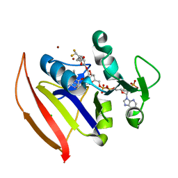
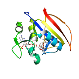
Crystal Structure of FK506-binding protein 1A from Aspergillus fumigatus Bound to Ascomycin
Descriptor: (3S,4R,5S,8R,9E,12S,14S,15R,16S,18R,19R,22R,26aS)-8-ethyl-5,19-dihydroxy-3-{(1E)-1-[(1R,3R,4R)-4-hydroxy-3-methoxycyclohexyl]prop-1-en-2-yl}-14,16-dimethoxy-4,10,12,18-tetramethyl-5,6,8,11,12,13,14,15,16,17,18,19,24,25,26,26a-hexadecahydro-3H-15,19-epoxypyrido[2,1-c][1,4]oxazacyclotricosine-1,7,20,21(4H,23H)-tetrone, ACETATE ION, FK506-binding protein 1A, ...
Authors:DeBouver, N.D, Fox III, D, Hoy, M.J, Heitman, J, Lorimer, D.D, Horanyi, P.S, Edwards, T.E, Seattle Structural Genomics Center for Infectious Disease (SSGCID)
Deposit date:2022-02-18
Release date:2022-07-27
Last modified:2023-10-18
Method:X-RAY DIFFRACTION (1.7 Å)
Cite:Structure-Guided Synthesis of FK506 and FK520 Analogs with Increased Selectivity Exhibit In Vivo Therapeutic Efficacy against Cryptococcus.
Mbio, 13, 2022
7U0U
DownloadVisualize
BU of 7u0u by Molmil
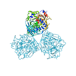
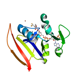
Crystal Structure of a Aspergillus fumigatus Calcineurin A - Calcineurin B fusion bound to FKBP12 and FK-506
Descriptor: 8-DEETHYL-8-[BUT-3-ENYL]-ASCOMYCIN, CALCIUM ION, PHOSPHATE ION, ...
Authors:Fox III, D, Abendroth, J, DeBouver, N.D, Hoy, M.J, Heitman, J, Lorimer, D.D, Horanyi, P.S, Edwards, T.E, Seattle Structural Genomics Center for Infectious Disease (SSGCID)
Deposit date:2022-02-18
Release date:2022-08-03
Last modified:2023-10-18
Method:X-RAY DIFFRACTION (1.9 Å)
Cite:Structure-Guided Synthesis of FK506 and FK520 Analogs with Increased Selectivity Exhibit In Vivo Therapeutic Efficacy against Cryptococcus.
Mbio, 13, 2022
7U0T
DownloadVisualize
BU of 7u0t by Molmil
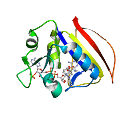
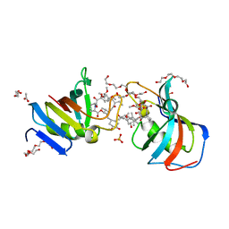
Crystal Structure of a human Calcineurin A - Calcineurin B fusion bound to FKBP12 and FK-520
Descriptor: (3S,4R,5S,8R,9E,12S,14S,15R,16S,18R,19R,22R,26aS)-8-ethyl-5,19-dihydroxy-3-{(1E)-1-[(1R,3R,4R)-4-hydroxy-3-methoxycyclohexyl]prop-1-en-2-yl}-14,16-dimethoxy-4,10,12,18-tetramethyl-5,6,8,11,12,13,14,15,16,17,18,19,24,25,26,26a-hexadecahydro-3H-15,19-epoxypyrido[2,1-c][1,4]oxazacyclotricosine-1,7,20,21(4H,23H)-tetrone, 1,2-ETHANEDIOL, CALCIUM ION, ...
Authors:Fox III, D, Mayclin, S.J, DeBouver, N.D, Hoy, M.J, Heitman, J, Lorimer, D.D, Horanyi, P.S, Edwards, T.E, Seattle Structural Genomics Center for Infectious Disease (SSGCID)
Deposit date:2022-02-18
Release date:2022-08-03
Last modified:2023-10-18
Method:X-RAY DIFFRACTION (2.45 Å)
Cite:Structure-Guided Synthesis of FK506 and FK520 Analogs with Increased Selectivity Exhibit In Vivo Therapeutic Efficacy against Cryptococcus.
Mbio, 13, 2022
6UDF
DownloadVisualize
BU of 6udf by Molmil
Crystal structure of Enoyl-[acyl-carrier-protein] reductase [NADH] (InhA) from Mycobacterium kansasii
Descriptor: Enoyl-[acyl-carrier-protein] reductase [NADH], FORMIC ACID
Authors:Seattle Structural Genomics Center for Infectious Disease (SSGCID)
Deposit date:2019-09-19
Release date:2019-10-16
Last modified:2023-10-11
Method:X-RAY DIFFRACTION (1.95 Å)
Cite:Crystal stucture of Enoyl-[acyl-carrier-protein] reductase [NADH] (InhA) from Mycobacterium kansasii
to be published
6UJ5
DownloadVisualize
BU of 6uj5 by Molmil
Crystal structure of CAB1 Pantothenate Kinase from Saccharomyces cerevisiae
Descriptor: MAGNESIUM ION, Pantothenate kinase CAB1
Authors:Seattle Structural Genomics Center for Infectious Disease (SSGCID)
Deposit date:2019-10-02
Release date:2020-10-07
Last modified:2024-03-13
Method:X-RAY DIFFRACTION (1.8 Å)
Cite:Crystal structure of CAB1 Pantothenate Kinase from Saccharomyces cerevisiae
to be published
6UJK
DownloadVisualize
BU of 6ujk by Molmil
Crystal Structure of a Probable short-chain type dehydrogenase/reductase (Rv1144) from Mycobacterium tuberculosis with bound NAD
Descriptor: 1,2-ETHANEDIOL, 2-AMINO-2-HYDROXYMETHYL-PROPANE-1,3-DIOL, NICOTINAMIDE-ADENINE-DINUCLEOTIDE, ...
Authors:Seattle Structural Genomics Center for Infectious Disease (SSGCID)
Deposit date:2019-10-03
Release date:2019-10-16
Last modified:2023-10-11
Method:X-RAY DIFFRACTION (1.2 Å)
Cite:Crystal Structure of a Probable short-chain type dehydrogenase/reductase (Rv1144) from Mycobacterium tuberculosis with bound NAD
to be published
6UK3
DownloadVisualize
BU of 6uk3 by Molmil
Crystal Structure of S-adenosyl-L-homocysteine hydrolase from Acanthamoeba castellanii with bound NAD and Adenosine
Descriptor: ACETATE ION, ADENOSINE, Adenosylhomocysteinase, ...
Authors:Seattle Structural Genomics Center for Infectious Disease (SSGCID)
Deposit date:2019-10-04
Release date:2019-10-16
Last modified:2023-10-11
Method:X-RAY DIFFRACTION (1.4 Å)
Cite:Crystal Structure of S-adenosyl-L-homocysteine hydrolase from Acanthamoeba castellanii with bound NAD and Adenosine
to be published
6ULO
DownloadVisualize
BU of 6ulo by Molmil
Structure of an N-terminally truncated uncharacterized protein from Leptospira interrogans serogroup Icterohaemorrhagiae serovar Copenhageni (strain Fiocruz L1-130)
Descriptor: CHLORIDE ION, Uncharacterized protein
Authors:Seattle Structural Genomics Center for Infectious Disease (SSGCID)
Deposit date:2019-10-08
Release date:2019-10-16
Last modified:2024-03-13
Method:X-RAY DIFFRACTION (1.3 Å)
Cite:Structure of an N-terminally truncated uncharacterized protein from Leptospira interrogans serogroup Icterohaemorrhagiae serovar Copenhageni (strain Fiocruz L1-130)
to be published
6TYJ
DownloadVisualize
BU of 6tyj by Molmil
Crystal structure of zinc-bound Hemerythrin HHE cation binding domain-containing protein (soak): Rv2633c homolog from Mycobacterium kansasii
Descriptor: Hemerythrin HHE cation binding domain protein, ZINC ION
Authors:Seattle Structural Genomics Center for Infectious Disease (SSGCID)
Deposit date:2019-08-09
Release date:2020-01-22
Last modified:2023-10-11
Method:X-RAY DIFFRACTION (1.6 Å)
Cite:Crystal structure of zinc-bound Hemerythrin HHE cation binding domain-containing protein (soak): Rv2633c homolog from Mycobacterium kansasii
to be published
6UH2
DownloadVisualize
BU of 6uh2 by Molmil
Crystal Structure of Short Chain Dehydrogenase from Leptospira borgpetersenii serovar Hardjo-bovis (Strain JB197) with bound NAD+
Descriptor: NICOTINAMIDE-ADENINE-DINUCLEOTIDE, Short chain dehydrogenase
Authors:Seattle Structural Genomics Center for Infectious Disease (SSGCID)
Deposit date:2019-09-26
Release date:2019-10-09
Last modified:2023-10-11
Method:X-RAY DIFFRACTION (1.9 Å)
Cite:TBD
TO BE PUBLISHED
6ULD
DownloadVisualize
BU of 6uld by Molmil
Crystal structure of serine hydroxymethyltransferase from Mycobacterium tuberculosis with bound PLP forming a Schiff base with substrate Serine in one monomer and PLP forming a Schiff base with product Glycine in the other monomer
Descriptor: (4S)-2-METHYL-2,4-PENTANEDIOL, GLYCINE, PYRIDOXAL-5'-PHOSPHATE, ...
Authors:Seattle Structural Genomics Center for Infectious Disease (SSGCID)
Deposit date:2019-10-07
Release date:2019-10-16
Last modified:2023-10-11
Method:X-RAY DIFFRACTION (1.5 Å)
Cite:Crystal structure of serine hydroxymethyltransferase from Mycobacterium tuberculosis with bound PLP forming a Schiff base with substrate Serine in one monomer and PLP forming a Schiff base with product Glycine in the other monomer
to be published
6UCZ
DownloadVisualize
BU of 6ucz by Molmil
Crystal structure of dihydropteroate synthase from Anaplasma phagocytophilum with bound 6-hydroxymethylpterin-monophosphate
Descriptor: 1,2-ETHANEDIOL, ACETATE ION, CITRIC ACID, ...
Authors:Seattle Structural Genomics Center for Infectious Disease (SSGCID)
Deposit date:2019-09-18
Release date:2019-10-16
Last modified:2023-10-11
Method:X-RAY DIFFRACTION (2 Å)
Cite:Crystal structure of dihydropteroate synthase from Anaplasma phagocytophilum with bound 6-hydroxymethylpterin-monophosphate
To be Published
6UDE
DownloadVisualize
BU of 6ude by Molmil
Crystal structure of Glycerol kinase from Elizabethkingia anophelis NUHP1 in complex with ADP and glycerol
Descriptor: ADENOSINE-5'-DIPHOSPHATE, GLYCEROL, Glycerol kinase, ...
Authors:Seattle Structural Genomics Center for Infectious Disease (SSGCID)
Deposit date:2019-09-19
Release date:2019-10-16
Last modified:2023-10-11
Method:X-RAY DIFFRACTION (1.95 Å)
Cite:Crystal structure of Glycerol kinase from Elizabethkingia anophelis NUHP1 in complex with ADP and glycerol
TO BE PUBLISHED
6UDG
DownloadVisualize
BU of 6udg by Molmil
Crystal structure of a Probable thiol peroxidase from Elizabethkingia anophelis NUHP1
Descriptor: Thiol peroxidase
Authors:Seattle Structural Genomics Center for Infectious Disease (SSGCID)
Deposit date:2019-09-19
Release date:2019-10-16
Last modified:2024-04-03
Method:X-RAY DIFFRACTION (2.65 Å)
Cite:Crystal structure of a Probable thiol peroxidase from Elizabethkingia anophelis NUHP1
TO BE PUBLISHED
6UYN
DownloadVisualize
BU of 6uyn by Molmil
Crystal structure of influenza A virus hemagglutinin from A/Ohio/09/2015 bound to the stalk-binding CR6261 antibody Fab
Descriptor: 2-acetamido-2-deoxy-beta-D-glucopyranose, 2-acetamido-2-deoxy-beta-D-glucopyranose-(1-4)-2-acetamido-2-deoxy-beta-D-glucopyranose, CHLORIDE ION, ...
Authors:Seattle Structural Genomics Center for Infectious Disease, Seattle Structural Genomics Center for Infectious Disease (SSGCID)
Deposit date:2019-11-13
Release date:2020-09-02
Last modified:2023-10-11
Method:X-RAY DIFFRACTION (2.85 Å)
Cite:Crystal structure of influenza A virus hemagglutinin from A/Ohio/09/2015 bound to the stalk-binding CR6261 antibody Fab
To Be Published
6UM4
DownloadVisualize
BU of 6um4 by Molmil
Crystal structure of malate dehydrogenase from Naegleria fowleri ATCC 30863
Descriptor: 1,2-ETHANEDIOL, Malate dehydrogenase
Authors:Seattle Structural Genomics Center for Infectious Disease (SSGCID)
Deposit date:2019-10-09
Release date:2019-10-23
Last modified:2024-04-03
Method:X-RAY DIFFRACTION (2.05 Å)
Cite:Crystal structure of malate dehydrogenase from Naegleria fowleri ATCC 30863
TO BE PUBLISHED
6UJD
DownloadVisualize
BU of 6ujd by Molmil
Crystal structure of Cysteine-tRNA ligase from Elizabethkingia sp.
Descriptor: 1,2-ETHANEDIOL, 1-METHOXY-2-[2-(2-METHOXY-ETHOXY]-ETHANE, CHLORIDE ION, ...
Authors:Seattle Structural Genomics Center for Infectious Disease (SSGCID)
Deposit date:2019-10-02
Release date:2019-10-16
Last modified:2023-10-11
Method:X-RAY DIFFRACTION (2.6 Å)
Cite:Crystal structure of Cysteine-tRNA ligase from Elizabethkingia sp.
TO BE PUBLISHED
6V45
DownloadVisualize
BU of 6v45 by Molmil
Crystal structure of a Probable carnitine operon oxidoreductase caia from Brucella melitensis
Descriptor: Probable carnitine operon oxidoreductase caia, SULFATE ION
Authors:Seattle Structural Genomics Center for Infectious Disease (SSGCID)
Deposit date:2019-11-27
Release date:2019-12-11
Last modified:2024-04-03
Method:X-RAY DIFFRACTION (2.6 Å)
Cite:Crystal structure of a Probable carnitine operon oxidoreductase caia from Brucella melitensis
to be published
6UYH
DownloadVisualize
BU of 6uyh by Molmil
Crystal structure of prolyl-tRNA synthetase from Naegleria fowleri in complex with halofuginone and AMPPNP
Descriptor: 1,2-ETHANEDIOL, 7-bromo-6-chloro-3-{3-[(2R,3S)-3-hydroxypiperidin-2-yl]-2-oxopropyl}quinazolin-4(3H)-one, MAGNESIUM ION, ...
Authors:Seattle Structural Genomics Center for Infectious Disease (SSGCID)
Deposit date:2019-11-13
Release date:2019-11-27
Last modified:2023-10-11
Method:X-RAY DIFFRACTION (1.75 Å)
Cite:Crystal structure of prolyl-tRNA synthetase from Naegleria fowleri in complex with halofuginone and AMPPNP
TO BE PUBLISHED

222926

PDB entries from 2024-07-24

PDB statisticsPDBj update infoContact PDBjnumon