Loading
PDBj
MenuPDBj@FacebookPDBj@TwitterPDBj@YouTubewwPDB FoundationwwPDB
RCSB PDBPDBeBMRBAdv. SearchSearch help
Search by PDB author
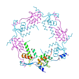
2OB9
DownloadVisualize
BU of 2ob9 by Molmil
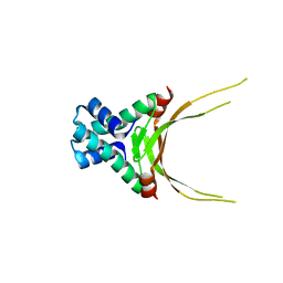
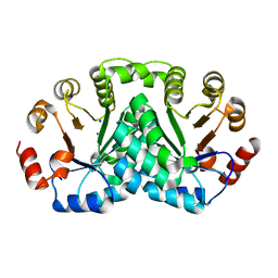
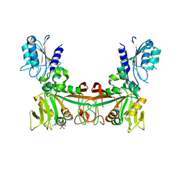
Structure of bacteriophage HK97 tail assembly chaperone
Descriptor: Tail assembly chaperone
Authors:McGrath, T.E, Tuite, A, Bona, D, Saridakis, V, Edwards, A.M, Maxwell, K, Chirgadze, N.Y.
Deposit date:2006-12-18
Release date:2007-12-18
Last modified:2024-10-16
Method:X-RAY DIFFRACTION (2.3 Å)
Cite:A conserved spiral structure for highly diverged phage tail assembly chaperones.
J.Mol.Biol., 425, 2013
6NTY
DownloadVisualize
BU of 6nty by Molmil
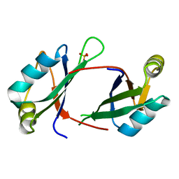
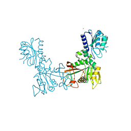
2.1 A resolution structure of the Musashi-2 (Msi2) RNA recognition motif 1 (RRM1) domain
Descriptor: PHOSPHATE ION, RNA-binding protein Musashi homolog 2
Authors:Lovell, S, Kashipathy, M.M, Battaile, K.P, Lan, L, Xiaoqing, W, Cooper, A, Gao, F.P, Xu, L.
Deposit date:2019-01-30
Release date:2019-10-23
Last modified:2023-10-11
Method:X-RAY DIFFRACTION (2.1 Å)
Cite:Crystal and solution structures of human oncoprotein Musashi-2 N-terminal RNA recognition motif 1.
Proteins, 88, 2020
5FBM
DownloadVisualize
BU of 5fbm by Molmil
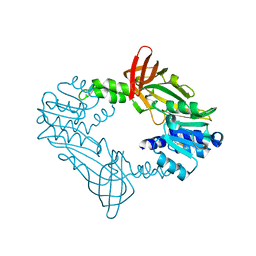
Crystal Structure of Histone Like Protein (HLP) from Streptococcus mutans Refined to 1.9 A Resolution
Descriptor: DNA-binding protein HU
Authors:Lovell, S, Battaile, K.P, Mehzabeen, N, O'Neil, P, Biswas, I.
Deposit date:2015-12-14
Release date:2016-04-06
Last modified:2023-09-27
Method:X-RAY DIFFRACTION (1.9 Å)
Cite:Crystal structure of histone-like protein from Streptococcus mutans refined to 1.9 angstrom resolution.
Acta Crystallogr F Struct Biol Commun, 72, 2016
3CXN
DownloadVisualize
BU of 3cxn by Molmil
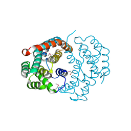
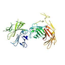
Structure of the Urease Accessory Protein UreF from Helicobacter pylori
Descriptor: GLYCEROL, Urease accessory protein ureF
Authors:Lam, R, Johns, K, Romanov, V, Dong, A, Wu-Brown, J, Guthrie, J, Dharamsi, A, Thambipillai, D, Mansoury, K, Edwards, A.M, Pai, E.F, Chirgadze, N.Y.
Deposit date:2008-04-24
Release date:2009-05-12
Last modified:2024-10-09
Method:X-RAY DIFFRACTION (1.55 Å)
Cite:Crystal structure of a truncated urease accessory protein UreF from Helicobacter pylori.
Proteins, 78, 2010
3T3S
DownloadVisualize
BU of 3t3s by Molmil
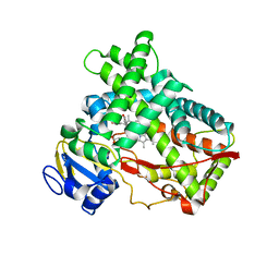
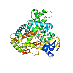
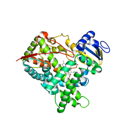
Human Cytochrome P450 2A13 in complex with Pilocarpine
Descriptor: (3S,4R)-3-ethyl-4-[(1-methyl-1H-imidazol-5-yl)methyl]dihydrofuran-2(3H)-one, Cytochrome P450 2A13, PROTOPORPHYRIN IX CONTAINING FE
Authors:DeVore, N.M, Scott, E.E.
Deposit date:2011-07-25
Release date:2011-12-07
Last modified:2023-09-13
Method:X-RAY DIFFRACTION (3 Å)
Cite:Structural comparison of cytochromes P450 2A6, 2A13, and 2E1 with pilocarpine.
Febs J., 279, 2012
3T3R
DownloadVisualize
BU of 3t3r by Molmil
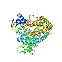
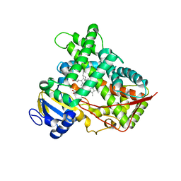
Human Cytochrome P450 2A6 in complex with Pilocarpine
Descriptor: (3S,4R)-3-ethyl-4-[(1-methyl-1H-imidazol-5-yl)methyl]dihydrofuran-2(3H)-one, Cytochrome P450 2A6, PROTOPORPHYRIN IX CONTAINING FE
Authors:DeVore, N.M, Scott, E.E.
Deposit date:2011-07-25
Release date:2011-12-07
Last modified:2023-09-13
Method:X-RAY DIFFRACTION (2.4 Å)
Cite:Structural comparison of cytochromes P450 2A6, 2A13, and 2E1 with pilocarpine.
Febs J., 279, 2012
3T3Q
DownloadVisualize
BU of 3t3q by Molmil
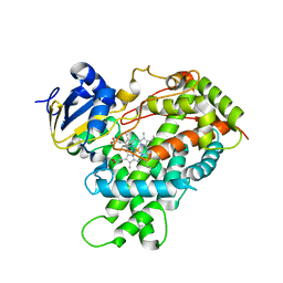
Human Cytochrome P450 2A6 I208S/I300F/G301A/S369G in complex with Pilocarpine
Descriptor: (3S,4R)-3-ethyl-4-[(1-methyl-1H-imidazol-5-yl)methyl]dihydrofuran-2(3H)-one, Cytochrome P450 2A6, PROTOPORPHYRIN IX CONTAINING FE
Authors:DeVore, N.M, Scott, E.E.
Deposit date:2011-07-25
Release date:2011-12-07
Last modified:2023-09-13
Method:X-RAY DIFFRACTION (2.1 Å)
Cite:Structural comparison of cytochromes P450 2A6, 2A13, and 2E1 with pilocarpine.
Febs J., 279, 2012
3T3Z
DownloadVisualize
BU of 3t3z by Molmil
Human Cytochrome P450 2E1 in complex with pilocarpine
Descriptor: (3S,4R)-3-ethyl-4-[(1-methyl-1H-imidazol-5-yl)methyl]dihydrofuran-2(3H)-one, Cytochrome P450 2E1, PROTOPORPHYRIN IX CONTAINING FE, ...
Authors:Meneely, K.M, DeVore, N.M, Scott, E.E.
Deposit date:2011-07-25
Release date:2011-12-07
Last modified:2023-11-29
Method:X-RAY DIFFRACTION (2.35 Å)
Cite:Structural comparison of cytochromes P450 2A6, 2A13, and 2E1 with pilocarpine.
Febs J., 279, 2012
6DE6
DownloadVisualize
BU of 6de6 by Molmil
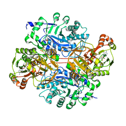
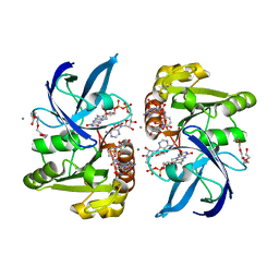
2.1 A resolution structure of histamine dehydrogenase from Rhizobium sp. 4-9
Descriptor: ADENOSINE-5'-DIPHOSPHATE, FLAVIN MONONUCLEOTIDE, Histamine dehydrogenase, ...
Authors:Goyal, P, Lovell, S, Richter, M.
Deposit date:2018-05-11
Release date:2019-05-15
Last modified:2024-10-23
Method:X-RAY DIFFRACTION (2.1 Å)
Cite:Structure of Rhizobium sp. 4-9 histamine dehydrogenase and analysis of the electron transfer pathway to an abiological electron acceptor.
Arch.Biochem.Biophys., 2023
3KOH
DownloadVisualize
BU of 3koh by Molmil
Cytochrome P450 2E1 with omega-imidazolyl octanoic acid
Descriptor: 8-(1H-imidazol-1-yl)octanoic acid, Cytochrome P450 2E1, PROTOPORPHYRIN IX CONTAINING FE
Authors:Scott, E.E, Porubsky, P.R.
Deposit date:2009-11-13
Release date:2010-05-12
Last modified:2023-09-06
Method:X-RAY DIFFRACTION (2.9 Å)
Cite:Human cytochrome P450 2E1 structures with fatty acid analogs reveal a previously unobserved binding mode.
J.Biol.Chem., 285, 2010
3LC4
DownloadVisualize
BU of 3lc4 by Molmil
Human Cytochrome P450 2E1 in Complex with Omega-Imidazolyl-Dodecanoic Acid
Descriptor: 12-(1H-imidazol-1-yl)dodecanoic acid, Cytochrome P450 2E1, PROTOPORPHYRIN IX CONTAINING FE
Authors:Scott, E.E, Porubsky, P.R.
Deposit date:2010-01-09
Release date:2010-05-12
Last modified:2023-09-06
Method:X-RAY DIFFRACTION (3.1 Å)
Cite:Human cytochrome P450 2E1 structures with fatty acid analogs reveal a previously unobserved binding mode.
J.Biol.Chem., 285, 2010
4DC7
DownloadVisualize
BU of 4dc7 by Molmil
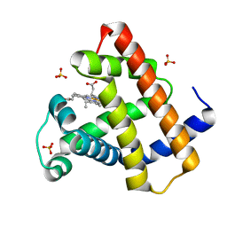
Crystal Structure of Myoglobin Exposed to Excessive SONICC Imaging Laser Dose.
Descriptor: Myoglobin, PROTOPORPHYRIN IX CONTAINING FE, SULFATE ION
Authors:Becker, M, Mulichak, A.M, Kissick, D.J, Fischetti, R.F, Keefe, L.J, Simpson, G.J.
Deposit date:2012-01-17
Release date:2013-01-23
Last modified:2024-02-28
Method:X-RAY DIFFRACTION (1.5 Å)
Cite:Towards protein-crystal centering using second-harmonic generation (SHG) microscopy.
Acta Crystallogr.,Sect.D, 69, 2013
8DOR
DownloadVisualize
BU of 8dor by Molmil
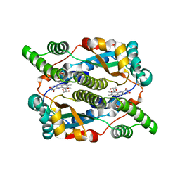
Crystal structure of Dihydropteridine reductase/oxygen-insensitive NAD(P)H nitroreductase from Klebsiella pneumoniae
Descriptor: Dihydropteridine reductase/oxygen-insensitive NAD(P)H nitroreductase, FLAVIN MONONUCLEOTIDE, GLYCEROL, ...
Authors:Seattle Structural Genomics Center for Infectious Disease (SSGCID)
Deposit date:2022-07-14
Release date:2022-07-27
Last modified:2024-08-21
Method:X-RAY DIFFRACTION (1.35 Å)
Cite:Crystal structures of NAD(P)H nitroreductases from Klebsiella pneumoniae.
Acta Crystallogr.,Sect.F, 80, 2024
8DOP
DownloadVisualize
BU of 8dop by Molmil
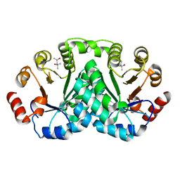
Crystal structure of 2,3-diketo-5-methylthiopentyl-1-phosphate enolase-phosphatase from Klebsiella aerogenes (P1 Form)
Descriptor: 2,3-diketo-5-methylthiopentyl-1-phosphate enolase-phosphatase, IODIDE ION, SODIUM ION
Authors:Seattle Structural Genomics Center for Infectious Disease (SSGCID)
Deposit date:2022-07-14
Release date:2022-07-27
Last modified:2023-11-22
Method:X-RAY DIFFRACTION (2 Å)
Cite:Crystal structure of 2,3-diketo-5-methylthiopentyl-1-phosphate enolase-phosphatase from Klebsiella aerogenes (P1 Form)
To be published
8DOS
DownloadVisualize
BU of 8dos by Molmil
Crystal structure of Ferredoxin (flavodoxin):NADP(+) oxidoreductase from Klebsiella pneumoniae
Descriptor: CALCIUM ION, FLAVIN-ADENINE DINUCLEOTIDE, Flavodoxin/ferredoxin--NADP reductase, ...
Authors:Seattle Structural Genomics Center for Infectious Disease (SSGCID)
Deposit date:2022-07-14
Release date:2022-07-27
Last modified:2023-11-22
Method:X-RAY DIFFRACTION (1.872 Å)
Cite:Crystal structure of Ferredoxin (flavodoxin):NADP(+) oxidoreductase from Klebsiella pneumoniae
To be published
8DQC
DownloadVisualize
BU of 8dqc by Molmil
Crystal structure of 3-dehydroquinate dehydratase I from Klebsiella oxytoca (I222 Form)
Descriptor: 3-dehydroquinate dehydratase I, ACETATE ION, ZINC ION
Authors:Seattle Structural Genomics Center for Infectious Disease, Seattle Structural Genomics Center for Infectious Disease (SSGCID)
Deposit date:2022-07-18
Release date:2022-07-27
Last modified:2023-11-22
Method:X-RAY DIFFRACTION (2.7 Å)
Cite:Crystal structure of 3-dehydroquinate dehydratase I from Klebsiella oxytoca (I222 Form)
To be published
8DOQ
DownloadVisualize
BU of 8doq by Molmil
Crystal structure of 2,3-diketo-5-methylthiopentyl-1-phosphate enolase-phosphatase from Klebsiella aerogenes (P21 Form)
Descriptor: (4S)-2-METHYL-2,4-PENTANEDIOL, 2,3-diketo-5-methylthiopentyl-1-phosphate enolase-phosphatase, BENZOIC ACID
Authors:Seattle Structural Genomics Center for Infectious Disease (SSGCID)
Deposit date:2022-07-14
Release date:2022-07-27
Last modified:2023-11-22
Method:X-RAY DIFFRACTION (1.65 Å)
Cite:Crystal structure of 2,3-diketo-5-methylthiopentyl-1-phosphate enolase-phosphatase from Klebsiella aerogenes (P1 Form)
To be published
8DQ9
DownloadVisualize
BU of 8dq9 by Molmil
Crystal structure of GDP bound 3-dehydroquinate dehydratase I from Klebsiella oxytoca
Descriptor: 3-dehydroquinate dehydratase I, CHLORIDE ION, GUANOSINE-5'-DIPHOSPHATE, ...
Authors:Seattle Structural Genomics Center for Infectious Disease, Seattle Structural Genomics Center for Infectious Disease (SSGCID)
Deposit date:2022-07-18
Release date:2022-07-27
Last modified:2023-11-22
Method:X-RAY DIFFRACTION (2.8 Å)
Cite:Crystal structure of GDP bound 3-dehydroquinate dehydratase I from Klebsiella oxytoca
To be published
8DQB
DownloadVisualize
BU of 8dqb by Molmil
Crystal structure of 3-dehydroquinate dehydratase I from Klebsiella oxytoca (I23 Form)
Descriptor: 3-dehydroquinate dehydratase I, ZINC ION
Authors:Seattle Structural Genomics Center for Infectious Disease, Seattle Structural Genomics Center for Infectious Disease (SSGCID)
Deposit date:2022-07-18
Release date:2022-07-27
Last modified:2023-11-22
Method:X-RAY DIFFRACTION (2.5 Å)
Cite:Crystal structure of 3-dehydroquinate dehydratase I from Klebsiella oxytoca (I23 Form)
To be published
8DUR
DownloadVisualize
BU of 8dur by Molmil
Crystal structure of apo protein arginine N-methyltransferase 1 (PRMT1) from Naegleria fowleri
Descriptor: Protein arginine N-methyltransferase 1 (PRMT1)
Authors:Seattle Structural Genomics Center for Infectious Disease (SSGCID)
Deposit date:2022-07-27
Release date:2022-08-10
Last modified:2024-10-30
Method:X-RAY DIFFRACTION (1.97 Å)
Cite:Crystal structure of apo protein arginine N-methyltransferase 1 (PRMT1) from Naegleria fowleri
To be published
6V02
DownloadVisualize
BU of 6v02 by Molmil
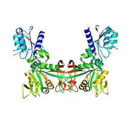
N-terminal 5 domains of CI-MPR
Descriptor: 2-acetamido-2-deoxy-beta-D-glucopyranose, Cation-independent mannose-6-phosphate receptor
Authors:Olson, L.J, Dahms, N.M, Kim, J.-J.P.
Deposit date:2019-11-18
Release date:2020-09-30
Last modified:2024-10-16
Method:X-RAY DIFFRACTION (2.46 Å)
Cite:Allosteric regulation of lysosomal enzyme recognition by the cation-independent mannose 6-phosphate receptor.
Commun Biol, 3, 2020
9B22
DownloadVisualize
BU of 9b22 by Molmil
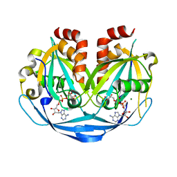
Crystal structure of ADP-ribose diphosphatase from Klebsiella pneumoniae (ADP Ribose and AMP bound)
Descriptor: ADENOSINE MONOPHOSPHATE, ADP-ribose pyrophosphatase, MAGNESIUM ION, ...
Authors:Seattle Structural Genomics Center for Infectious Disease, Seattle Structural Genomics Center for Infectious Disease (SSGCID)
Deposit date:2024-03-14
Release date:2024-03-27
Method:X-RAY DIFFRACTION (1.3 Å)
Cite:Crystal structure of ADP-ribose diphosphatase from Klebsiella pneumoniae (ADP Ribose and AMP bound)
To be published
9B21
DownloadVisualize
BU of 9b21 by Molmil
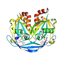
Crystal structure of ADP-ribose diphosphatase from Klebsiella pneumoniae (ADP Ribose bound, Orthorhombic P form)
Descriptor: ADP-ribose pyrophosphatase, MAGNESIUM ION, [(2R,3S,4R,5R)-5-(6-AMINOPURIN-9-YL)-3,4-DIHYDROXY-OXOLAN-2-YL]METHYL [HYDROXY-[[(2R,3S,4R,5S)-3,4,5-TRIHYDROXYOXOLAN-2-YL]METHOXY]PHOSPHORYL] HYDROGEN PHOSPHATE
Authors:Seattle Structural Genomics Center for Infectious Disease, Seattle Structural Genomics Center for Infectious Disease (SSGCID)
Deposit date:2024-03-14
Release date:2024-03-27
Method:X-RAY DIFFRACTION (1.6 Å)
Cite:Crystal structure of ADP-ribose diphosphatase from Klebsiella pneumoniae (ADP Ribose bound, Orthorhombic P form)
To be published
9B1Z
DownloadVisualize
BU of 9b1z by Molmil
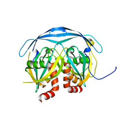
Crystal structure of ADP-ribose diphosphatase from Klebsiella pneumoniae (Apo)
Descriptor: ADP-ribose pyrophosphatase, SODIUM ION
Authors:Seattle Structural Genomics Center for Infectious Disease, Seattle Structural Genomics Center for Infectious Disease (SSGCID)
Deposit date:2024-03-14
Release date:2024-03-27
Method:X-RAY DIFFRACTION (1.25 Å)
Cite:Crystal structure of ADP-ribose diphosphatase from Klebsiella pneumoniae (Apo)
To be published
9AV6
DownloadVisualize
BU of 9av6 by Molmil
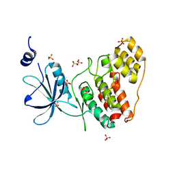
Crystal structure of CMGC family protein kinase from Trichomonas vaginalis (Apo)
Descriptor: SULFATE ION, non-specific serine/threonine protein kinase
Authors:Seattle Structural Genomics Center for Infectious Disease, Seattle Structural Genomics Center for Infectious Disease (SSGCID)
Deposit date:2024-03-01
Release date:2024-03-13
Method:X-RAY DIFFRACTION (2 Å)
Cite:Crystal structure of CMGC family protein kinase from Trichomonas vaginalis (Apo)
To be published

226707

PDB entries from 2024-10-30

PDB statisticsPDBj update infoContact PDBjnumon