Loading
PDBj
MenuPDBj@FacebookPDBj@TwitterPDBj@YouTubewwPDB FoundationwwPDB
RCSB PDBPDBeBMRBAdv. SearchSearch help
Search by PDB author
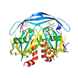
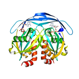
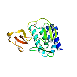
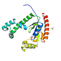
6MB0
DownloadVisualize
BU of 6mb0 by Molmil
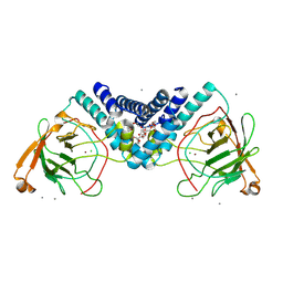
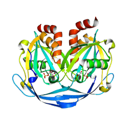
Crystal structure of N-myristoyl transferase (NMT) G386E mutant from Plasmodium vivax in complex with inhibitor IMP-1002
Descriptor: 1,2-ETHANEDIOL, 1-(5-{4-fluoro-2-[2-(1,3,5-trimethyl-1H-pyrazol-4-yl)ethoxy]phenyl}-1-methyl-1H-indazol-3-yl)-N,N-dimethylmethanamine, CHLORIDE ION, ...
Authors:Seattle Structural Genomics Center for Infectious Disease (SSGCID)
Deposit date:2018-08-29
Release date:2019-06-05
Last modified:2024-03-13
Method:X-RAY DIFFRACTION (1.55 Å)
Cite:Structure-Guided Identification of Resistance Breaking Antimalarial N‐Myristoyltransferase Inhibitors.
Cell Chem Biol, 26, 2019
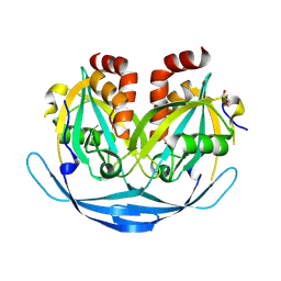
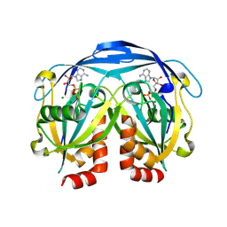
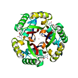
9DUG
DownloadVisualize
BU of 9dug by Molmil
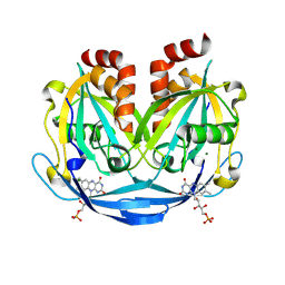
Crystal Structure of Dephospho-CoA kinase from Klebsiella aerogenes (Sulfate bound)
Descriptor: Dephospho-CoA kinase, SULFATE ION
Authors:Seattle Structural Genomics Center for Infectious Disease, Seattle Structural Genomics Center for Infectious Disease (SSGCID)
Deposit date:2024-10-02
Release date:2024-10-16
Method:X-RAY DIFFRACTION (1.5 Å)
Cite:Crystal Structure of Dephospho-CoA kinase from Klebsiella aerogenes (Sulfate bound)
To be published
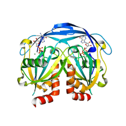
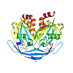
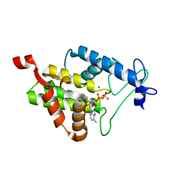
9DR6
DownloadVisualize
BU of 9dr6 by Molmil
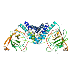
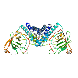
Crystal structure of Catechol 1,2-dioxygenase from Burkholderia multivorans (Zinc bound)
Descriptor: CALCIUM ION, CHLORIDE ION, Catechol 1,2-dioxygenase, ...
Authors:Seattle Structural Genomics Center for Infectious Disease, Seattle Structural Genomics Center for Infectious Disease (SSGCID)
Deposit date:2024-09-25
Release date:2024-10-02
Method:X-RAY DIFFRACTION (1.75 Å)
Cite:Crystal structure of Catechol 1,2-dioxygenase from Burkholderia multivorans (Zinc bound)
To be published
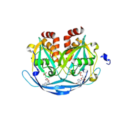
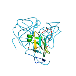
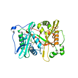
9DUF
DownloadVisualize
BU of 9duf by Molmil
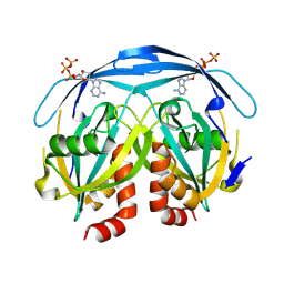
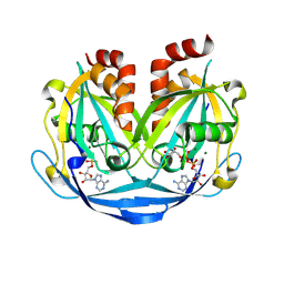
Crystal structure of ADP-ribose diphosphatase from Klebsiella pneumoniae (FMN bound)
Descriptor: ADP-ribose pyrophosphatase, FLAVIN MONONUCLEOTIDE, MAGNESIUM ION
Authors:Seattle Structural Genomics Center for Infectious Disease, Seattle Structural Genomics Center for Infectious Disease (SSGCID)
Deposit date:2024-10-02
Release date:2024-10-16
Method:X-RAY DIFFRACTION (1.32 Å)
Cite:Crystal structure of ADP-ribose diphosphatase from Klebsiella pneumoniae (FMN bound)
To be published
9DU6
DownloadVisualize
BU of 9du6 by Molmil
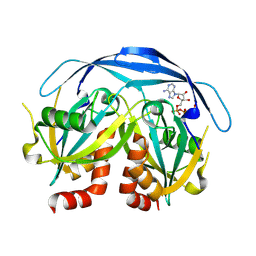
Crystal structure of ADP-ribose diphosphatase from Klebsiella pneumoniae (ATP bound)
Descriptor: ADENOSINE-5'-TRIPHOSPHATE, ADP-ribose pyrophosphatase
Authors:Seattle Structural Genomics Center for Infectious Disease, Seattle Structural Genomics Center for Infectious Disease (SSGCID)
Deposit date:2024-10-02
Release date:2024-10-16
Method:X-RAY DIFFRACTION (1.55 Å)
Cite:Crystal structure of ADP-ribose diphosphatase from Klebsiella pneumoniae (ATP bound)
To be published
9DRF
DownloadVisualize
BU of 9drf by Molmil
Crystal structure of ADP-ribose diphosphatase from Klebsiella pneumoniae (DTP bound, P21 form)
Descriptor: 2'-DEOXYADENOSINE 5'-TRIPHOSPHATE, ADP-ribose pyrophosphatase, MAGNESIUM ION
Authors:Seattle Structural Genomics Center for Infectious Disease, Seattle Structural Genomics Center for Infectious Disease (SSGCID)
Deposit date:2024-09-25
Release date:2024-10-02
Method:X-RAY DIFFRACTION (1.78 Å)
Cite:Crystal structure of ADP-ribose diphosphatase from Klebsiella pneumoniae (DTP bound, P21 form)
To be published
9DRE
DownloadVisualize
BU of 9dre by Molmil
Crystal structure of ADP-ribose diphosphatase from Klebsiella pneumoniae (ADP bound, P21 form)
Descriptor: ADENOSINE-5'-DIPHOSPHATE, ADP-ribose pyrophosphatase, MAGNESIUM ION
Authors:Seattle Structural Genomics Center for Infectious Disease, Seattle Structural Genomics Center for Infectious Disease (SSGCID)
Deposit date:2024-09-25
Release date:2024-10-02
Method:X-RAY DIFFRACTION (1.69 Å)
Cite:Crystal structure of ADP-ribose diphosphatase from Klebsiella pneumoniae (ADP bound, P21 form)
To be published
9DRD
DownloadVisualize
BU of 9drd by Molmil
Crystal structure of ADP-ribose diphosphatase from Klebsiella pneumoniae (Apo, monoclinic P form 2)
Descriptor: ADP-ribose pyrophosphatase, DI(HYDROXYETHYL)ETHER
Authors:Seattle Structural Genomics Center for Infectious Disease, Seattle Structural Genomics Center for Infectious Disease (SSGCID)
Deposit date:2024-09-25
Release date:2024-10-02
Method:X-RAY DIFFRACTION (1.65 Å)
Cite:Crystal structure of ADP-ribose diphosphatase from Klebsiella pneumoniae (Apo, monoclinic P form 2)
To be published
9DU9
DownloadVisualize
BU of 9du9 by Molmil
Crystal structure of ADP-ribose diphosphatase from Klebsiella pneumoniae (GDP/Mg bound)
Descriptor: ADP-ribose pyrophosphatase, GUANOSINE-5'-DIPHOSPHATE, MAGNESIUM ION
Authors:Seattle Structural Genomics Center for Infectious Disease, Seattle Structural Genomics Center for Infectious Disease (SSGCID)
Deposit date:2024-10-02
Release date:2024-10-16
Method:X-RAY DIFFRACTION (1.35 Å)
Cite:Crystal structure of ADP-ribose diphosphatase from Klebsiella pneumoniae (GDP/Mg bound)
To be published
9DUA
DownloadVisualize
BU of 9dua by Molmil
Crystal structure of ADP-ribose diphosphatase from Klebsiella pneumoniae (GMP bound)
Descriptor: ADP-ribose pyrophosphatase, GUANOSINE-5'-MONOPHOSPHATE, MAGNESIUM ION
Authors:Seattle Structural Genomics Center for Infectious Disease, Seattle Structural Genomics Center for Infectious Disease (SSGCID)
Deposit date:2024-10-02
Release date:2024-10-16
Method:X-RAY DIFFRACTION (1.4 Å)
Cite:Crystal structure of ADP-ribose diphosphatase from Klebsiella pneumoniae (GMP bound)
To be published
9DT7
DownloadVisualize
BU of 9dt7 by Molmil
Crystal structure of ADP-ribose diphosphatase from Klebsiella pneumoniae (5-O-phosphono-alpha-D-ribofuranose bound)
Descriptor: 5-O-phosphono-alpha-D-ribofuranose, ADP-ribose pyrophosphatase, MAGNESIUM ION
Authors:Seattle Structural Genomics Center for Infectious Disease, Seattle Structural Genomics Center for Infectious Disease (SSGCID)
Deposit date:2024-09-30
Release date:2024-10-09
Method:X-RAY DIFFRACTION (1.15 Å)
Cite:Crystal structure of ADP-ribose diphosphatase from Klebsiella pneumoniae (5-O-phosphono-alpha-D-ribofuranose bound)
To be published
9DR8
DownloadVisualize
BU of 9dr8 by Molmil
Crystal structure of Catechol 1,2-dioxygenase from Burkholderia multivorans (Iron bound)
Descriptor: CALCIUM ION, Catechol 1,2-dioxygenase, DODECAETHYLENE GLYCOL, ...
Authors:Seattle Structural Genomics Center for Infectious Disease, Seattle Structural Genomics Center for Infectious Disease (SSGCID)
Deposit date:2024-09-25
Release date:2024-10-09
Method:X-RAY DIFFRACTION (1.42 Å)
Cite:Crystal structure of Catechol 1,2-dioxygenase from Burkholderia multivorans (Iron bound)
To be published
9DRA
DownloadVisualize
BU of 9dra by Molmil
Crystal structure of Catechol 1,2-dioxygenase from Burkholderia multivorans (Iron and 4,5-dichloro-1,2-catechol bound)
Descriptor: (6R)-3,4-dichloro-6-hydroxycyclohex-3-en-1-one, CALCIUM ION, Catechol 1,2-dioxygenase, ...
Authors:Seattle Structural Genomics Center for Infectious Disease, Seattle Structural Genomics Center for Infectious Disease (SSGCID)
Deposit date:2024-09-25
Release date:2024-10-09
Method:X-RAY DIFFRACTION (1.62 Å)
Cite:Crystal structure of Catechol 1,2-dioxygenase from Burkholderia multivorans (Iron and 4,5-dichloro-1,2-catechol bound)
To be published
9DTC
DownloadVisualize
BU of 9dtc by Molmil
Crystal structure of ADP-ribose diphosphatase from Klebsiella pneumoniae (ADP Ribose bound, orthrhombic form2)
Descriptor: ADENOSINE MONOPHOSPHATE, ADP-ribose pyrophosphatase, MAGNESIUM ION, ...
Authors:Seattle Structural Genomics Center for Infectious Disease, Seattle Structural Genomics Center for Infectious Disease (SSGCID)
Deposit date:2024-09-30
Release date:2024-10-09
Method:X-RAY DIFFRACTION (1.4 Å)
Cite:Crystal structure of ADP-ribose diphosphatase from Klebsiella pneumoniae (ADP Ribose bound, orthrhombic form2)
To be published
9DSZ
DownloadVisualize
BU of 9dsz by Molmil
Crystal structure of ADP-ribose diphosphatase from Klebsiella pneumoniae (DTP bound)
Descriptor: 2'-DEOXYADENOSINE 5'-TRIPHOSPHATE, ADP-ribose pyrophosphatase, MAGNESIUM ION
Authors:Seattle Structural Genomics Center for Infectious Disease, Seattle Structural Genomics Center for Infectious Disease (SSGCID)
Deposit date:2024-09-30
Release date:2024-10-09
Method:X-RAY DIFFRACTION (1.9 Å)
Cite:Crystal structure of ADP-ribose diphosphatase from Klebsiella pneumoniae (DTP bound)
To be published
9DU8
DownloadVisualize
BU of 9du8 by Molmil
Crystal structure of ADP-ribose diphosphatase from Klebsiella pneumoniae (ADP/Mg bound)
Descriptor: ADENOSINE-5'-DIPHOSPHATE, ADP-ribose pyrophosphatase, MAGNESIUM ION
Authors:Seattle Structural Genomics Center for Infectious Disease, Seattle Structural Genomics Center for Infectious Disease (SSGCID)
Deposit date:2024-10-02
Release date:2024-10-16
Method:X-RAY DIFFRACTION (1.35 Å)
Cite:Crystal structure of ADP-ribose diphosphatase from Klebsiella pneumoniae (ADP/Mg bound)
To be published
9DT8
DownloadVisualize
BU of 9dt8 by Molmil
Crystal structure of ADP-ribose diphosphatase from Klebsiella pneumoniae (ADP bound, Orthorhombic P)
Descriptor: ACETATE ION, ADENOSINE-5'-DIPHOSPHATE, ADP-ribose pyrophosphatase
Authors:Seattle Structural Genomics Center for Infectious Disease, Seattle Structural Genomics Center for Infectious Disease (SSGCID)
Deposit date:2024-09-30
Release date:2024-10-09
Method:X-RAY DIFFRACTION (1.15 Å)
Cite:Crystal structure of ADP-ribose diphosphatase from Klebsiella pneumoniae (ADP bound, Orthorhombic P)
To be published
6MAI
DownloadVisualize
BU of 6mai by Molmil
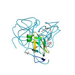
Crystal structure of Deoxyuridine 5'-triphosphate nucleotidohydrolase from Legionella pneumophila Philadelphia 1
Descriptor: 1,2-ETHANEDIOL, Deoxyuridine 5'-triphosphate nucleotidohydrolase
Authors:Seattle Structural Genomics Center for Infectious Disease (SSGCID)
Deposit date:2018-08-27
Release date:2018-09-05
Last modified:2023-10-11
Method:X-RAY DIFFRACTION (1.8 Å)
Cite:Crystal structure of Deoxyuridine 5'-triphosphate nucleotidohydrolase from Legionella pneumophila Philadelphia 1
TO BE PUBLISHED
6MAO
DownloadVisualize
BU of 6mao by Molmil
Crystal structure of Deoxyuridine 5'-triphosphate nucleotidohydrolase from Legionella pneumophila Philadelphia 1 in complex with dUMP (Deoxyuridine 5'-monophosphate)
Descriptor: (4S)-2-METHYL-2,4-PENTANEDIOL, 2'-DEOXYURIDINE 5'-MONOPHOSPHATE, Deoxyuridine 5'-triphosphate nucleotidohydrolase, ...
Authors:Seattle Structural Genomics Center for Infectious Disease (SSGCID)
Deposit date:2018-08-28
Release date:2018-09-12
Last modified:2023-10-11
Method:X-RAY DIFFRACTION (1.95 Å)
Cite:Crystal structure of Deoxyuridine 5'-triphosphate nucleotidohydrolase from Legionella pneumophila Philadelphia 1 in complex with dUMP (Deoxyuridine 5'-monophosphate)
TO BE PUBLISHED
3JS5
DownloadVisualize
BU of 3js5 by Molmil
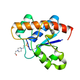
Crystal structure of protein tyrosine phosphatase from Entamoeba histolytica with Hepes in the active site. High resolution, alternative crystal form with 1 molecule in asymmetric unit
Descriptor: 4-(2-HYDROXYETHYL)-1-PIPERAZINE ETHANESULFONIC ACID, Protein tyrosine phosphatase, SODIUM ION
Authors:Seattle Structural Genomics Center for Infectious Disease (SSGCID)
Deposit date:2009-09-09
Release date:2009-09-22
Last modified:2023-09-06
Method:X-RAY DIFFRACTION (1.94 Å)
Cite:Crystal structure and putative substrate identification for the Entamoeba histolytica low molecular weight tyrosine phosphatase.
Mol.Biochem.Parasitol., 193, 2014
7RHE
DownloadVisualize
BU of 7rhe by Molmil
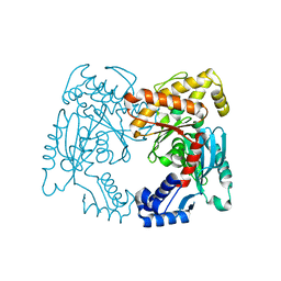
Crystal Structure of ROK family protein from Burkholderia vietnamiensis G4
Descriptor: ROK family protein
Authors:Seattle Structural Genomics Center for Infectious Disease (SSGCID)
Deposit date:2021-07-16
Release date:2021-11-17
Last modified:2024-04-03
Method:X-RAY DIFFRACTION (2.3 Å)
Cite:Crystal Structure of ROK family protein from Burkholderia vietnamiensis G4
to be published
3KC6
DownloadVisualize
BU of 3kc6 by Molmil
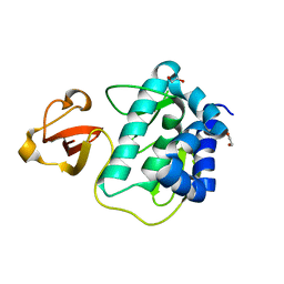
Crystal structure of the large c-terminal domain of polymerase basic protein 2 from influenza virus a/viet nam/1203/2004 (h5n1)
Descriptor: 1,2-ETHANEDIOL, Polymerase PB2
Authors:Seattle Structural Genomics Center for Infectious Disease (SSGCID)
Deposit date:2009-10-20
Release date:2010-03-09
Last modified:2023-09-06
Method:X-RAY DIFFRACTION (2.05 Å)
Cite:Biological and structural characterization of a host-adapting amino acid in influenza virus.
Plos Pathog., 6, 2010
3KHW
DownloadVisualize
BU of 3khw by Molmil
Crystal structure of the large c-terminal domain of polymerase basic protein 2 from influenza virus a/mexico/indre4487/2009(h1n1)
Descriptor: GLYCEROL, Polymerase PB2
Authors:Seattle Structural Genomics Center for Infectious Disease (SSGCID)
Deposit date:2009-10-31
Release date:2010-03-09
Last modified:2023-09-06
Method:X-RAY DIFFRACTION (2.1 Å)
Cite:Biological and structural characterization of a host-adapting amino acid in influenza virus.
Plos Pathog., 6, 2010
3KE1
DownloadVisualize
BU of 3ke1 by Molmil
Crystal structure of 2C-methyl-D-erythritol 2,4-cyclodiphosphate synthase from Burkholderia pseudomallei in complex with a fragment-nucleoside fusion D000161829
Descriptor: 2-C-methyl-D-erythritol 2,4-cyclodiphosphate synthase, 5'-deoxy-5'-[(pyridin-4-ylcarbonyl)amino]cytidine, POTASSIUM ION, ...
Authors:Seattle Structural Genomics Center for Infectious Disease (SSGCID)
Deposit date:2009-10-23
Release date:2009-11-10
Last modified:2023-09-06
Method:X-RAY DIFFRACTION (2.05 Å)
Cite:Cytidine derivatives as IspF inhibitors of Burkolderia pseudomallei.
Bioorg.Med.Chem.Lett., 8, 2013
4N67
DownloadVisualize
BU of 4n67 by Molmil
Crystal Structure of the N-terminal Fic Domain of a Putative Cell Filamentation protein (VirB-translocated Bep effector protein) with bound ADP from Bartonella quintana
Descriptor: ADENOSINE-5'-DIPHOSPHATE, MAGNESIUM ION, Putative cell filamentation protein
Authors:Seattle Structural Genomics Center for Infectious Disease (SSGCID)
Deposit date:2013-10-11
Release date:2014-09-24
Last modified:2023-09-20
Method:X-RAY DIFFRACTION (1.55 Å)
Cite:Evolutionary Diversification of Host-Targeted Bartonella Effectors Proteins Derived from a Conserved FicTA Toxin-Antitoxin Module.
Microorganisms, 9, 2021

226707

PDB entries from 2024-10-30

PDB statisticsPDBj update infoContact PDBjnumon