Loading
PDBj
MenuPDBj@FacebookPDBj@TwitterPDBj@YouTubewwPDB FoundationwwPDB
RCSB PDBPDBeBMRBAdv. SearchSearch help
Search by PDB author
4ECW
DownloadVisualize
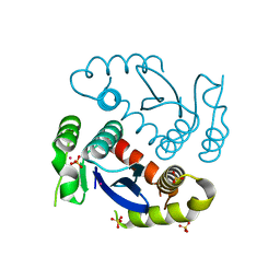
BU of 4ecw by Molmil
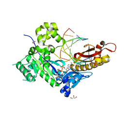
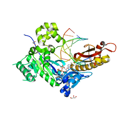
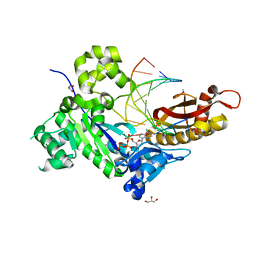
Human DNA polymerase eta - DNA ternary complex: Reaction in the AT crystal at pH 7.0 for 250 sec
Descriptor: 2'-DEOXYADENOSINE 5'-TRIPHOSPHATE, CALCIUM ION, DIPHOSPHATE, ...
Authors:Nakamura, T, Zhao, Y, Yang, W.
Deposit date:2012-03-26
Release date:2012-07-11
Last modified:2024-03-20
Method:X-RAY DIFFRACTION (1.896 Å)
Cite:Watching DNA polymerase eta make a phosphodiester bond
Nature, 487, 2012
4ECQ
DownloadVisualize
BU of 4ecq by Molmil
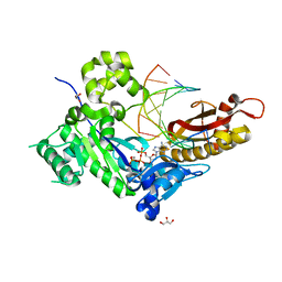
Human DNA polymerase eta- DNA ternary complex: AT crystal at pH6.8(K+ MES) with 1 Ca2+ ion
Descriptor: 2'-DEOXYADENOSINE 5'-TRIPHOSPHATE, CALCIUM ION, DNA (5'-D(*AP*GP*CP*GP*TP*CP*AP*TP*A)-3'), ...
Authors:Nakamura, T, Zhao, Y, Yang, W.
Deposit date:2012-03-26
Release date:2012-07-11
Last modified:2024-03-20
Method:X-RAY DIFFRACTION (1.5 Å)
Cite:Watching DNA polymerase eta make a phosphodiester bond
Nature, 487, 2012
4ED1
DownloadVisualize
BU of 4ed1 by Molmil
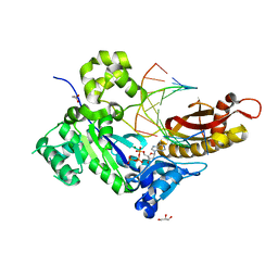
Human DNA polymerase eta - DNA ternary complex: AT crystal at pH 7.0 (Na+ MES) with 1 Ca2+ ion
Descriptor: 2'-DEOXYADENOSINE 5'-TRIPHOSPHATE, CALCIUM ION, DNA (5'-D(*AP*GP*CP*GP*TP*CP*AP*T)-3'), ...
Authors:Nakamura, T, Zhao, Y, Yang, W.
Deposit date:2012-03-26
Release date:2012-07-11
Last modified:2024-03-20
Method:X-RAY DIFFRACTION (1.806 Å)
Cite:Watching DNA polymerase eta make a phosphodiester bond
Nature, 487, 2012
4ECR
DownloadVisualize
BU of 4ecr by Molmil
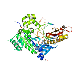
Human DNA polymerase eta - DNA ternary complex: Reaction in the AT crystal at pH 7.0 for 40 sec
Descriptor: 2'-DEOXYADENOSINE 5'-TRIPHOSPHATE, CALCIUM ION, DNA (5'-D(*AP*GP*CP*GP*TP*CP*AP*T)-3'), ...
Authors:Nakamura, T, Zhao, Y, Yang, W.
Deposit date:2012-03-26
Release date:2012-07-11
Last modified:2024-03-20
Method:X-RAY DIFFRACTION (1.892 Å)
Cite:Watching DNA polymerase eta make a phosphodiester bond
Nature, 487, 2012
4ECY
DownloadVisualize
BU of 4ecy by Molmil
Human DNA polymerase eta - DNA ternary complex: AT crystal at pH 6.0 (Na+ MES) with 1 Ca2+ ion
Descriptor: 2'-DEOXYADENOSINE 5'-TRIPHOSPHATE, CALCIUM ION, DNA (5'-D(*AP*GP*CP*GP*TP*CP*AP*T)-3'), ...
Authors:Nakamura, T, Zhao, Y, Yang, W.
Deposit date:2012-03-26
Release date:2012-07-11
Last modified:2024-03-20
Method:X-RAY DIFFRACTION (1.943 Å)
Cite:Watching DNA polymerase eta make a phosphodiester bond
Nature, 487, 2012
4ECZ
DownloadVisualize
BU of 4ecz by Molmil
Human DNA polymerase eta - DNA ternary complex: AT crystal at pH 6.5 (Na+ MES) with 1 Ca2+ ion
Descriptor: 2'-DEOXYADENOSINE 5'-TRIPHOSPHATE, CALCIUM ION, DNA (5'-D(*AP*GP*CP*GP*TP*CP*AP*T)-3'), ...
Authors:Nakamura, T, Zhao, Y, Yang, W.
Deposit date:2012-03-26
Release date:2012-07-11
Last modified:2024-03-20
Method:X-RAY DIFFRACTION (1.834 Å)
Cite:Watching DNA polymerase eta make a phosphodiester bond
Nature, 487, 2012
1MPG
DownloadVisualize
BU of 1mpg by Molmil
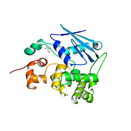
3-METHYLADENINE DNA GLYCOSYLASE II FROM ESCHERICHIA COLI
Descriptor: 3-METHYLADENINE DNA GLYCOSYLASE II, GLYCEROL
Authors:Labahn, J, Schaerer, O.D, Long, A, Ezaz-Nikpay, K, Verdine, G.L, Ellenberger, T.E.
Deposit date:1997-10-28
Release date:1998-01-28
Last modified:2024-02-14
Method:X-RAY DIFFRACTION (1.8 Å)
Cite:Structural basis for the excision repair of alkylation-damaged DNA.
Cell(Cambridge,Mass.), 86, 1996
6LFW
DownloadVisualize
BU of 6lfw by Molmil
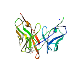
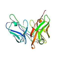
Crystal structure of PCB4scFv(hN56D) in complex with PCB#126
Descriptor: 1,2,3-tris(chloranyl)-5-(3-chloranyl-4-methoxy-phenyl)benzene, PCB4scFv(hN56D), SODIUM ION
Authors:Nakamura, T, Yamagata, Y, Morioka, H.
Deposit date:2019-12-03
Release date:2020-12-09
Last modified:2024-10-16
Method:X-RAY DIFFRACTION (1.41 Å)
Cite:Crystal structure of PCB4scFv(hN56D) in complex with PCB#126
To Be Published
6LFV
DownloadVisualize
BU of 6lfv by Molmil
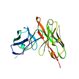
Crystal structure of PCB4scFv(hN56D)
Descriptor: PCB4scFv(hN56D)
Authors:Nakamura, T, Yamagata, Y, Morioka, H.
Deposit date:2019-12-03
Release date:2020-12-09
Last modified:2024-10-30
Method:X-RAY DIFFRACTION (1.9 Å)
Cite:Crystal structure of PCB4scFv(hN56D)
To Be Published
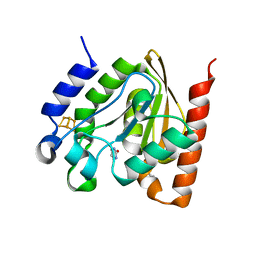
4ZBY
DownloadVisualize
BU of 4zby by Molmil
Family 4 uracil-DNA glycosylase from Sulfolobus tokodaii (uracil complex form)
Descriptor: 2-(N-MORPHOLINO)-ETHANESULFONIC ACID, IRON/SULFUR CLUSTER, URACIL, ...
Authors:Kawai, A, Miyamoto, S.
Deposit date:2015-04-15
Release date:2015-09-09
Last modified:2023-11-08
Method:X-RAY DIFFRACTION (1.7 Å)
Cite:Crystal structure of family 4 uracil-DNA glycosylase from Sulfolobus tokodaii and a function of tyrosine 170 in DNA binding
Febs Lett., 589, 2015
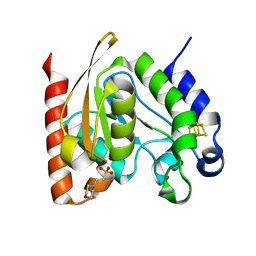
4ZBX
DownloadVisualize
BU of 4zbx by Molmil
Family 4 uracil-DNA glycosylase from Sulfolobus tokodaii (free form, X-ray wavelength=0.9000)
Descriptor: 2-(N-MORPHOLINO)-ETHANESULFONIC ACID, IRON/SULFUR CLUSTER, Uracil-DNA glycosylase
Authors:Kawai, A, Miyamoto, S.
Deposit date:2015-04-15
Release date:2015-09-09
Last modified:2023-11-08
Method:X-RAY DIFFRACTION (1.7 Å)
Cite:Crystal structure of family 4 uracil-DNA glycosylase from Sulfolobus tokodaii and a function of tyrosine 170 in DNA binding
Febs Lett., 589, 2015
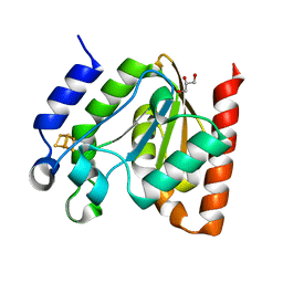
4ZBZ
DownloadVisualize
BU of 4zbz by Molmil
Family 4 uracil-DNA glycosylase from Sulfolobus tokodaii (free form, X-ray wavelength=1.5418)
Descriptor: 2-(N-MORPHOLINO)-ETHANESULFONIC ACID, GLYCEROL, IRON/SULFUR CLUSTER, ...
Authors:Kawai, A, Miyamoto, S.
Deposit date:2015-04-15
Release date:2015-09-09
Last modified:2023-11-08
Method:X-RAY DIFFRACTION (1.9 Å)
Cite:Crystal structure of family 4 uracil-DNA glycosylase from Sulfolobus tokodaii and a function of tyrosine 170 in DNA binding
Febs Lett., 589, 2015
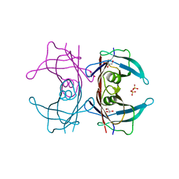
3A4D
DownloadVisualize
BU of 3a4d by Molmil
Crystal structure of Human Transthyretin (wild-type)
Descriptor: GLYCEROL, SULFATE ION, Transthyretin
Authors:Miyata, M.
Deposit date:2009-07-06
Release date:2009-12-22
Last modified:2023-11-01
Method:X-RAY DIFFRACTION (2 Å)
Cite:Role of the Glutamic Acid 54 Residue in Transthyretin Stability and Thyroxine Binding
Biochemistry, 2009
6L0C
DownloadVisualize
BU of 6l0c by Molmil
Crystal structure of HIV-1 Integrase catalytic core domain (A128T/K173Q/F185K)
Descriptor: ARSENIC, Integrase, SULFATE ION
Authors:Nakamura, T, Nakamura, T.
Deposit date:2019-09-26
Release date:2020-07-01
Last modified:2023-11-22
Method:X-RAY DIFFRACTION (2.101 Å)
Cite:A Conformational Escape Reaction of HIV-1 against an Allosteric Integrase Inhibitor.
J.Virol., 94, 2020
1WSI
DownloadVisualize
BU of 1wsi by Molmil
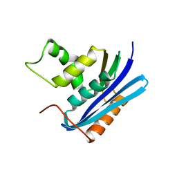
Crystal structure of E.coli RNase HI active site mutant (E48A/K87A/D134N)
Descriptor: Ribonuclease HI
Authors:Tsunaka, Y, Takano, K, Matsumura, H, Yamagata, Y, Kanaya, S.
Deposit date:2004-11-07
Release date:2004-11-23
Last modified:2024-05-29
Method:X-RAY DIFFRACTION (2 Å)
Cite:

1WSH
DownloadVisualize
BU of 1wsh by Molmil
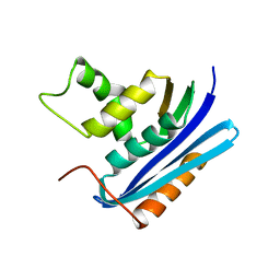
Crystal structure of E.coli RNase HI active site mutant (E48A/K87A)
Descriptor: Ribonuclease HI
Authors:Tsunaka, Y, Takano, K, Matsumura, H, Yamagata, Y, Kanaya, S.
Deposit date:2004-11-07
Release date:2004-11-23
Last modified:2024-05-29
Method:X-RAY DIFFRACTION (1.9 Å)
Cite:

1WSJ
DownloadVisualize
BU of 1wsj by Molmil
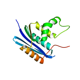
Crystal structure of E.coli RNase HI active site mutant (K87A/H124A)
Descriptor: Ribonuclease HI
Authors:Tsunaka, Y, Takano, K, Matsumura, H, Yamagata, Y, Kanaya, S.
Deposit date:2004-11-07
Release date:2004-11-23
Last modified:2024-05-29
Method:X-RAY DIFFRACTION (2 Å)
Cite:

2LAO
DownloadVisualize
BU of 2lao by Molmil
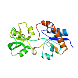
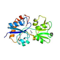
THREE-DIMENSIONAL STRUCTURES OF THE PERIPLASMIC LYSINE-, ARGININE-, ORNITHINE-BINDING PROTEIN WITH AND WITHOUT A LIGAND
Descriptor: LYSINE, ARGININE, ORNITHINE-BINDING PROTEIN
Authors:Kim, S.-H, Oh, B.-H, Kang, C.-H.
Deposit date:1993-02-25
Release date:1994-06-22
Last modified:2017-11-29
Method:X-RAY DIFFRACTION (1.9 Å)
Cite:Three-dimensional structures of the periplasmic lysine/arginine/ornithine-binding protein with and without a ligand.
J.Biol.Chem., 268, 1993
1LST
DownloadVisualize
BU of 1lst by Molmil
THREE-DIMENSIONAL STRUCTURES OF THE PERIPLASMIC LYSINE-, ARGININE-, ORNITHINE-BINDING PROTEIN WITH AND WITHOUT A LIGAND
Descriptor: LYSINE, ARGININE, ORNITHINE-BINDING PROTEIN
Authors:Kim, S.-H, Oh, B.-H.
Deposit date:1993-02-25
Release date:1994-06-22
Last modified:2024-10-23
Method:X-RAY DIFFRACTION (1.8 Å)
Cite:Three-dimensional structures of the periplasmic lysine/arginine/ornithine-binding protein with and without a ligand.
J.Biol.Chem., 268, 1993
6LFX
DownloadVisualize
BU of 6lfx by Molmil
Crystal structure of PCB4scFv(hN56D) in complex with PCB#77
Descriptor: 1,2-bis(chloranyl)-4-(3-chloranyl-4-methoxy-phenyl)benzene, PCB4scFv(hN56D)
Authors:Nakamura, T, Yamagata, Y, Morioka, H.
Deposit date:2019-12-03
Release date:2020-12-09
Last modified:2023-11-22
Method:X-RAY DIFFRACTION (1.43 Å)
Cite:Crystal structure of PCB4scFv(hN56D) in complex with PCB#77
To Be Published

226707

PDB entries from 2024-10-30

PDB statisticsPDBj update infoContact PDBjnumon