






 9Y80
9Y80
Homomeric Glycine Receptor alpha2 with PTX in a Desensitized State
Summary for 9Y80
| Entry DOI | 10.2210/pdb9y80/pdb |
| EMDB information | 72676 |
| Descriptor | Glycine receptor subunit alpha-2, 2-acetamido-2-deoxy-beta-D-glucopyranose, GLYCINE, ... (5 entities in total) |
| Functional Keywords | homopentamer, ion channel, ligand gated ion channel, transport protein |
| Biological source | Homo sapiens (human) |
| Total number of polymer chains | 5 |
| Total formula weight | 281978.00 |
| Authors | Klemm, E.,Gibbs, E.,Chakrapani, S. (deposition date: 2025-09-11, release date: 2026-03-18, Last modification date: 2026-05-20) |
| Primary citation | Klemm, E.,Gibbs, E.,Stauffer, M.,Mohapatra, D.,Meyer, C.,Chakrapani, S. Human GlyR alpha 2 pore dynamics in gating and inhibition. Structure, 34:798-, 2026 Cited by PubMed Abstract: Glycine receptors (GlyRs) mediate inhibitory neurotransmission in the central nervous system. The GlyRα2 subtype contributes to critical neural circuitry in early neurodevelopment and is also found in adults. GlyRα2 dysfunctions are implicated in neurodevelopmental disorders, including autism, epilepsy, and cognitive delays. GlyRα2 functional properties and pharmacology are distinct from GlyRα1, but the structural basis for these differences remains poorly defined. Here, we report cryo-electron microscopy structures of full-length, human GlyRα2 reconstituted in peptidiscs captured in multiple conformational states. In addition to symmetric resting and desensitized states, we resolved an asymmetric open state, previously observed only in heteromeric GlyRs. This suggests that asymmetry is intrinsic to GlyRα2, independent of β-subunit incorporation. Furthermore, we identified distinct conformations of GlyRα2 with the pore-blocker picrotoxin, providing new insights into allosteric interactions. These findings uncover the structural basis of GlyRα2 function, providing a foundation for understanding its role in development and in GlyRα2-associated disorders. PubMed: 41856109DOI: 10.1016/j.str.2026.02.013 PDB entries with the same primary citation |
| Experimental method | ELECTRON MICROSCOPY (3.55 Å) |
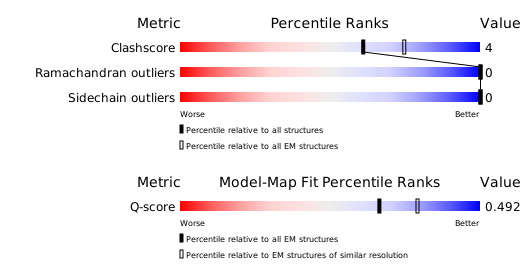
Structure validation
Download full validation report


