






 9TH8
9TH8
Cryo-EM structure of MCM2-7 DH bound to Sld3-Sld7, Cdc45 and DNA
This is a non-PDB format compatible entry.
Summary for 9TH8
| Entry DOI | 10.2210/pdb9th8/pdb |
| EMDB information | 55918 |
| Descriptor | dsDNA (60-MER), Cell division control protein 45, Mitochondrial morphogenesis protein SLD7, ... (13 entities in total) |
| Functional Keywords | helicase, activation, phosphorylation, replication |
| Biological source | Saccharomyces cerevisiae More |
| Total number of polymer chains | 17 |
| Total formula weight | 1435146.17 |
| Authors | Saleh, A.,Noguchi, Y.,Schneider, S.,Aramayo, R.,Speck, C. (deposition date: 2025-12-03, release date: 2026-08-26, Last modification date: 2026-09-23) |
| Primary citation | Noguchi, Y.,Saleh, A.,Schneider, S.,Ivanova, M.E.,Chen, Z.A.,Ranjha, L.,Aramayo, R.,Tognetti, S.,Faull, S.V.,Rappsilber, J.,Speck, C. Structural insights into Sld3-Sld7-dependent Cdc45 loading during replication initiation. Nat Commun, 17:-, 2026 Cited by PubMed Abstract: Regulated helicase activation by DDK kinase is central for genome stability. However, how DDK phosphorylation primes the MCM2-7 double hexamer (DH) for Sld3-Sld7 binding and Cdc45 loading remained unclear. We define this mechanism through cryo-EM structures of MCM2-7 DH-Sld3-Sld7 (MS) and MCM2-7 DH-Sld3-Sld7-Cdc45 (MSC). We reveal that the autoinhibitory Mcm4 tail engages not only Mcm4 but also Mcm6. Upon DDK-dependent phosphorylation, both of these sites become accessible. In the context of the MS structure, we identify that two short Sld3 motifs that contact Mcm4 and Mcm6 read out the DH phosphorylation state, while the Sld3 Treslin domain (STD) binds to Mcm2. In the MSC structure, Cdc45 dislodges the Sld3 STD from Mcm2, allowing Sld3 to position Cdc45 at the Mcm2/Mcm5 interface. Mutagenesis of the Sld3 STD-Cdc45 interface disrupts Cdc45 loading, validating this interaction. Together, our data reveal a phosphorylation-encoded mechanism coupling DDK-activated Mcm4/Mcm6 surfaces to distal Cdc45 placement, explaining how firing factors choreograph the DH-to-CMG transition. PubMed: 42733061DOI: 10.1038/s41467-026-76309-6 PDB entries with the same primary citation |
| Experimental method | ELECTRON MICROSCOPY (2.9 Å) |
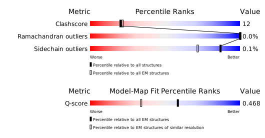
Structure validation
Download full validation report


