






 9PM9
9PM9
PhuZ Tubulin from phage Goslar (locally refined monomer)
Summary for 9PM9
| Entry DOI | 10.2210/pdb9pm9/pdb |
| EMDB information | 71729 |
| Descriptor | Tubulin/FtsZ GTPase domain-containing protein, PHOSPHOMETHYLPHOSPHONIC ACID GUANYLATE ESTER (2 entities in total) |
| Functional Keywords | phage, tubulin, phuz, viral protein |
| Biological source | Goslarvirus |
| Total number of polymer chains | 1 |
| Total formula weight | 36184.17 |
| Authors | Basu, D.,Gu, Y.,Corbett, K.D. (deposition date: 2025-07-16, release date: 2025-12-24, Last modification date: 2026-07-08) |
| Primary citation | Basu, D.,Chen, S.,Li, Y.X.,Klusch, N.,Inlow, K.,Pogliano, J.,Villa, E.,Corbett, K.D. A bacteriophage tubulin forms microtubule-like assemblies with nine protofilaments. Structure, 34:394-403.e3, 2026 Cited by PubMed Abstract: Tubulin family proteins play central roles in the organization and dynamics of cytoskeletal systems across the Tree of Life. In one family of bacteriophages (phages), tubulin-like proteins called PhuZ (Phage tubulin/FtsZ) form dynamic filaments that position and rotate an intracellular compartment, the "phage nucleus," in which the phage genome is replicated. PhuZ filaments also mediate trafficking of nascent capsids from the cell periphery to the phage nucleus for genome packaging. PhuZ from the Pseudomonas-infecting phages 201Phi2-1 and PhiKZ form assemblies with three protofilaments. Here, we determine a 2.8 Å resolution structure of PhuZ from the E. coli-infecting phage Goslar, which forms an elaborate "cytoskeletal vortex" in infected cells. We find that in vitro-assembled Goslar PhuZ forms rigid tubes with nine nearly-straight protofilaments. The lateral interactions mediating this assembly are fundamentally different from eukaryotic tubulin, leading to a distinctive overall architecture for Goslar PhuZ filaments. PubMed: 41468894DOI: 10.1016/j.str.2025.12.002 PDB entries with the same primary citation |
| Experimental method | ELECTRON MICROSCOPY (2.75 Å) |
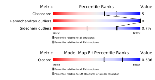
Structure validation
Download full validation report


