






 9NON
9NON
Cryo-EM structure of amyloid fibril isolated from the kidney of a variant Alect2-I40V amyloidosis patient
Summary for 9NON
| Entry DOI | 10.2210/pdb9non/pdb |
| EMDB information | 49601 |
| Descriptor | Leukocyte cell-derived chemotaxin-2 (1 entity in total) |
| Functional Keywords | leukocyte cell-derived chemotaxin-2, lect2, systemic amyloidosis, alect2, systemic, amyloid, kidney., protein fibril |
| Biological source | Homo sapiens (human) |
| Total number of polymer chains | 5 |
| Total formula weight | 72968.63 |
| Authors | Nguyen, B.,Saelices, L. (deposition date: 2025-03-10, release date: 2026-02-11, Last modification date: 2026-06-17) |
| Primary citation | Afrin, S.,Nguyen, B.A.,Singh, V.,Singh, P.,Bassett, P.,Pekala, M.,Evers, B.,Lopez, C.,Ahmed, Y.,Li, L.,Kallem, R.R.,Lemoff, A.,Argyropoulos, C.,Kluve-Beckerman, B.,Saelices, L. Structural polymorphism of ex-vivo ALECT2 amyloid fibrils revealed by cryo-EM. Nat Commun, 17:-, 2026 Cited by PubMed Abstract: ALECT2 amyloidosis is a rare systemic disease characterized by the pathological deposition of leukocyte cell-derived chemotaxin-2 (LECT2) as amyloid fibrils, primarily affecting the kidneys and liver. The molecular mechanisms underlying LECT2 aggregation remain poorly defined, hindering diagnostic and therapeutic development. Here, we present cryo-electron microscopy structures of ex-vivo ALECT2 fibrils extracted from a patient's kidney. We identified three fibril polymorphs: a predominant single-protofilament morphology and two minor double-protofilament morphologies. The dominant single-protofilament morphology comprises the full-length 133-residue LECT2 protein and retains all three native disulfide bonds. Low-resolution reconstructions of double-protofilament morphologies suggest they adopt a similar fold to the single protofilament morphology, but form paired assemblies with different inter-filament interfaces. Mass spectrometry also reveals acetylation within the fibrils. These findings offer critical insights into the structural basis of ALECT2 amyloid formation and identify molecular features that could inform future diagnostic and therapeutic approaches. PubMed: 41965342DOI: 10.1038/s41467-026-71223-3 PDB entries with the same primary citation |
| Experimental method | ELECTRON MICROSCOPY (2.4 Å) |
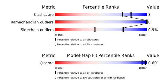
Structure validation
Download full validation report


