






 9JV8
9JV8
Crystal structure of M. tuberculosis EccCb1-D2 domain
Summary for 9JV8
| Entry DOI | 10.2210/pdb9jv8/pdb |
| Descriptor | ESX-1 secretion system protein EccCb1, ADENOSINE-5'-TRIPHOSPHATE, MAGNESIUM ION, ... (4 entities in total) |
| Functional Keywords | m. tuberculosis esx-1, eccc1, esxab, molecular docking, dynamics simulation, transport protein |
| Biological source | Mycobacterium tuberculosis H37Rv |
| Total number of polymer chains | 1 |
| Total formula weight | 34949.61 |
| Authors | Saxena, A.K. (deposition date: 2024-10-08, release date: 2025-10-08, Last modification date: 2026-04-22) |
| Primary citation | Saxena, A.K.,Chandra, A.,Srivastava, S.,Kumar, R. Structural analysis of M. tuberculosis EccC1 and its complex with EsxAB virulence factor using X-ray crystallography, molecular docking, and dynamics simulation techniques. Int.J.Biol.Macromol., 319:145279-145279, 2025 Cited by PubMed Abstract: M. tuberculosis ESX-1 system secretes virulence factors into host macrophages during infection, however, the mechanism of secretion is currently unknown. Here, we have determined the crystal structure of MtbEccCb1-D2 protein (Leu34-Ser313 residues, Mw ∼ 31.4 kDa) in complex with ATPγS and Mg, which adopts a classical Ftsk/SpoEIII type fold. The EccCb1-D2 showed two melting temperatures, Tm1 at 37.64 ± 0.08 °C and Tm2 at 65.85 ± 0.12 °C, during the unfolding pathway. Modeled ∆EccC1 and ∆EccC1 + EsxAB hexamers showed a channel (∼34 Å) involved in EsxAB (∼29 Å) translocation toward the inner membrane. At the entrance gate of the channel, the LxxxMxF motif of the EsxB export arm binds to the substrate binding pocket of the EccCb1-D3 protein. Inside the channel, the PL-1 and PL-2 pore loops, close to the α7-helix and the loop between β8-β9 strands in EccCa1-D1, EccCb1-D2, and EccCb1-D3 may be involved in EsxAB factor translocation. Stability, fluctuation, and compactness parameters in 100 ns dynamics simulation analysis showed the highest flexibility in ΔEccCa1, ∆EccC1, and ∆EccC1 + EsxAB hexamers and stability in ΔEccCb1 hexamer. Our EccCb1-D2 structure and dynamics simulation analysis on four modeled systems have revealed the mechanism involved in EsxAB translocation, a key target for the development of antivirulence inhibitors against M. tuberculosis. PubMed: 40541887DOI: 10.1016/j.ijbiomac.2025.145279 PDB entries with the same primary citation |
| Experimental method | X-RAY DIFFRACTION (2.2 Å) |
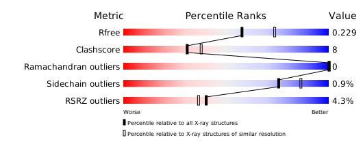
Structure validation
Download full validation report


