






 7ZPR
7ZPR
KtrAB complex with N-terminal deletion of KtrB 1-19
Summary for 7ZPR
| Entry DOI | 10.2210/pdb7zpr/pdb |
| Related | 7zp9 7zpo |
| EMDB information | 14851 14859 14862 |
| Descriptor | Ktr system potassium uptake protein A, Ktr system potassium uptake protein B, MAGNESIUM ION, ... (5 entities in total) |
| Functional Keywords | potassium transport, membrane protein, transporter |
| Biological source | Vibrio alginolyticus More |
| Total number of polymer chains | 12 |
| Total formula weight | 384108.18 |
| Authors | Vonck, J.,Stautz, J. (deposition date: 2022-04-28, release date: 2023-05-10, Last modification date: 2026-05-20) |
| Primary citation | Stautz, J.,Griwatz, D.,Kaltwasser, S.,Mehdipour, A.R.,Ketter, S.,Thiel, C.,Wunnicke, D.,Schrecker, M.,Mills, D.J.,Hummer, G.,Vonck, J.,Hanelt, I. A short intrinsically disordered region at KtrB's N-terminus facilitates allosteric regulation of K + channel KtrAB. Nat Commun, 16:4252-4252, 2025 Cited by PubMed Abstract: K homeostasis is crucial for bacterial survival. The bacterial K+ channel KtrAB is regulated by the binding of ADP and ATP to the cytosolic RCK subunits KtrA. While the ligand-induced conformational changes in KtrA are well described, the transmission to the gating regions within KtrB is not understood. Here, we present a cryo-EM structure of the ADP-bound, inactive KtrAB complex from Vibrio alginolyticus, which resolves part of KtrB's N termini. They are short intrinsically disordered regions (IDRs) located at the interface of KtrA and KtrB. We reveal that these IDRs play a decisive role in ATP-mediated channel opening, while the closed ADP-bound state does not depend on the N-termini. We propose an allosteric mechanism, in which ATP-induced conformational changes within KtrA trigger an interaction of KtrB's N-terminal IDRs with the membrane, stabilizing the active and conductive state of KtrAB. PubMed: 40335548DOI: 10.1038/s41467-025-59546-z PDB entries with the same primary citation |
| Experimental method | ELECTRON MICROSCOPY (3.56 Å) |
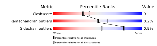
Structure validation
Download full validation report


