 Movie
Movie Controller


+ Open data

- Basic information
| Entry |  | |||||||||
|---|---|---|---|---|---|---|---|---|---|---|
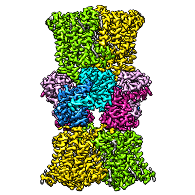
| Title | KtrAB complex | |||||||||
 Map data Map data | KtrAB complex of KtrA8 ring with KtrB dimer on each side | |||||||||
 Sample Sample |
| |||||||||
 Keywords Keywords | potassium transporter / membrane transport protein / MEMBRANE PROTEIN | |||||||||
| Function / homology |  Function and homology information Function and homology informationpotassium:chloride symporter activity / monoatomic cation transmembrane transporter activity / potassium ion transport / ATP binding / plasma membrane Similarity search - Function | |||||||||
| Biological species |  Vibrio alginolyticus (bacteria) Vibrio alginolyticus (bacteria) | |||||||||
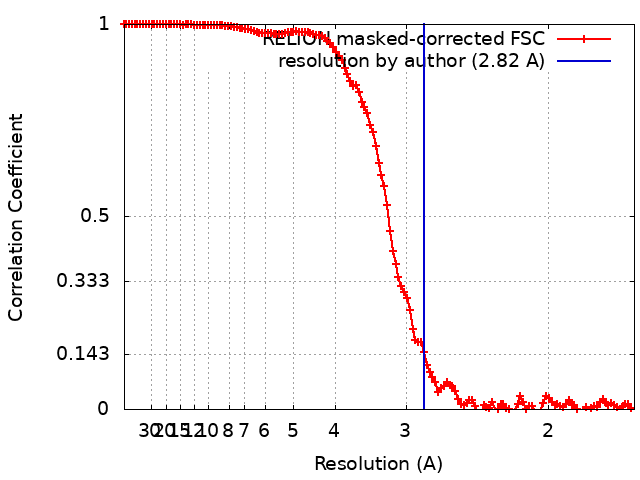
| Method | single particle reconstruction / cryo EM / Resolution: 2.82 Å | |||||||||
 Authors Authors | Vonck J / Stautz J | |||||||||
| Funding support |  Germany, 2 items Germany, 2 items
| |||||||||
 Citation Citation | Journal: To Be Published Title: KtrAB complex Authors: Stautz J | |||||||||
| History |
|
- Structure visualization
| Supplemental images |
|---|
- Downloads & links
-EMDB archive
| Map data | emd_14851.map.gz | 161.6 MB | EMDB map data format | |
|---|---|---|---|---|
| Header (meta data) | emd-14851-v30.xmlemd-14851.xml | 20.2 KB 20.2 KB | Display Display | EMDB header |
| FSC (resolution estimation) | emd_14851_fsc.xml | 12.8 KB | Display | FSC data file |
| Images |  emd_14851.png emd_14851.png | 176.9 KB | ||
| Masks | emd_14851_msk_1.map | 178 MB | Mask map | |
| Filedesc metadata | emd-14851.cif.gz | 6.4 KB | ||
| Others | emd_14851_additional_1.map.gzemd_14851_half_map_1.map.gzemd_14851_half_map_2.map.gz | 165.9 MB 139.5 MB 139.5 MB | ||
| Archive directory |  http://ftp.pdbj.org/pub/emdb/structures/EMD-14851ftp://ftp.pdbj.org/pub/emdb/structures/EMD-14851 http://ftp.pdbj.org/pub/emdb/structures/EMD-14851ftp://ftp.pdbj.org/pub/emdb/structures/EMD-14851 | HTTPS FTP |
-Related structure data
| Related structure data |  7zp9MC M: atomic model generated by this map C: citing same article ( |
|---|---|
| Similar structure data |


-Links
| EMDB pages | EMDB (EBI/PDBe) / EMDataResource |
|---|---|
| Related items in Molecule of the Month |

-Map
| File | Download / File: emd_14851.map.gz / Format: CCP4 / Size: 178 MB / Type: IMAGE STORED AS FLOATING POINT NUMBER (4 BYTES) | ||||||||||||||||||||||||||||||||||||
|---|---|---|---|---|---|---|---|---|---|---|---|---|---|---|---|---|---|---|---|---|---|---|---|---|---|---|---|---|---|---|---|---|---|---|---|---|---|
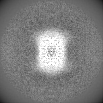
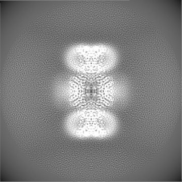
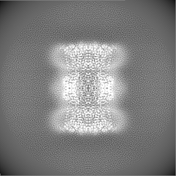
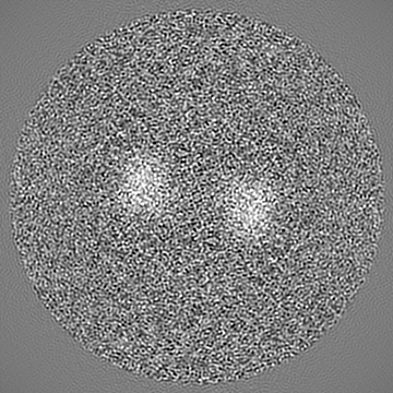
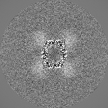
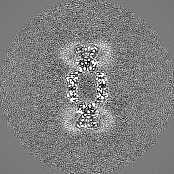
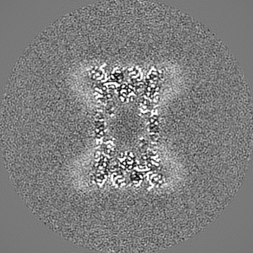
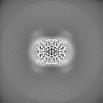
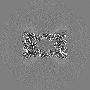
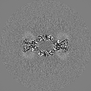
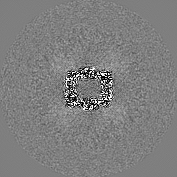
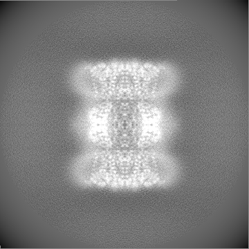
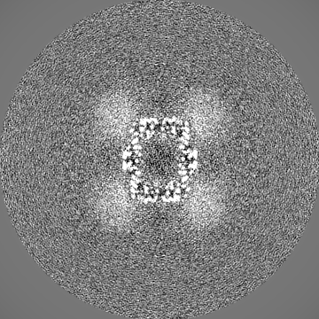
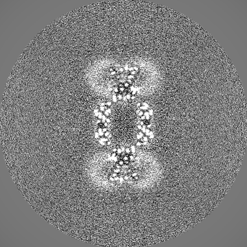
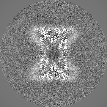
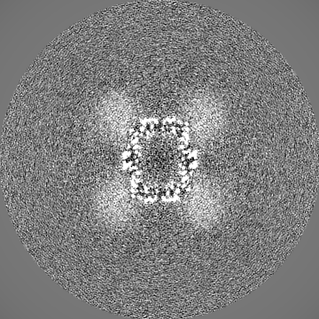
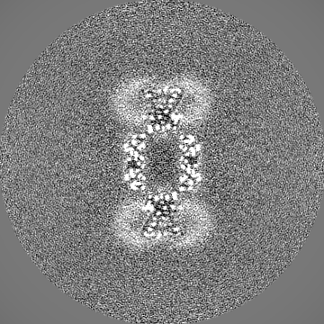
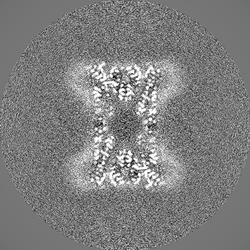
| Annotation | KtrAB complex of KtrA8 ring with KtrB dimer on each side | ||||||||||||||||||||||||||||||||||||
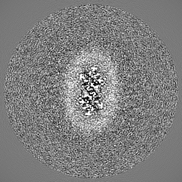
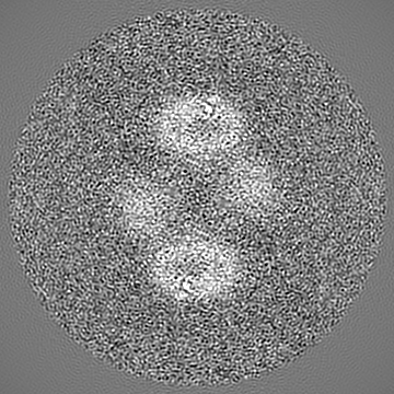
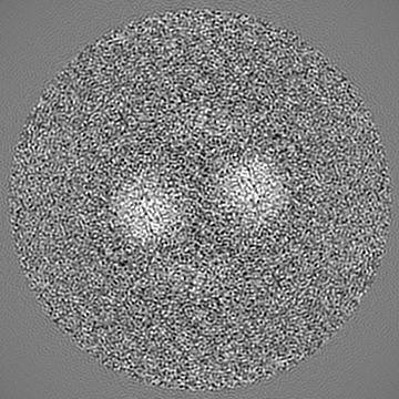
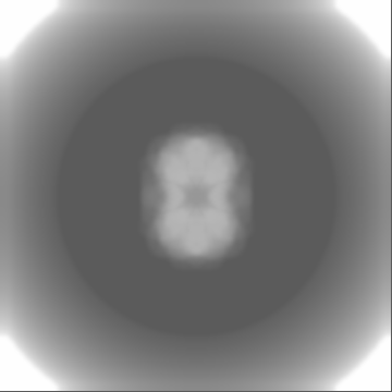
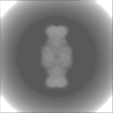
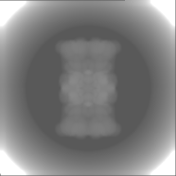
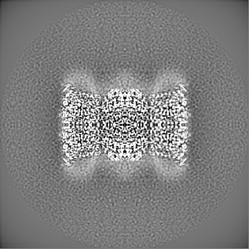
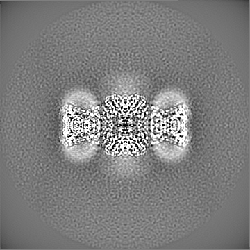
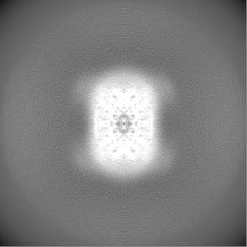
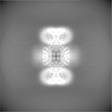
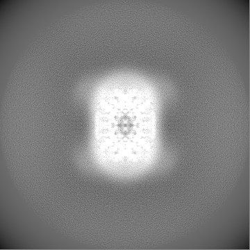
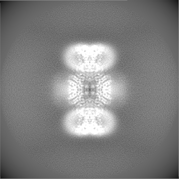
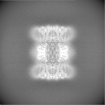
| Projections & slices | Image control
Images are generated by Spider. | ||||||||||||||||||||||||||||||||||||
| Voxel size | X=Y=Z: 0.831 Å | ||||||||||||||||||||||||||||||||||||
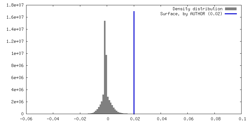
| Density |
| ||||||||||||||||||||||||||||||||||||
| Symmetry | Space group: 1 | ||||||||||||||||||||||||||||||||||||
| Details | EMDB XML:
|
 Z (Sec.)
 Y (Row.)
 X (Col.)


-Supplemental data
-Mask #1
| File | emd_14851_msk_1.map | ||||||||||||
|---|---|---|---|---|---|---|---|---|---|---|---|---|---|
| Projections & Slices |
| ||||||||||||
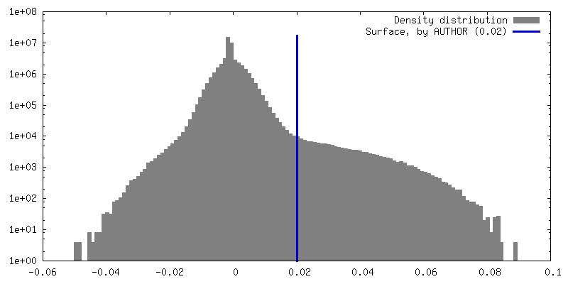
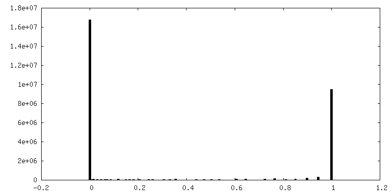
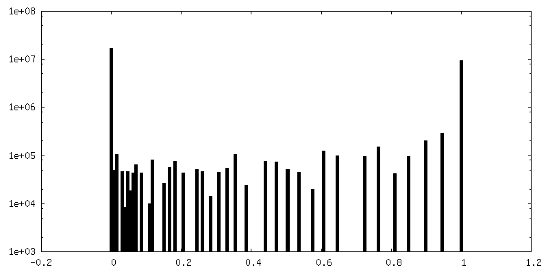
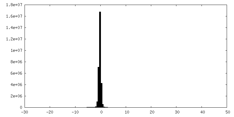
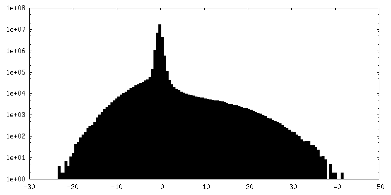
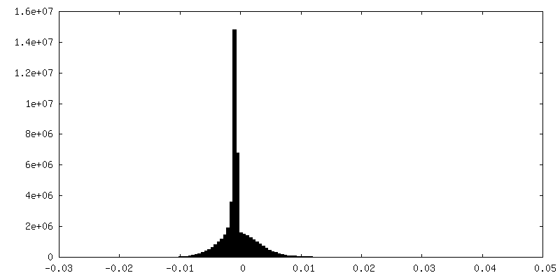
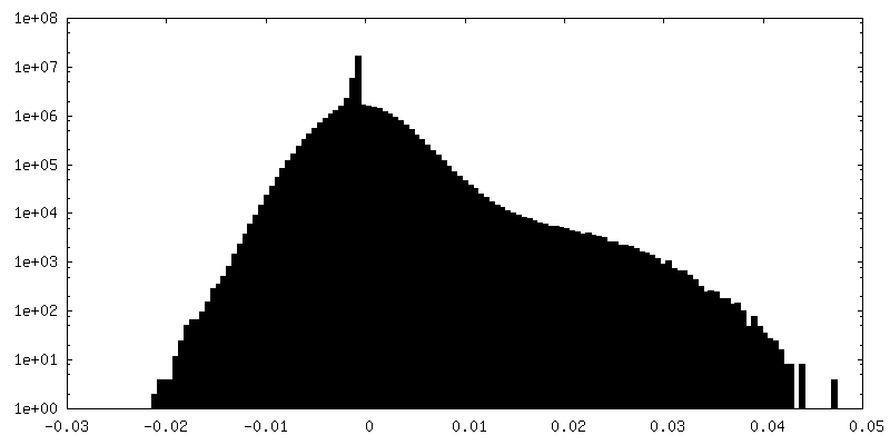
| Density Histograms |


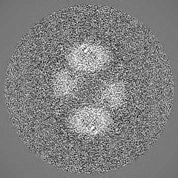
-Additional map: Map at 2.54 Angstrom resolution after density modification in Phenix
| File | emd_14851_additional_1.map | ||||||||||||
|---|---|---|---|---|---|---|---|---|---|---|---|---|---|
| Annotation | Map at 2.54 Angstrom resolution after density modification in Phenix | ||||||||||||
| Projections & Slices |
| ||||||||||||
| Density Histograms |
-Half map: #2
| File | emd_14851_half_map_1.map | ||||||||||||
|---|---|---|---|---|---|---|---|---|---|---|---|---|---|
| Projections & Slices |
| ||||||||||||
| Density Histograms |

-Half map: #1
| File | emd_14851_half_map_2.map | ||||||||||||
|---|---|---|---|---|---|---|---|---|---|---|---|---|---|
| Projections & Slices |
| ||||||||||||
| Density Histograms |

- Sample components
-Entire : KtrAB complex with a second KtrB dimer attached
| Entire | Name: KtrAB complex with a second KtrB dimer attached |
|---|---|
| Components |
|
-Supramolecule #1: KtrAB complex with a second KtrB dimer attached
| Supramolecule | Name: KtrAB complex with a second KtrB dimer attached / type: complex / ID: 1 / Parent: 0 / Macromolecule list: #1-#2 |
|---|---|
| Source (natural) | Organism: Vibrio alginolyticus (bacteria) |
| Molecular weight | Theoretical: 390 KDa |
-Macromolecule #1: Ktr system potassium uptake protein A
| Macromolecule | Name: Ktr system potassium uptake protein A / type: protein_or_peptide / ID: 1 / Number of copies: 8 / Enantiomer: LEVO |
|---|---|
| Source (natural) | Organism: Vibrio alginolyticus (bacteria) |
| Molecular weight | Theoretical: 23.83692 KDa |
| Recombinant expression | Organism: |
| Sequence | String: MKTGDKQFAV IGLGRFGLAV CKELQDSGSQ VLAVDINEDR VKEAAGFVSQ AIVANCTHEE TVAELKLDDY DMVMIAIGAD VNASILATL IAKEAGVKSV WVKANDRFQA RVLQKIGADH IIMPERDMGI RVARKMLDKR VLEFHPLGSG LAMTEFVVGS R LMGKTLSD ...String: MKTGDKQFAV IGLGRFGLAV CKELQDSGSQ VLAVDINEDR VKEAAGFVSQ AIVANCTHEE TVAELKLDDY DMVMIAIGAD VNASILATL IAKEAGVKSV WVKANDRFQA RVLQKIGADH IIMPERDMGI RVARKMLDKR VLEFHPLGSG LAMTEFVVGS R LMGKTLSD LALCKVEGVQ VLGYKRGPEI IKAPDMSTTL EIGDLIIVVG PQDKLANKLK SL UniProtKB: Ktr system potassium uptake protein A |
-Macromolecule #2: Ktr system potassium uptake protein B
| Macromolecule | Name: Ktr system potassium uptake protein B / type: protein_or_peptide / ID: 2 / Number of copies: 4 / Enantiomer: LEVO |
|---|---|
| Source (natural) | Organism: Vibrio alginolyticus (bacteria) |
| Molecular weight | Theoretical: 49.707715 KDa |
| Recombinant expression | Organism: |
| Sequence | String: MTQFHQRGVF YVPDGKRDKA KGGEPRIILL SFLGVLLPSA VLLTLPVFSV SGLSITDALF TATSAISVTG LGVVDTGQHF TLAGKILLM CLMQIGGLGQ MTLSAVLLYM FGVRLSLRQQ ALAKEALGQE RQVNLRRLVK KIVTFALVAE AIGFVFLSYR W VPEMGWQT ...String: MTQFHQRGVF YVPDGKRDKA KGGEPRIILL SFLGVLLPSA VLLTLPVFSV SGLSITDALF TATSAISVTG LGVVDTGQHF TLAGKILLM CLMQIGGLGQ MTLSAVLLYM FGVRLSLRQQ ALAKEALGQE RQVNLRRLVK KIVTFALVAE AIGFVFLSYR W VPEMGWQT GMFYALFHSI SAFNNAGFAL FSDSMMSFVN DPLVSFTLAG LFIFGGLGFT VIGDVWRHWR KGFHFLHIHT KI MLIATPL LLLVGTVLFW LLERHNPNTM GSLTTGGQWL AAFFQSASAR TAGFNSVDLT QFTQPALLIM IVLMLIGAGS TST GGGIKV STFAVAFMAT WTFLRQKKHV VMFKRTVNWP TVTKSLAIIV VSGAILTTAM FLLMLTEKAS FDKVMFETIS AFAT VGLTA GLTAELSEPG KYIMIVVMII GRIGPLTLAY MLARPEPTLI KYPEDTVLTG UniProtKB: Ktr system potassium uptake protein B |
-Macromolecule #3: MAGNESIUM ION
| Macromolecule | Name: MAGNESIUM ION / type: ligand / ID: 3 / Number of copies: 8 / Formula: MG |
|---|---|
| Molecular weight | Theoretical: 24.305 Da |
-Macromolecule #4: ADENOSINE-5'-DIPHOSPHATE
| Macromolecule | Name: ADENOSINE-5'-DIPHOSPHATE / type: ligand / ID: 4 / Number of copies: 8 / Formula: ADP |
|---|---|
| Molecular weight | Theoretical: 427.201 Da |
| Chemical component information |  ChemComp-ADP: |
-Macromolecule #5: DODECYL-BETA-D-MALTOSIDE
| Macromolecule | Name: DODECYL-BETA-D-MALTOSIDE / type: ligand / ID: 5 / Number of copies: 36 / Formula: LMT |
|---|---|
| Molecular weight | Theoretical: 510.615 Da |
| Chemical component information |  ChemComp-LMT: |
-Macromolecule #6: POTASSIUM ION
| Macromolecule | Name: POTASSIUM ION / type: ligand / ID: 6 / Number of copies: 4 / Formula: K |
|---|---|
| Molecular weight | Theoretical: 39.098 Da |
-Macromolecule #7: water
| Macromolecule | Name: water / type: ligand / ID: 7 / Number of copies: 43 / Formula: HOH |
|---|---|
| Molecular weight | Theoretical: 18.015 Da |
| Chemical component information |  ChemComp-HOH: |
-Experimental details
-Structure determination
| Method | cryo EM |
|---|---|
 Processing Processing | single particle reconstruction |
| Aggregation state | particle |
-Sample preparation
| Buffer | pH: 8 |
|---|---|
| Grid | Model: C-flat-1.2/1.3 / Material: COPPER / Pretreatment - Type: GLOW DISCHARGE |
| Vitrification | Cryogen name: ETHANE / Instrument: FEI VITROBOT MARK IV |
- Electron microscopy
| Microscope | FEI TITAN KRIOS |
|---|---|
| Image recording | Film or detector model: GATAN K3 BIOQUANTUM (6k x 4k) / Number grids imaged: 1 / Number real images: 2716 / Average electron dose: 40.0 e/Å2 |
| Electron beam | Acceleration voltage: 300 kV / Electron source:  FIELD EMISSION GUN FIELD EMISSION GUN |
| Electron optics | Calibrated defocus max: 2.5 µm / Calibrated defocus min: 1.0 µm / Calibrated magnification: 60168 / Illumination mode: FLOOD BEAM / Imaging mode: BRIGHT FIELD / Cs: 2.7 mm / Nominal defocus max: 2.3000000000000003 µm / Nominal defocus min: 1.2 µm / Nominal magnification: 105000 |
| Sample stage | Specimen holder model: FEI TITAN KRIOS AUTOGRID HOLDER / Cooling holder cryogen: NITROGEN |
| Experimental equipment |  Model: Titan Krios / Image courtesy: FEI Company |
+Image processing

-Atomic model buiding 1
| Details | The model was refined by phenix.real-space-refine |
|---|---|
| Refinement | Space: REAL / Protocol: OTHER |
| Output model | PDB-7zp9: |
