






 12EM
12EM
Crystal structure of a B1,3-Glucosyltransferase (B3GLCT) reveals an unusual mode of substrate recognition by a two-domain GT-A fold glycosyltransferase
Summary for 12EM
| Entry DOI | 10.2210/pdb12em/pdb |
| Descriptor | Beta-1,3-glucosyltransferase, Thrombospondin-1, MANGANESE (II) ION, ... (9 entities in total) |
| Functional Keywords | o-fucosylation, glycosyltransferase, peters plus syndrome, thrombospondin type 1 repeats, carbohydrate |
| Biological source | Homo sapiens (human) More |
| Total number of polymer chains | 2 |
| Total formula weight | 56938.39 |
| Authors | Berardinelli, S.J.,Kadirvelraj, R.,Wood, Z.A. (deposition date: 2026-03-30, release date: 2026-08-26) |
| Primary citation | Berardinelli, S.J.,Kadirvelraj, R.,Luther, K.B.,Gao, Z.,Chapla, D.,Huang, C.,Tehrani, D.M.,Zhang, A.,Moremen, K.W.,Wood, Z.A.,Haltiwanger, R.S. Crystal structure of a beta 1,3-Glucosyltransferase reveals an unusual substrate recognition by a two-domain GT-A fold glycosyltransferase. J.Biol.Chem., :113418-113418, 2026 Cited by PubMed Abstract: β1,3-Glucosyltransferase (B3GLCT) adds a glucose onto O-linked fucose on thrombospondin type 1 repeats (TSRs). Protein O-fucosyltransferase 2 (POFUT2) first transfers a fucose to properly folded TSRs containing a consensus sequence for O-fucosylation. This uncommon O-fucose modification is then extended to a glucose-fucose disaccharide by B3GLCT. B3GLCT is a GT-A fold glycosyltransferase and pathogenic variants cause Peters Plus Syndrome (PTRPLS, OMIM #261540), a Congenital Disorder of Glycosylation (CDG). Most GT-A fold family members have a single GT-A domain, but B3GLCT contains an additional GT-A domain. To assess the function of the additional GT-A domain and binding of TSR substrates, we determined the crystal structure of an O-fucosylated TSR (Fuc-O-TSR3) from Thrombospondin-1 bound to B3GLCT. The additional GT-A domain is essential for substrate binding but is catalytically inactive: it does not bind UDP or Mn and is not a β1,3-glucosyltransferase. It also creates a deep pocket with a vestigial active site located on the opposite face of the Fuc-O-TSR3 substrate binding site. The Fuc-O-TSR3 acceptor substrate binds in a cleft between the two GT-A domains, each of which has evolved hypervariable regions for Fuc-O-TSR recognition. This structure is an unusual example of a glycosyltransferase with a catalytically inactive extra GT-A domain, providing insight into the binding of B3GLCT's diverse Fuc-O-TSR substrates. We also discuss how B3GLCT mimics the two-domain structure of GT-B fold glycosyltransferases. All GT-B fold glycosyltransferases contain two bilobal Rossmann-like fold domains, like B3GLCT. Our data also explains how PTRPLS-associated variants and predicted pathogenic mutations disrupt B3GLCT function. PubMed: 42575440DOI: 10.1016/j.jbc.2026.113418 PDB entries with the same primary citation |
| Experimental method | X-RAY DIFFRACTION (2.07 Å) |
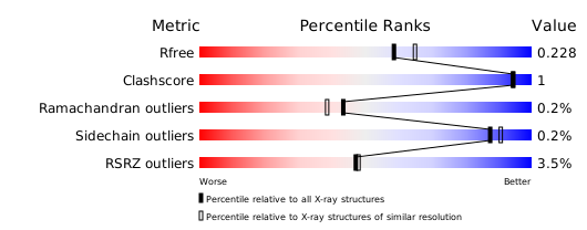
Structure validation
Download full validation report


