






 10GI
10GI
Structure of Glyceraldehyde-3-Phosphate Dehydrogenase from Babesia bovis
Summary for 10GI
| Entry DOI | 10.2210/pdb10gi/pdb |
| Descriptor | Glyceraldehyde-3-phosphate dehydrogenase, NICOTINAMIDE-ADENINE-DINUCLEOTIDE (3 entities in total) |
| Functional Keywords | bovine babesiosis, babesia bovis, gapdh, structural genomics, seattle structural genomics center for infectious disease, ssgcid, oxidoreductase |
| Biological source | Babesia bovis |
| Total number of polymer chains | 2 |
| Total formula weight | 78334.42 |
| Authors | Ukrainski, B.,Galvao, E.B.,Silva, M.,Iulek, J.,Seattle Structural Genomics Center for Infectious Disease (SSGCID) (deposition date: 2026-01-19, release date: 2026-08-19, Last modification date: 2026-08-26) |
| Primary citation | Ukrainski, B.,Galvao, E.B.,Silva, M.,Iulek, J. Expression, purification, crystallization and structure solution of glyceraldehyde-3-phosphate dehydrogenase from the babesiosis infective agent Babesia bovis. Protein Expr.Purif., 243:106991-106991, 2026 Cited by PubMed Abstract: Babesia bovis is a species of apicomplexan hemoparasitic protozoa that can be transmitted by ticks, causing a global cattle disease. As it depends mainly on the glycolytic pathway for energy production and life cycle maintenance, glycolytic enzymes are possible targets for drug development against Babesia. Glyceraldehyde-3-Phosphate Dehydrogenase (GAPDH) has been one of such targets, against several parasitic organisms. It performs the reversible oxidative phosphorylation of glyceraldehyde-3-phosphate to 1,3-bisphospho-D-glycerate in the presence of nicotinamide adenine dinucleotide. The protocol for Babesia bovis Glyceraldehyde-3-Phosphate Dehydrogenase (BbGAPDH) expression and purification has been developed to yields of 28 mg of pure protein per liter of culture medium, with a specific activity of 55.5 ± 6.99 U mg after His-tag removal, indicating preservation of enzymatic activity. For crystallization, the His-tag removal proved essential. Crystals diffracted to 3.12 Å resolution in the P321 space group; NAD cofactor molecules are observed in their respective sites. Comparisons to the theoretical model indicate a number of side chain conformation differences. These results provide support for future enzyme inhibition assays, in addition to crystallization assays with potential inhibitors. PubMed: 42600714DOI: 10.1016/j.pep.2026.106991 PDB entries with the same primary citation |
| Experimental method | X-RAY DIFFRACTION (3.12 Å) |
Structure validation
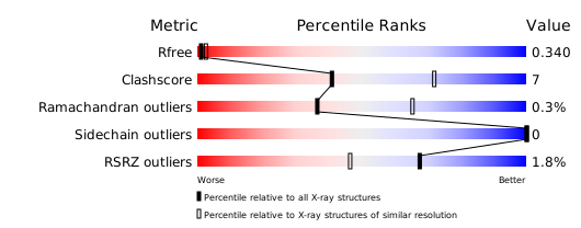
Download full validation report


