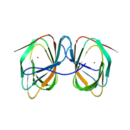
9JEW
 
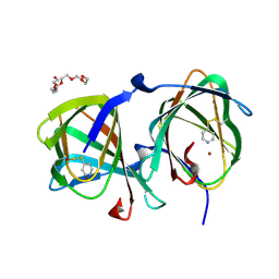
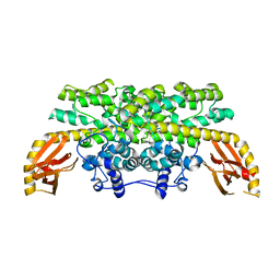
 | | Crystal structure of a cupin protein (tm1459, C106V mutant) in iron (Fe) substituted form | | Descriptor: | Cupin type-2 domain-containing protein, FE (III) ION | | Authors: | Fujieda, N, Ichihashi, H, Kurisu, G, Itoh, S. | | Deposit date: | 2024-09-03 | | Release date: | 2025-05-07 | | Last modified: | 2025-07-02 | | Method: | X-RAY DIFFRACTION (1.08 Å) | | Cite: | Unusual Self-Hydroxylation in 4-Histidine Tetrad-Supporting Nonheme Iron Center.
Chem Asian J, 20, 2025
|
|
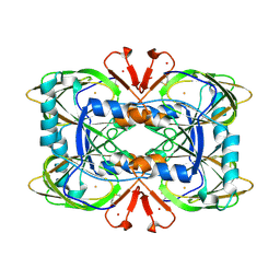
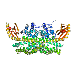
9JEV
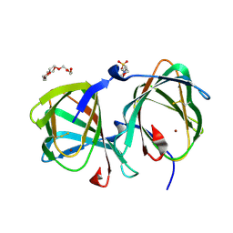
 | | Crystal structure of a cupin protein (tm1459) in zinc (Zn) substituted form | | Descriptor: | Cupin type-2 domain-containing protein, ZINC ION | | Authors: | Fujieda, N, Ichihashi, H, Kurisu, G, Itoh, S. | | Deposit date: | 2024-09-03 | | Release date: | 2025-05-07 | | Last modified: | 2025-07-02 | | Method: | X-RAY DIFFRACTION (1.12 Å) | | Cite: | Unusual Self-Hydroxylation in 4-Histidine Tetrad-Supporting Nonheme Iron Center.
Chem Asian J, 20, 2025
|
|
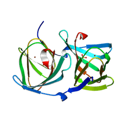
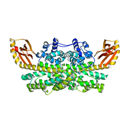
9JEU
 | | Crystal structure of a cupin protein (tm1459) in iron (Fe) substituted form | | Descriptor: | Cupin type-2 domain-containing protein, FE (III) ION | | Authors: | Fujieda, N, Ichihashi, H, Kurisu, G, Itoh, S. | | Deposit date: | 2024-09-03 | | Release date: | 2025-05-07 | | Last modified: | 2025-07-02 | | Method: | X-RAY DIFFRACTION (1.19 Å) | | Cite: | Unusual Self-Hydroxylation in 4-Histidine Tetrad-Supporting Nonheme Iron Center.
Chem Asian J, 20, 2025
|
|
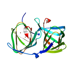
9JET
 | | Crystal structure of a cupin protein (tm1459) in manganese (Mn) substituted form | | Descriptor: | Cupin type-2 domain-containing protein, MANGANESE (II) ION | | Authors: | Fujieda, N, Ichihashi, H, Kurisu, G, Itoh, S. | | Deposit date: | 2024-09-03 | | Release date: | 2025-05-07 | | Last modified: | 2025-07-02 | | Method: | X-RAY DIFFRACTION (1.19 Å) | | Cite: | Unusual Self-Hydroxylation in 4-Histidine Tetrad-Supporting Nonheme Iron Center.
Chem Asian J, 20, 2025
|
|
8ZYH
 | | Crystal structure of a cupin protein (tm1459, I49C-4py/H52A/H54A/C106D mutant) in copper (Cu) substituted form | | Descriptor: | 4-THIOPYRIDINE, COPPER (II) ION, Cupin type-2 domain-containing protein, ... | | Authors: | Morita, Y, Kubo, H, Matsumoto, R, Fujieda, N. | | Deposit date: | 2024-06-17 | | Release date: | 2024-09-04 | | Last modified: | 2025-03-19 | | Method: | X-RAY DIFFRACTION (1.07 Å) | | Cite: | A thiopyridine-bound mirror-image copper center in an artificial non-heme metalloenzyme.
J.Inorg.Biochem., 260, 2024
|
|
8ZYG
 | | Crystal structure of a cupin protein (tm1459, I49C-4py/H52A/C106D mutant) in copper (Cu) substituted form | | Descriptor: | 2-(N-MORPHOLINO)-ETHANESULFONIC ACID, COPPER (II) ION, Cupin type-2 domain-containing protein, ... | | Authors: | Morita, Y, Kubo, H, Matsumoto, R, Fujieda, N. | | Deposit date: | 2024-06-17 | | Release date: | 2024-09-04 | | Last modified: | 2025-03-19 | | Method: | X-RAY DIFFRACTION (1.08 Å) | | Cite: | A thiopyridine-bound mirror-image copper center in an artificial non-heme metalloenzyme.
J.Inorg.Biochem., 260, 2024
|
|
8OZ8
 | | Crystal Structure of an Hydroxynitrile lyase variant (H96A) from Granulicella tundricola | | Descriptor: | 1-METHOXY-2-[2-(2-METHOXY-ETHOXY]-ETHANE, 3-CARBOXY-N,N,N-TRIMETHYLPROPAN-1-AMINIUM, BROMIDE ION, ... | | Authors: | Bento, I, Coloma, J, Hagedoorn, P.-L, Hanefeld, U. | | Deposit date: | 2023-05-08 | | Release date: | 2024-04-10 | | Method: | X-RAY DIFFRACTION (1.85 Å) | | Cite: | Can a Hydroxynitrile Lyase Catalyze an Oxidative Cleavage?
Acs Catalysis, 13, 2023
|
|
8HLF
 | | Crystal structure of DddK-DMSOP complex | | Descriptor: | 3-[dimethyl(oxidanyl)-$l^{4}-sulfanyl]propanoic acid, MANGANESE (II) ION, Novel protein with potential Cupin domain | | Authors: | Peng, M, Li, C.Y, Zhang, Y.Z. | | Deposit date: | 2022-11-30 | | Release date: | 2023-10-04 | | Last modified: | 2023-12-20 | | Method: | X-RAY DIFFRACTION (1.62 Å) | | Cite: | DMSOP-cleaving enzymes are diverse and widely distributed in marine microorganisms.
Nat Microbiol, 8, 2023
|
|
8E8W
 | |
8CH4
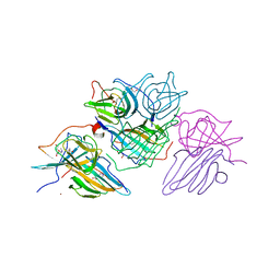
 | | Crystal structure of the ring cleaving dioxygenase 5-nitrosalicylate 1,2-dioxygenase from Bradyrhizobium sp. | | Descriptor: | 5-nitrosalicylic acid 1,2-dioxygenase, FE (III) ION | | Authors: | Ferraroni, M, Stolz, A, Eppinger, E. | | Deposit date: | 2023-02-07 | | Release date: | 2023-07-05 | | Last modified: | 2024-11-13 | | Method: | X-RAY DIFFRACTION (2.1 Å) | | Cite: | Crystal structure of the monocupin ring-cleaving dioxygenase 5-nitrosalicylate 1,2-dioxygenase from Bradyrhizobium sp.
Acta Crystallogr D Struct Biol, 79, 2023
|
|
8HJZ
 | |
8HJX
 | |
8HJY
 | |
7ZYB
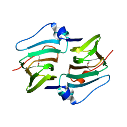
 | | BeKdgF with Ca | | Descriptor: | CALCIUM ION, Cupin, GLYCEROL | | Authors: | Fredslund, F, Teze, D, Welner, D.H. | | Deposit date: | 2022-05-24 | | Release date: | 2023-06-14 | | Last modified: | 2024-02-07 | | Method: | X-RAY DIFFRACTION (1.5 Å) | | Cite: | BeKdgF with Ca
To Be Published
|
|
7ZYC
 | | BeKdgF with Zn | | Descriptor: | Cupin, GLYCEROL, ZINC ION | | Authors: | Fredslund, F, Teze, D, Welner, D.H. | | Deposit date: | 2022-05-24 | | Release date: | 2023-06-14 | | Last modified: | 2024-02-07 | | Method: | X-RAY DIFFRACTION (2 Å) | | Cite: | BeKdgF with Ca
To Be Published
|
|
8AWN
 | | Crystal structure of a manganese-containing cupin (tm1459) from Thermotoga maritima, variant C106Q | | Descriptor: | CHLORIDE ION, Cupin_2 domain-containing protein | | Authors: | Grininger, C, Steiner, K, Gruber, K, Pavkov-Keller, T. | | Deposit date: | 2022-08-30 | | Release date: | 2023-03-08 | | Last modified: | 2024-02-07 | | Method: | X-RAY DIFFRACTION (1.45 Å) | | Cite: | Engineering TM1459 for Stabilisation against Inactivation by Amino Acid Oxidation
Chem Ing Tech, 2023
|
|
8AWP
 | |
8AWO
 | |
8HFB
 | | Evolved variant of quercetin 2,4-dioxygenase from Bacillus subtilis | | Descriptor: | 1,2-ETHANEDIOL, GLYCEROL, NICKEL (II) ION, ... | | Authors: | Eom, H, Song, W.J. | | Deposit date: | 2022-11-10 | | Release date: | 2023-03-08 | | Last modified: | 2024-05-22 | | Method: | X-RAY DIFFRACTION (2.24 Å) | | Cite: | Underlying Role of Hydrophobic Environments in Tuning Metal Elements for Efficient Enzyme Catalysis.
J.Am.Chem.Soc., 145, 2023
|
|
7ZVM
 | |
7ZVY
 | | Thermococcus kadokarensis phosphomannose isomerase | | Descriptor: | Cupin_2 domain-containing protein, ZINC ION | | Authors: | Hoh, f, Calio, A. | | Deposit date: | 2022-05-17 | | Release date: | 2022-08-24 | | Last modified: | 2024-05-01 | | Method: | X-RAY DIFFRACTION (2.16 Å) | | Cite: | Unravelling the Adaptation Mechanisms to High Pressure in Proteins.
Int J Mol Sci, 23, 2022
|
|
6VZY
 | |
6XCV
 | |
6L2F
 | | Crystal structure of a cupin protein (tm1459, H54AH58A mutant) in copper (Cu) substituted form | | Descriptor: | 2-(N-MORPHOLINO)-ETHANESULFONIC ACID, 3,6,9,12,15,18-HEXAOXAICOSANE-1,20-DIOL, COPPER (II) ION, ... | | Authors: | Fujieda, N, Ichihashi, H, Nishikawa, Y, Kurisu, G, Itoh, S. | | Deposit date: | 2019-10-03 | | Release date: | 2020-04-01 | | Last modified: | 2024-10-30 | | Method: | X-RAY DIFFRACTION (1.23 Å) | | Cite: | Cupin Variants as a Macromolecular Ligand Library for Stereoselective Michael Addition of Nitroalkanes.
Angew.Chem.Int.Ed.Engl., 59, 2020
|
|
6L2D
 | | Crystal structure of a cupin protein (tm1459) in copper (Cu) substituted form | | Descriptor: | COPPER (II) ION, Cupin_2 domain-containing protein | | Authors: | Fujieda, N, Ichihashi, H, Nishikawa, Y, Kurisu, G, Itoh, S. | | Deposit date: | 2019-10-03 | | Release date: | 2020-04-01 | | Last modified: | 2024-11-20 | | Method: | X-RAY DIFFRACTION (1.198 Å) | | Cite: | Cupin Variants as a Macromolecular Ligand Library for Stereoselective Michael Addition of Nitroalkanes.
Angew.Chem.Int.Ed.Engl., 59, 2020
|
|


