100D
 
 | |
101M
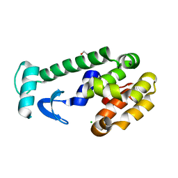
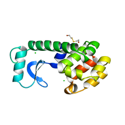
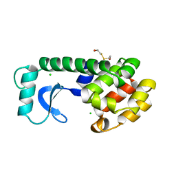
 | | SPERM WHALE MYOGLOBIN F46V N-BUTYL ISOCYANIDE AT PH 9.0 | | Descriptor: | MYOGLOBIN, N-BUTYL ISOCYANIDE, PROTOPORPHYRIN IX CONTAINING FE, ... | | Authors: | Smith, R.D, Olson, J.S, Phillips Jr, G.N. | | Deposit date: | 1997-12-13 | | Release date: | 1998-04-08 | | Last modified: | 2024-04-03 | | Method: | X-RAY DIFFRACTION (2.07 Å) | | Cite: | Correlations between Bound N-Alkyl Isocyanide Orientations and Pathways for Ligand Binding in Recombinant Myoglobins
Thesis, Rice, 1999
|
|
102D
 | |
102L
 | |
102M
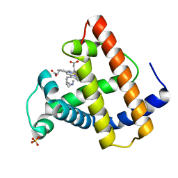
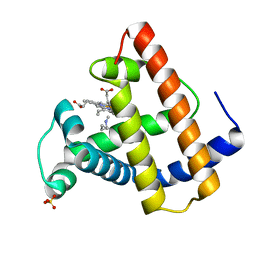
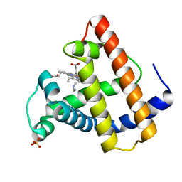
 | | SPERM WHALE MYOGLOBIN H64A AQUOMET AT PH 9.0 | | Descriptor: | MYOGLOBIN, PROTOPORPHYRIN IX CONTAINING FE, SULFATE ION | | Authors: | Smith, R.D, Olson, J.S, Phillips Jr, G.N. | | Deposit date: | 1997-12-15 | | Release date: | 1998-04-08 | | Last modified: | 2024-04-03 | | Method: | X-RAY DIFFRACTION (1.84 Å) | | Cite: | Correlations between Bound N-Alkyl Isocyanide Orientations and Pathways for Ligand Binding in Recombinant Myoglobins
Thesis, Rice, 1999
|
|
103L
 | |
103M
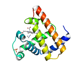
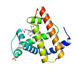
 | | SPERM WHALE MYOGLOBIN H64A N-BUTYL ISOCYANIDE AT PH 9.0 | | Descriptor: | MYOGLOBIN, N-BUTYL ISOCYANIDE, PROTOPORPHYRIN IX CONTAINING FE, ... | | Authors: | Smith, R.D, Olson, J.S, Phillips Jr, G.N. | | Deposit date: | 1997-12-16 | | Release date: | 1998-04-08 | | Last modified: | 2024-04-03 | | Method: | X-RAY DIFFRACTION (2.07 Å) | | Cite: | Correlations between Bound N-Alkyl Isocyanide Orientations and Pathways for Ligand Binding in Recombinant Myoglobins
Thesis, Rice, 1999
|
|
104L
 | |
104M
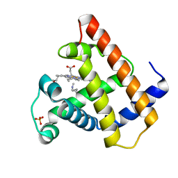
 | | SPERM WHALE MYOGLOBIN N-BUTYL ISOCYANIDE AT PH 7.0 | | Descriptor: | MYOGLOBIN, N-BUTYL ISOCYANIDE, PROTOPORPHYRIN IX CONTAINING FE, ... | | Authors: | Smith, R.D, Olson, J.S, Phillips Jr, G.N. | | Deposit date: | 1997-12-18 | | Release date: | 1998-04-08 | | Last modified: | 2024-04-03 | | Method: | X-RAY DIFFRACTION (1.71 Å) | | Cite: | Correlations between Bound N-Alkyl Isocyanide Orientations and Pathways for Ligand Binding in Recombinant Myoglobins
Thesis, Rice, 1999
|
|
105M
 | | SPERM WHALE MYOGLOBIN N-BUTYL ISOCYANIDE AT PH 9.0 | | Descriptor: | MYOGLOBIN, N-BUTYL ISOCYANIDE, PROTOPORPHYRIN IX CONTAINING FE, ... | | Authors: | Smith, R.D, Olson, J.S, Phillips Jr, G.N. | | Deposit date: | 1997-12-18 | | Release date: | 1998-04-08 | | Last modified: | 2024-04-03 | | Method: | X-RAY DIFFRACTION (2.02 Å) | | Cite: | Correlations between Bound N-Alkyl Isocyanide Orientations and Pathways for Ligand Binding in Recombinant Myoglobins
Thesis, Rice, 1999
|
|
106M
 | | SPERM WHALE MYOGLOBIN V68F ETHYL ISOCYANIDE AT PH 9.0 | | Descriptor: | ETHYL ISOCYANIDE, MYOGLOBIN, PROTOPORPHYRIN IX CONTAINING FE, ... | | Authors: | Smith, R.D, Olson, J.S, Phillips Jr, G.N. | | Deposit date: | 1997-12-21 | | Release date: | 1998-04-08 | | Last modified: | 2024-04-03 | | Method: | X-RAY DIFFRACTION (1.99 Å) | | Cite: | Correlations between Bound N-Alkyl Isocyanide Orientations and Pathways for Ligand Binding in Recombinant Myoglobins
Thesis, Rice, 1999
|
|
107L
 | |
107M
 | | SPERM WHALE MYOGLOBIN V68F N-BUTYL ISOCYANIDE AT PH 9.0 | | Descriptor: | MYOGLOBIN, N-BUTYL ISOCYANIDE, PROTOPORPHYRIN IX CONTAINING FE, ... | | Authors: | Smith, R.D, Olson, J.S, Phillips Jr, G.N. | | Deposit date: | 1997-12-22 | | Release date: | 1998-04-08 | | Last modified: | 2024-04-03 | | Method: | X-RAY DIFFRACTION (2.09 Å) | | Cite: | Correlations between Bound N-Alkyl Isocyanide Orientations and Pathways for Ligand Binding in Recombinant Myoglobins
Thesis, Rice, 1999
|
|
108L
 | |
108M
 | | SPERM WHALE MYOGLOBIN V68F N-BUTYL ISOCYANIDE AT PH 7.0 | | Descriptor: | MYOGLOBIN, N-BUTYL ISOCYANIDE, PROTOPORPHYRIN IX CONTAINING FE, ... | | Authors: | Smith, R.D, Olson, J.S, Phillips Jr, G.N. | | Deposit date: | 1997-12-23 | | Release date: | 1998-05-20 | | Last modified: | 2024-04-03 | | Method: | X-RAY DIFFRACTION (2.67 Å) | | Cite: | Correlations between Bound N-Alkyl Isocyanide Orientations and Pathways for Ligand Binding in Recombinant Myoglobins
Thesis, Rice, 1999
|
|
109D
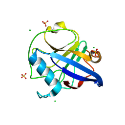
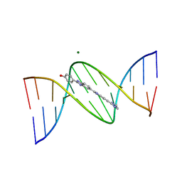 | | VARIABILITY IN DNA MINOR GROOVE WIDTH RECOGNISED BY LIGAND BINDING: THE CRYSTAL STRUCTURE OF A BIS-BENZIMIDAZOLE COMPOUND BOUND TO THE DNA DUPLEX D(CGCGAATTCGCG)2 | | Descriptor: | 5-(2-IMIDAZOLINYL)-2-[2-(4-HYDROXYPHENYL)-5-BENZIMIDAZOLYL]BENZIMIDAZOLE, DNA (5'-D(*CP*GP*CP*GP*AP*AP*TP*TP*CP*GP*CP*G)-3'), MAGNESIUM ION | | Authors: | Czarny, A, Boykin, D.W, Wood, A.A, Nunn, C.M, Neidle, S, Zhao, M, Wilson, W.D. | | Deposit date: | 1995-02-15 | | Release date: | 1995-05-08 | | Last modified: | 2024-02-07 | | Method: | X-RAY DIFFRACTION (2 Å) | | Cite: | Variability in DNA minor groove width recognised by ligand binding: the crystal structure of a bis-benzimidazole compound bound to the DNA duplex d(CGCGAATTCGCG)2.
Nucleic Acids Res., 23, 1995
|
|
109L
 | |
109M
 | | SPERM WHALE MYOGLOBIN D122N ETHYL ISOCYANIDE AT PH 9.0 | | Descriptor: | ETHYL ISOCYANIDE, MYOGLOBIN, PROTOPORPHYRIN IX CONTAINING FE, ... | | Authors: | Smith, R.D, Olson, J.S, Phillips Jr, G.N. | | Deposit date: | 1997-12-22 | | Release date: | 1998-04-08 | | Last modified: | 2024-04-03 | | Method: | X-RAY DIFFRACTION (1.83 Å) | | Cite: | Correlations between Bound N-Alkyl Isocyanide Orientations and Pathways for Ligand Binding in Recombinant Myoglobins
Thesis, Rice, 1999
|
|
10AF
 | |
10AH
 | |
10AI
 | |
10AJ
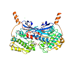
 | | Crystal Structure of Human WRN helicase with compound 1 | | Descriptor: | 1,2-ETHANEDIOL, 4-(4-methyl-4H-1,2,4-triazol-3-yl)-1-[4-(1H-pyrrol-1-yl)benzene-1-sulfonyl]piperidine, Bifunctional 3'-5' exonuclease/ATP-dependent helicase WRN, ... | | Authors: | Toms, A.V, Caravella, J.A, Sitnikov, N, Bartels, F, Svensson, R, Jacques O'Hagan, S, Borthwick, J, Campos, S, Yin, Y, Zhao, X, Li, L, Talbot, E, Kong, H, Freund, R.R.A, Browning, B, Genung, N.E, Carreiro, S, Brennan, D, Graves, A.P, Loh, C, Tummino, P, Edmonson, S.E, Li, D. | | Deposit date: | 2026-01-08 | | Release date: | 2026-05-13 | | Last modified: | 2026-06-10 | | Method: | X-RAY DIFFRACTION (2.42 Å) | | Cite: | Design of Cyclic Vinyl Sulfones as WRN Covalent Inhibitors from Noncovalent Binders.
J.Med.Chem., 69, 2026
|
|
10AK
 | | Crystal Structure of Human WRN helicase with compound 4 | | Descriptor: | 1,2-ETHANEDIOL, Bifunctional 3'-5' exonuclease/ATP-dependent helicase WRN, CHLORIDE ION, ... | | Authors: | Toms, A.V, Caravella, J.A, Sitnikov, N, Bartels, F, Svensson, R, Jacques O'Hagan, S, Borthwick, J, Yin, Y, Zhoa, X, Li, L, Liu, R, Talbot, E, Kong, H, Freund, R.R.A, Browning, B, Genung, N, Carreiro, S, Brennan, D, Graves, A.P, Loh, C, Tummino, P, Edmondson, S.D, Li, D. | | Deposit date: | 2026-01-08 | | Release date: | 2026-05-13 | | Last modified: | 2026-06-10 | | Method: | X-RAY DIFFRACTION (1.37 Å) | | Cite: | Design of Cyclic Vinyl Sulfones as WRN Covalent Inhibitors from Noncovalent Binders.
J.Med.Chem., 69, 2026
|
|
10AP
 | | Crystal Structure of Human WRN helicase with compound 26 | | Descriptor: | (2R)-N-[(3R)-1,1-dioxo-1lambda~6~-thiolan-3-yl]-N-{[2-(2-hydroxypropan-2-yl)pyridin-4-yl]methyl}-2-methoxy-2-[(1M)-3,3',4'-trifluoro[1,1'-biphenyl]-4-yl]acetamide, 1,2-ETHANEDIOL, ADENOSINE-5'-TRIPHOSPHATE, ... | | Authors: | Toms, A.V, Caravella, J.A, Sitnikov, N, Bartels, F, Svensson, R, Jacques O'Hagan, S, Borthwick, J, Campos, S, Yin, Y, Zhao, X, Li, L, Talbot, E, Kong, H, Freund, R.R.A, Browning, B, Genung, N.E, Carreiro, S, Brennan, D, Graves, A.P, Loh, C, Tummino, P, Edmonson, S.D, Li, D. | | Deposit date: | 2026-01-08 | | Release date: | 2026-05-13 | | Last modified: | 2026-06-10 | | Method: | X-RAY DIFFRACTION (2.58 Å) | | Cite: | Design of Cyclic Vinyl Sulfones as WRN Covalent Inhibitors from Noncovalent Binders.
J.Med.Chem., 69, 2026
|
|
10BL
 | |


